Open treatment
1. Emergency treatment
Emergency treatment in midfacial fractures may be indicated in the following cases:
- Partial or complete visual loss due to direct or indirect optic nerve trauma
- Severely increased intraocular pressure
- Acute space-occupying lesion (eg, retrobulbar hematoma, emphysema)
- Severe shift of orbital content
- Entrapment of eye muscle (particularly in pediatric patients)
- Severe nasal and/or oral bleeding
Globe rupture and intraocular trauma
There are a variety of possible injuries affecting the globe that may require immediate ophthalmological treatment.
Retrobulbar hematoma
Pressure increase in the periorbital region due to retrobulbar hematoma can cause significant injury of the neurovascular structures with eventual loss of vision.
More details on retrobulbar hemmorage can be found here.
Note: Intraorbital bleeding in patients taking anticoagulation drugs requires special attention.
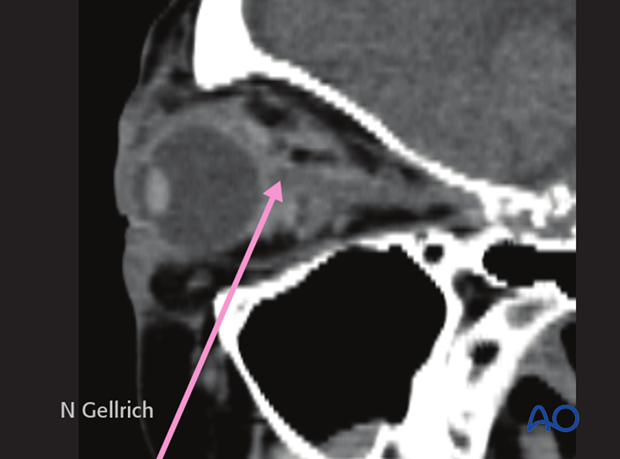
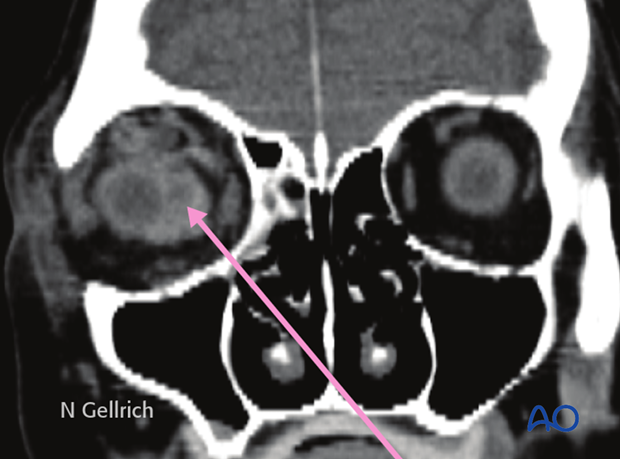
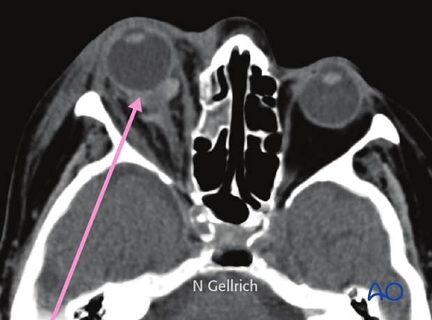
If a retrobulbar hematoma leads to a tense, proptotic globe, emergency decompression should be considered.
If a retrobulbar hematoma in the cooperative patient results in blindness, the time window to release the intraorbital pressure is limited to around one hour measured from the onset of blindness. This could mean urgent treatment under local anesthesia even in the emergency room prior to further imaging.
Transcutaneous transseptal incisions may help evacuate the hematoma and release the periorbital pressure. Alternative methods such as transconjunctival pressure release and/or lateral canthotomy and inferior cantholysis should be considered according to patient condition.
An exception may be where there is a pulsating exophthalmos which may be a sign of carotid-cavernous sinus fistula. A fistula of this nature requires appropriate preoperative imaging.
Note: retrobulbar hematoma is one of the most severe postoperative complications in patients who have undergone orbital trauma and/or surgery. This is one reason why the surgeon has to assess appropriate vision as soon as possible after injury and/or surgery.
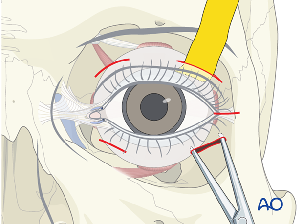
Emphysema
Severe emphysema might significantly raise intraorbital pressure. If this compromises visual function or endangers the orbital contents, orbital decompression has to be considered. Drug protocol may include antibiotics and decongestive nasal drops.
Patients with sinus fractures in the periorbital region should not blow their nose in order to avoid additional emphysema due to acute pressure rise. Should sneezing occur, maintaining an open mouth posture minimizes increasing intranasal/intrasinus pressure.
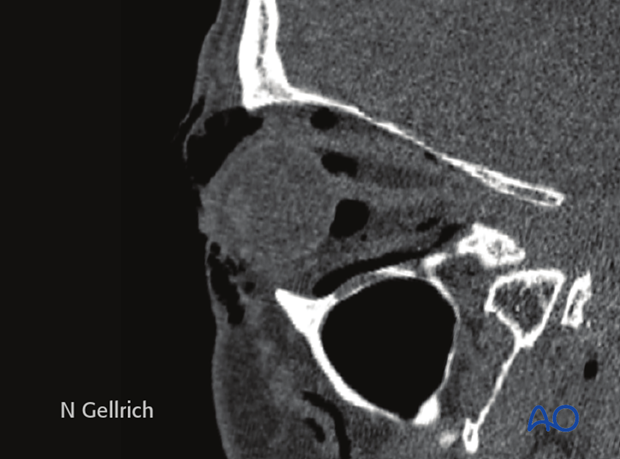
Usually there is no need for emergency treatment in orbital floor/medial wall fractures unless there is severe ongoing hemorrhage in the orbital cavity, the paranasal, or nasal cavity.
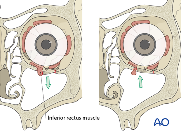
In some younger patients, the so-called trap-door phenomenon can occur in which there is danger of necrosis of the entrapped rectus muscle within a few hours; immediate release of entrapped tissues is necessary.
Entrapment of eye muscle (especially in children)
The inferior rectus muscle is the most common ocular muscle to become entrapped with an orbital floor fracture (trap-door phenomenon) and this may not be visible on conventional x-rays. Entrapment requires urgent freeing of the muscle to prevent necrosis of the incarcerated muscle. Clinical examination should give evidence on impaired ocular muscle function. Entrapment is often associated with severe ocular pain on attempted range of motion, as well as nausea and vomiting, especially in children.
Note: In children, entrapment of the eye muscles is more common than in adults; this might be due to the higher elasticity of the bony structures (green-stick fractures are more common).
Bone fragments affecting the optic nerve
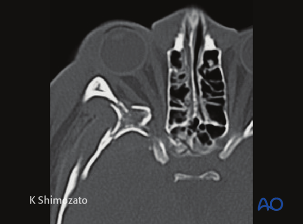
Special attention has to be paid to the posterior third of the orbit and the bony optic canal. Bony dislocations in these anatomical areas are more likely to be associated with traumatic optic nerve lesions.
Axial CT scan in the plane of the optic nerve shows multiple fractures of the lateral orbital wall and the greater wing of the sphenoid of the right side. Additional stretching of the right optic nerve is present. Fractures where fragments involve the posterior third of the orbit are susceptible to subsequent optic nerve disorders.
Severe bleeding
In case of severe bleeding the following options should be considered:
- Assessment of current medical treatment for anticoagulation such as Coumadin, Aspirin, or other anti-platelet medication.
- Compression: either by nasal packing, balloon tamponade, or direct compression.
- Electrocautery: if a clear bleeding source can be evaluated.
- Control and adjustment of blood pressure.
- Interventional embolization when other methods fail.
- In some cases fracture reduction may be required to reduce bleeding.
Note: following the emergency treatment, thorough diagnostic examination may be performed to find the source of bleeding:
- Intranasal inspection
- Angiography with possible superselective interventional embolization.
2. Selection of approach
Exposure of orbital roof fractures is normally via preexisting lacerations, upper blepharoplasty incisions or probably most often via coronal approach.
Once the orbital floor is exposed, periorbital dissection is performed.
The following pages provide general information regarding orbital anatomy and dissection
- Preoperative considerations
- Anatomy of the bony orbit
- Correlation of surface and cross sectional anatomy
- Introduction to periorbital dissection
- Orbital floor dissection
- Medial orbital wall dissection
- Lateral orbital wall dissection
- Orbital roof dissection
- Adjunctive access procedures (orbitotomies)
- Retrobulbar hemorrhage
3. General considerations
Visualization of fracture
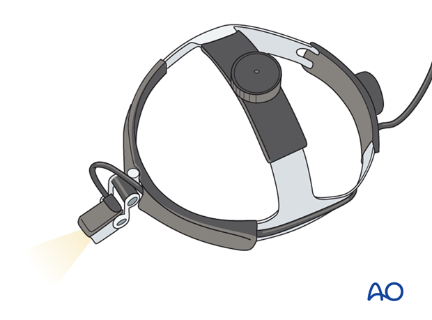
Appropriate exposure of the orbit requires adequate visualization. This requires appropriate retraction of the soft tissues and lighting. Regular OR lights serve this purpose for the midorbit. The posterior orbit is better visualized using headlights or illuminated retractors.
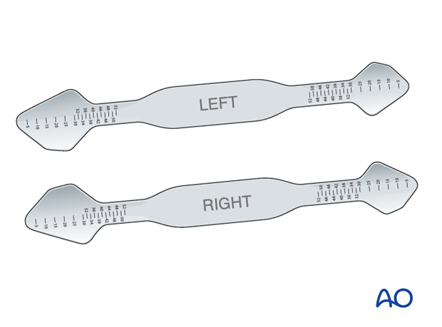
Special malleable orbital retractors (straight or anatomically formed) are available with metric markings providing the surgeon with additional information regarding the extent of the fracture and the depth of the orbital dissection. Specific orbital retractors have been developed to improve orbital retraction and minimize prolapse of soft tissue during insertion of implants.

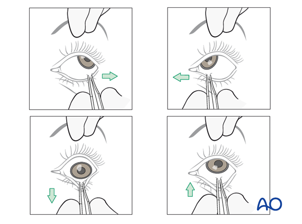
Forced duction test
The forced duction test should be performed to determine ocular motility as soon as general anesthesia is induced.
As soon as the periorbita has been dissected another forced duction test should be performed to determine ocular motility.
After the insertion of any implant material, it is imperative to perform a forced duction test to assure that the implant has not created a decrease in ocular motility.
Click here for details on the forced duction test.
4. Choice of reconstruction material
Reduction of the fractured, inferiorly displaced orbital roof fragment may be sufficient obviating the need for internal fixation hardware.
General considerations
The unique and complex anatomy of the orbit requires significant contouring of the implants to restore the proper anatomy.
The majority of cases require reconstruction of the orbital roof to support the reduced bone fragments and restore the shape of the orbit. The reason for this is that the bony walls are comminuted and/or bone fragments are missing. Therefore, one is reconstructing missing bone rather than reducing bone fragments. This can be accomplished using various materials.
There is hardly any anatomic region in the human body that is so controversial in terms of appropriate material used for fracture repair:
- Nonresorbable versus resorbable
- Autogenous/allogenous/xenogenous versus alloplastic material
- Standard versus custom-made plates
- Nonporous versus porous material
- Noncoated versus coated plates
Many surgeons recommend using materials that allow bending to an anatomical shape, that are radiopaque (to allow for intra- or postoperative radiologic confirmation of placement), and stable over time.
Reconstruction of the dislocated orbital walls is demanding. Secondary changes to this contour are undesirable. This is why critical consideration of the use of resorbable materials is necessary.
There are different preferences of implant material depending on regional differences, variations in schools of teaching, and socio-economic factors. There is a paucity of evidence to support the ideal choice for an orbital implant. Modern imaging analysis offers a unique chance to quantitatively asses the surgical result and stability over the time. This can provide valuable information for future recommendation.
In case of an intra- or subcranial approach, fixation of the orbital roof might be accomplished from inside the anterior cranial fossa.
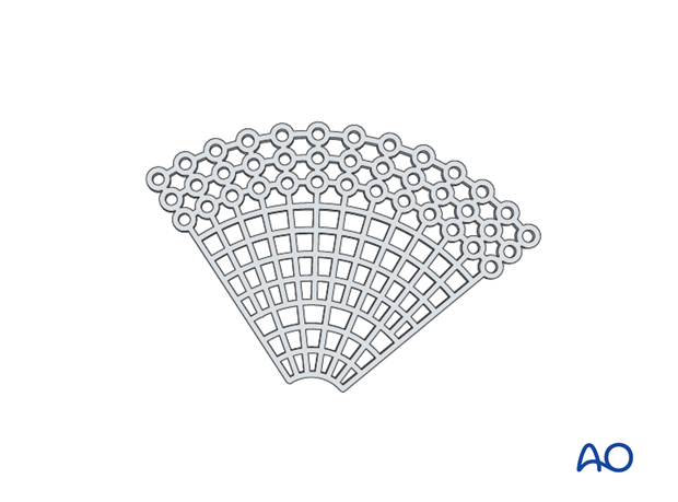
Titanium meshes
Advantages:
- Availability
- Stability
- Contouring (eased by the artificial sterile skull)
- Adequate in large three-wall fractures (the pre-bent plate is limited to medial wall and orbital wall fractures only).
- Radiopacity
- Spaces within the mesh to allow dissipation of fluids
- No donor site needed
- Tissue incorporation may occur
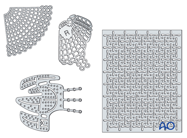
Disadvantages:
- Costs
- Possible sharp edges if not properly trimmed
Note: We do not have long term experience in a large series regarding the use of metallic mesh in young children. In several pediatric cases above the age of four it proved to be safe.
Note: The extensive use of mesh for orbital reconstruction in traumatized orbits has been shown to be safe, with few problems due to infections and no problems with secondary displacement due to additional trauma, on condition of proper handling, contouring, and fixation of the mesh.
Bone graft
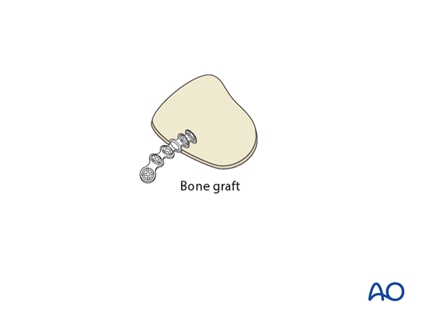
Illustration shows individual calvarial bone graft.
Advantages:
- Low material costs
- Smooth surface
- Variability in thickness
- Radiopacity
- Maximal biocompatibility
- Periorbita readily dissects off of the bone in secondary reconstructions
Disadvantages:
- Additional donor site needed (necessitating an additional surgical site for time of harvest, pain, scar, and possible surgical complications)
- Possible contour and dimensional changes due to remodeling
- Difficult to shape according to patients anatomy
- Less drainage from the orbit than with titanium mesh
Composite of porous polyethylene and titanium mesh
By combining titanium mesh with porous polyethylene the material becomes radiopaque, and more rigid than porous polyethylene of a similar thickness. Some surgeons also believe that there is less risk of having retained sharp barbs, which can lead to an entrapment of soft tissues during placement.
Advantages:
- Availability
- Stability
- Contouring (eased by the artificial sterile skull)
- Adequate in large three-wall fractures (the pre-bent plate is limited to medial wall and orbital wall fractures only).
- Radiopacity
- No donor site needed
- Tissue incorporation may occur
Disadvantages:
- Less drainage from the orbit than with titanium mesh
Resorbable materials
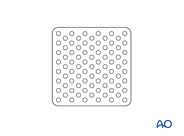
Thermoplastic and non-thermoplastic materials
Advantages:
- Availability
- Handling/contourability (only for thermoplastics)
- Smooth surface and smooth edges
Disadvantages:
- No radiopacity
- Degradation of material with possible contour loss
- Sterile infection / inflammatory response
- Difficult to shape according to patients anatomy (only for non-thermoplastics)
- Less drainage from the orbit than with uncovered titanium mesh (in case when nonperforated material is used)
Implant fixation
Fixation of orbital reconstruction material varies with the type and nature of the fracture.
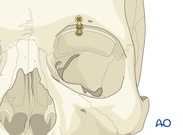
Fixation of most materials in the orbital roof is achieved by the use of one or more screws. The diameter depends on anatomical requirements but will normally vary between 1.0 1.3, or 1.5 mm. Alternatively Matrix Midface screws can be used.
5. Surgical exposure
Periorbital dissection
After exposure periorbital dissection is performed.
Retraction
Adequate exposure and illumination (headlights, illuminated retractors) of the fractured area is imperative prior to fixation.
If visualization of the orbital roof is difficult, a marginotomy of the supraorbital rim may be performed.
Adequate hemostasis has to be achieved.
Appropriate retraction of the intraorbital soft tissues has to be performed.
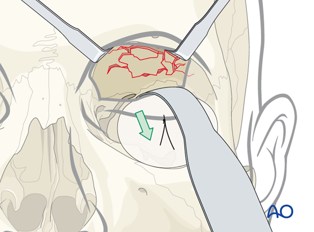
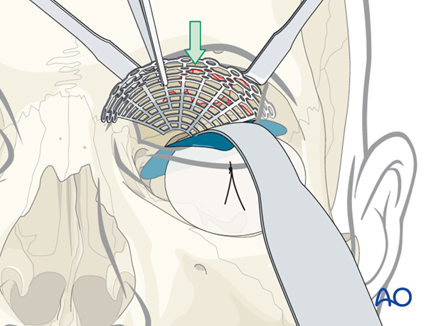
A foil (or sheet of other material) may help with a retractor to avoid prolapse of soft tissues, to improve visualization, and prevent entrapment of soft tissues during implant placement.
The illustration shows the insertion of this foil above the retractor.
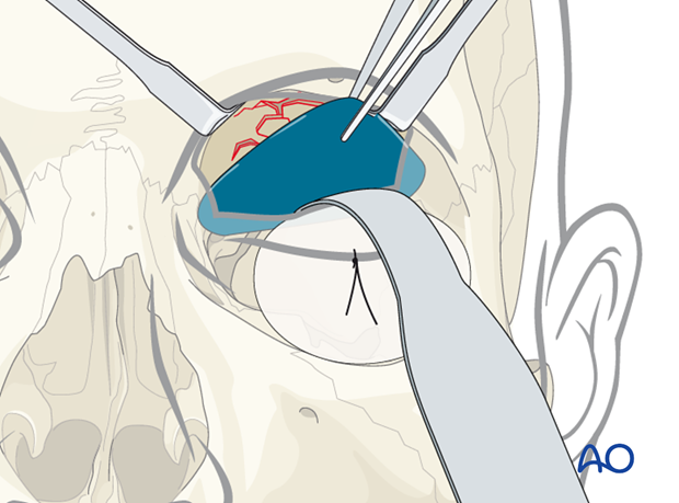
The retractor is then removed, placed above the foil, and the orbital soft tissues are properly retracted.
Note: Do not forget to remove the foil following implant placement.
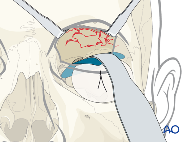
This illustration shows the foil larger than the size of the retractor.
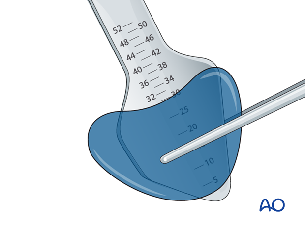
Helpful devices
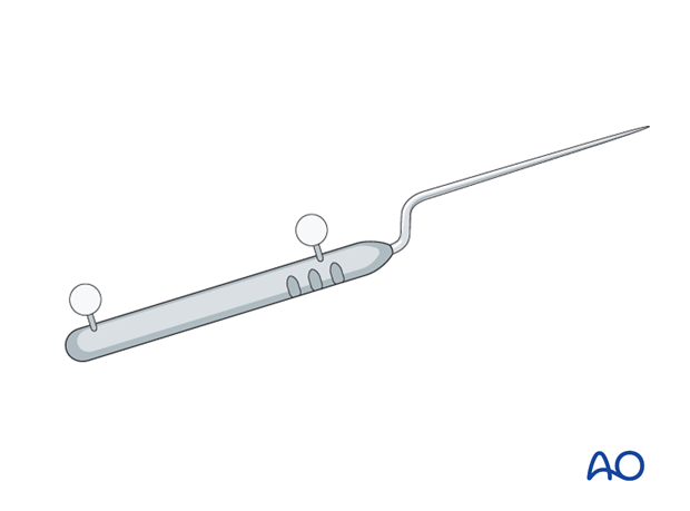
Many different devices have been used to facilitate retraction of the orbital contents, including malleable retractors, spoons, and special orbital retractors designed for the globe (as illustrated).
6. Reduction only
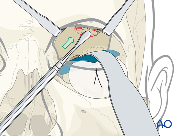
In some cases the orbital roof may be reduced and the fracture segment may be stable. Fixation may not be needed. In these cases, the patient should have close clinical and radiographical follow-up.
7. Reconstruction
Measurement of defect size and determination of implant size
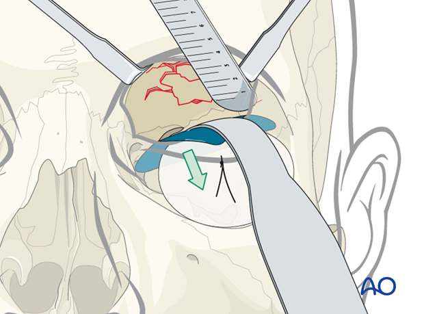
The defect size can be measured by the reading on the orbital retractor. Alternatively, any other instrument can help for detection of the defect size.
It is controversial whether or not to extend the anterior part of the mesh over the supraorbital rim. Whatever method (fixation anterior or posterior to the rim) is chosen, the upper eyelid function must not be compromised.
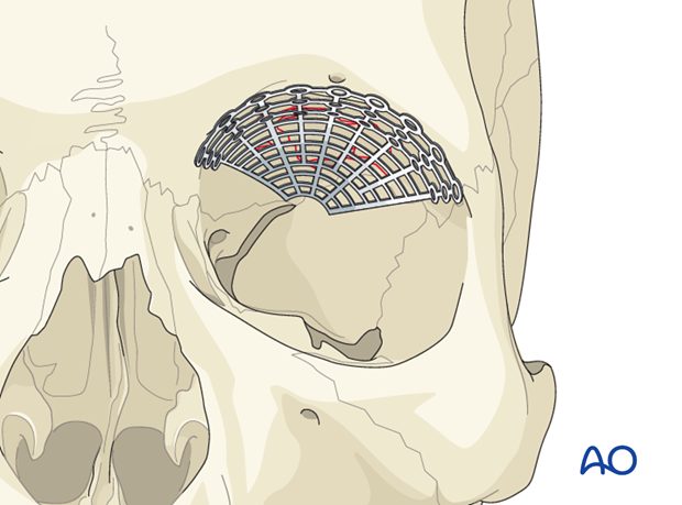
Example of reconstruction with titanium mesh
The mesh is cut, ...
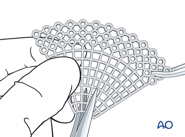
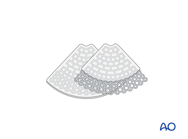
… all sharp edges of the plate are trimmed off to protect the soft tissues (note the shape of the fan has only a minimum number of screw holes), …

… and contoured to achieve the required shape, and accommodate key anatomical structures (nasolacrimal duct, infraorbital nerve, and optic nerve)
It is advisable not to extend the implant further posterior than 1 cm anterior to the optic canal entrance (if the posterior support to the orbit can be reached).
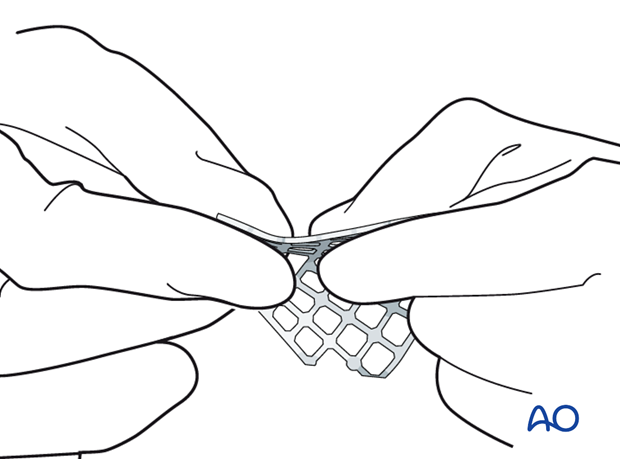
A sterile artificial skull allows proper anatomical contour of the implant.
Note:
- When using the fan-shaped plate, the outer circumference of the mesh is widest in the area of the supraorbital rim. The mesh should be trimmed so that the outer circumference is as small as possible but still provides enough width to cover the defect.
- The necessity for screw fixation varies with the type of material used and nature of the fracture.
- It is important to use a plate large enough to span the entire defect.
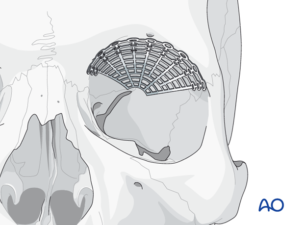
Regardless of the implant chosen, the insertion process must prevent deformation of the mesh. Under adequate retraction of the intraorbital soft tissues the mesh has to be positioned so that proper and stable reconstruction of the orbital roof results. Care has to be taken that neither orbital fat nor muscles are entrapped. During the insertion process, the mesh may require rotation in order to be properly positioned.
Navigation
Additionally, navigation may serve for intraoperative control of implant or fragment position. Modern 3-D C-arm technology will further improve intraoperative quality control of fracture reduction and implant positioning. Endoscopic modalities may be helpful in confirming proper implant placement.
8. Fixation
Mesh fixation
Orbital roof implants must always be stabilized. Fixation is provided by at least one screw. The screw size has to be appropriate to the chosen mesh type.
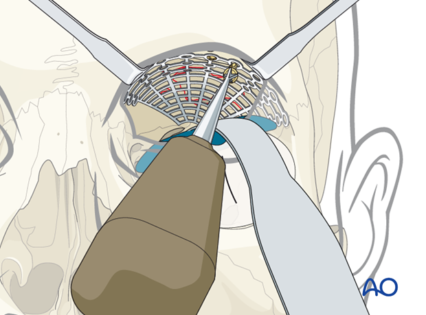
Forced duction test
After the insertion of any implant material, it is imperative to perform a forced duction test to assure that the implant has not created a decrease in ocular motility. Click here for details on the forced duction test.
Note: Pupils should be checked; however pupillary function might be compromised by drugs or mechanical force to the orbital contents.

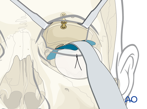
Alternative: Bone graft
If bone is harvested from a donor site, eg, cranial vault (parietal area), iliac crest, mandible, rib, etc. there may be donor site morbidity.
According to the dimension of the defect the bone has to be adapted and may be fixed either with screw fixation only or with plate and screw fixation (as illustrated).
9. Aftercare following open treatment of orbital fractures
Evaluation of the patients vision is performed as soon as they are awakened from anesthesia and then at regular intervals until they are discharged from the hospital.
A swinging flashlight test may serve in the unconscious and/or noncooperative patient; alternatively electrophysiological examination has to be performed but is dependent on the appropriate equipment (VEP).
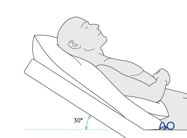
Postoperative positioning
Keeping the patient’s head in an upright position both preoperatively and postoperatively may significantly improve periorbital edema and pain.
Nose-blowing
To prevent orbital emphysema, nose-blowing should be avoided for at least 10 days following orbital fracture repair.
Medication
The use of the following perioperative medication is controversial. There is little evidence to make strong recommendations for postoperative care.
- No aspirin or nonsteroidal antiinflammatory drugs (NSAIDs) for 7 days
- Analgesia as necessary
- Antibiotics (many surgeons use perioperative antibiotics. There is no clear advantage of any one antibiotic, and the recommended duration of treatment is debatable.)
- Nasal decongestant may be helpful for symptomatic improvement in some patients.
- Steroids, in cases of severe orbital trauma, may help with postoperative edema. Some surgeons have noted increased complications with perioperative steroids.
- Ophthalmic ointment should follow local and approved protocol. This is not generally required in case of periorbital edema. Some surgeons prefer it. Some ointments have been found to cause significant conjunctival irritation.
- Regular perioral and oral wound care has to include disinfectant mouth rinse, lip care, etc.
Ophthalmological examination
Postoperative examination by an ophthalmologist may be requested. The following signs and symptoms are usually evaluated:
- Vision (except for alveolar ridge fracture, palatal fracture)
- Extraocular motion (motility) (except alveolar ridge fracture, palatal fracture)
- Diplopia (except Le Fort I, alveolar ridge fracture, palatal fracture)
- Globe position (except Le Fort I, alveolar ridge fracture, palatal fracture)
- Perimetric examination (except Le Fort I, alveolar ridge fracture, palatal fracture)
- Lid position
- If the patient complains of epiphora (tear overflow), the lacrimal duct must be checked.
Note: In case of postoperative double vision, ophthalmological assessment has to clarify the cause. Use of prism foils on existing glasses may be helpful as an early aid.
Postoperative imaging
Postoperative imaging has to be performed within the first days after surgery. 3-D imaging (CT, cone beam) is recommended to assess complex fracture reductions. An exception may be made for centers capable of intraoperative imaging.
Especially in fractures involving the alveolar area, orthopantomograms (OPG) are helpful.
Wound care
Remove sutures from skin after approximately 5 days if nonresorbable sutures have been used.
Apply ice packs (may be effective in a short term to minimize edema).
Avoid sun exposure and tanning to skin incisions for several months.
Diet
Diet depends on the fracture pattern.
Soft diet can be taken as tolerated until there has been adequate healing of the maxillary vestibular incision.
Intranasal feeding may be considered in cases with oral bone exposure and soft-tissue defects.
Clinical follow-up
Clinical follow-up depends on the complexity of the surgery, and whether the patient has any postoperative problems.
With patients having fracture patterns including periorbital trauma, issues to consider are the following:
- Globe position
- Double vision
- Other vision problems
Other issues to consider are:
- Facial deformity (incl. asymmetry)
- Sensory nerve compromise
- Problems of scar formation
Issues to consider with Le Fort fractures, palatal fractures and alveolar ridge fractures include:
- Problems of dentition and dental sensation
- Problems of occlusion
- Problems of the temporomandibular joint (TMJ), (lack of range of motion, pain)
Eye movement exercises
Following orbital fractures, eye movement exercises should be considered.
Implant removal
Implant removal is rarely required. It is possible that this may be requested by patients if the implant becomes palpable or visible. In some countries it will be more commonly requested. There have been cases where patients have complained of cold sensitivity in areas of plate placement. It is controversial whether this cold sensitivity is a result of the plate, a result of nerve injury from the original trauma, or from nerve injury due to trauma of the surgery. Issues of cold sensitivity generally improve or resolve with time without removal of the hardware.
Generally, orbital implant removal is not necessary except in the event of infection or exposure. Readmission might be indicated if long term stability of the orbital volume has not been maintained.
Follow-up
The patient needs to be examined and reassessed regularly and often. Additionally, ophthalmological, ENT, and neurological/neurosurgical examination may be necessary. If any clinical signs for meningitis or mental disturbances develop, professional help has to be sought. A regular follow-up CT scan is recommended 3-6 months after the trauma to assure proper pneumatization of the sinuses (particularly, mucocele formation has to be ruled out), sealing of the skull base and stability of fragment position.
Note: Posttrauma meningitis can occur even decades following trauma.
Special considerations for orbital fractures
Travel in commercial airlines is permitted following orbital fractures. Commercial airlines pressurize their cabins. Mild pain on descent may be noticed. However, flying in military aircraft should be avoided for a minimum of six weeks.
No scuba diving should be permitted for at least six weeks.


