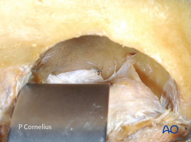
Periorbital dissection of Orbital Roof
1. Introduction
The roof of the orbit is triangular in shape. It has a distinct anterior concavity being greatest about 1.5 cm posterior to the supraorbital rim, which corresponds to the equator of the globe.
The orbital roof is thin and fragile except for the posterior convergence, where it consists of the lesser wing of the sphenoid.
The roof can be significantly pneumatized due to the extension of the frontal sinus and the ethmoid air cells.
Preoperative CT imaging needs to be checked for unusual pneumatization of the orbital roof and possible weak spots.
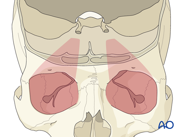
The orbital roof largely consists of the orbital process of the frontal bone.
Laterally, the lacrimal gland fossa is located medial to the zygomatic process and enlarges the post rim concavity. The trochlear fovea is a small depression superomedially. Above the fossa the frontal sinus extends postero-laterally commonly reaching the mid-point of the orbital roof.
Medially, the ethmoid air cells make up the orbital roof. Here, the roof appears to have multiple pits (foveolae ethmoidales) with bony ridges in between. The anterior and posterior ethmoidal foramina are located along the frontoethmoidal suture line in the transition zone from the roof to the medial wall of the orbit.
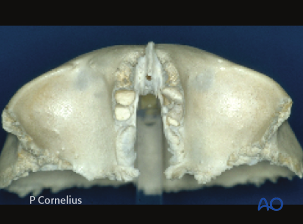
Fractures of the roof of the orbit are essentially anterior skull base fractures. They are associated with frontal sinus fractures or fractures of the squamous portion of the frontal bone.
Exposure of the roof of the orbit is normally done via preexisting lacerations or most often via a coronal approach. The roof is entered over the edge of the supraorbital rim.
The periosteum over the supraorbital rim is densely adherent to the bone and great care is taken not to tear to prevent herniation of the contents of the periorbita.
Elevation of the periorbita begins superiotemporally from the frontozygomatic suture line in the area of the periorbital sac where the periorbita is condensed and more resistant to injury If the periorbita is violated, the lacrimal gland will prolapse into the surgical field.
The elevation is then continued medially, to detach the periorbita along the entire supraorbital rim. The supraorbital neurovascular bundle must be released from its notch or canal to allow the dissection of the medial orbital roof to proceed.
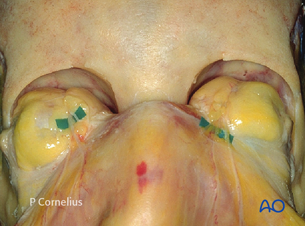
The dissection behind the supraorbital rim must take into account the post rim concavity. The periosteal elevators must be directed superiorly maintaining contact with the bone of the orbital roof to remain in a subperiosteal plane and avoid injury to the contents of the periorbita.
In addition one must be aware of any fractures or dehiscences in the bone to avoid injury to the dura and the resulting cerebrospinal fluid leak.
The frontal nerve and its terminal branches (supraorbital and supratrochlear nerve) is embedded within the periorbita along the whole roof. Posteromedially the trochlear nerve lies in direct contact with the periorbita. Both nerves can serve as a landmark in the dissection.
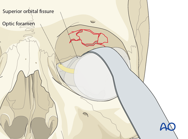
Schematic view demonstrating the periorbital dissection along the orbital roof to expose an isolated fracture site at the level of the midorbit. The orbital contents are retracted inferiorly and laterally. The posterior anatomic limits of the roof are indicated by the silhouettes of the superior orbital fissure and the optic foramen.
2. Deep Dissection
A deep dissection of the periorbita along the roof of the orbit will lead to the posterior end of the bony triangle, where the periorbital sac spans the optic foramen and the upper medial outline of the superior orbital fissure. Another suspension arises from around the recurrent meningeal artery foramen, which is located more anterolaterally.
The recurrent meningeal artery will be encountered when the periorbital dissection follows the zygomaticofrontal and the sphenofrontal suture line, which both make up the lateral border of the orbital roof.
