Correlation of surface and cross sectional anatomy
1. Introduction
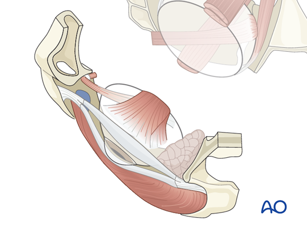
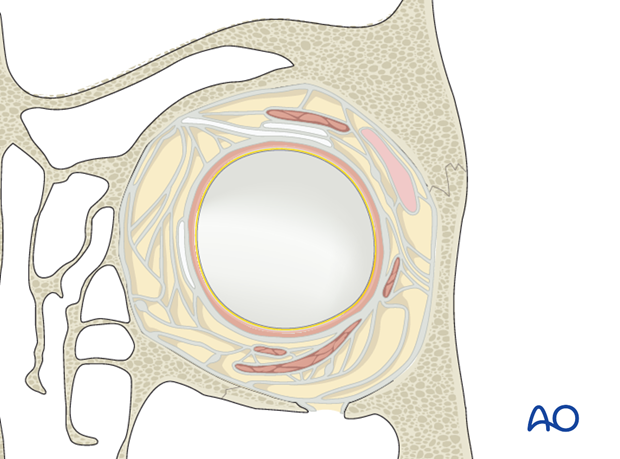
Periorbital dissection must be correlated to the cross sectional planes. This facilitates understanding of the topographical relationships and provides a reliable reference to CT imaging planes.
The outline and shape of the coronal cross sections changes continuously as one progresses from the rim to the apex.
The orbit can be divided into three zones to aid in the conceptualization of the complex orbital anatomy and landmarks.
2. Anterior orbit
The anterior orbit is the zone in front of the anterior ethmoidal foramen and the anterior loop of the inferior orbital fissure.
The largest internal orbital diameter is located just behind the anterior aperture (orbital rim) where it is almost circular with an inferomedial angulation. Here, the orbital floor is contiguous with the lateral wall.
The following landmarks determine this level: Inferior oblique muscle insertion, lacrimal fossa, trochlear fossa, Whitnall’s tubercle.

3. Midorbit
The midorbit corresponds to the sector between the anterior and posterior ethmoidal foramina.

Throughout the midorbit, the cross section is nearly four sided with a convexity of the medio lateral walls.
The floor and the lateral walls are separated by the inferior orbital fissure.
The posterior limit of the midorbit is at the level of the orbital process of the palatine bone. At this level the cross section narrows dramatically and transforms into a triangular shape.
The thickness of the lateral wall varies considerably. The greater wing of the sphenoid makes up the lateral wall. It is thinned out near the zygomatico sphenoid- suture line at the level of the anterior loop of the orbital fissure and appears thickest at the trigone over the temporal lobe extension of the middle cranial fossa. The lateral wall thins out again at the level of the superior orbital fissure.
The following landmarks determine this level: infraorbital groove, zygomaticosphenoid suture at the anterolateral circumference, ethmoidal foramina, lamina papyracea, internal orbital buttress.

4. Posterior orbit
The posterior orbit or apex begins behind the level of the posterior ethmoidal foramen. Here the orbital cross section is definitively triangular.
The main structures contained in the orbital apex are the superior orbital fissure and the optic foramen with the optic strut in between these two structures. The inferior orbital fissure narrows at the confluence above the foramen rotundum. The lateral wall leads to the tip of the middle cranial fossa.


