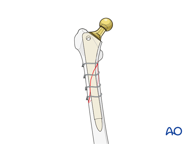
Diaphyseal fixation using long cylindrical stem
1. Principles
The most critical criterium is to achieve rigid stem fixation in the distal fragment.
Proper reduction of the two major fracture fragments for rotation and length is equally important.
2. Approach
The surgeon should use the surgical approach that is the most familiar to him/her for any total hip arthroplasty, such as:
- Anterolateral approach
- Direct anterior approach
- Iliofemoral (Smith-Petersen) approach
- Posterolateral approach
- Trochanteric osteotomy
- Extended trochanteric osteotomy
An extensile approach is necessary to access the fracture site for reduction and placement of additional fixation devices, such as cerclage wires, cables, strut graft, and/or plates.
These approaches can be performed with the patient in a lateral or supine position.
3. Implant removal
Acetabular cup assessment
Following adequate exposure into the hip joint, the surgeon should assess the position and the fixation stability of the acetabular cup.
The accepted "safe zone" is:
- cup inclination 40° to 55° (a)
- cup anteversion 20° to 40° (b)
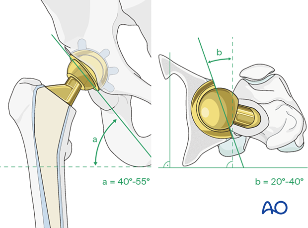
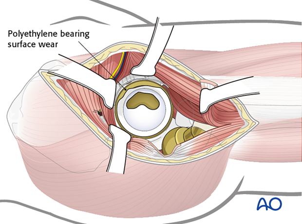
The surgeon should also assess for liner wear. If there is significant wear, liner revision should be done.
Femoral stem removal
The surgeon must perform adequate soft tissue releases around the proximal femur in order to reduce further fracture displacement or comminution.
It is critical to maintain the integrity of the hip abductor mechanism, in order to avoid abductor insufficiency.
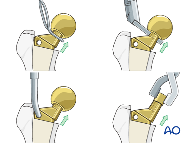
The femoral component is removed. Details on how to remove a femoral stem are described in the basic technique for femoral stem removal.
The surgeon must minimize any additional damage to the host bone during the stem removal process.
4. Provisional fracture reduction and fixation
General considerations
If the proximal femur fragment is comminuted, the surgeon can initially prepare the distal fragment. The proximal comminuted fracture fragments can later be reduced to the new femoral stem.
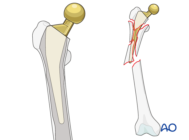
If the proximal and the distal fracture fragments are intact, provisional reduction and fixation of the two fragments can be performed prior to reaming and broaching for the new femoral stem.
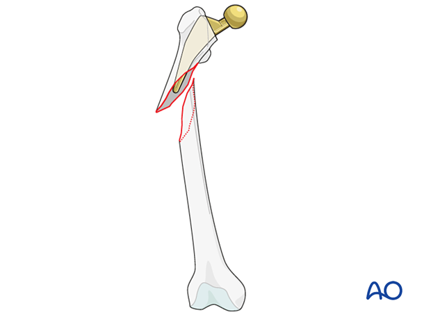
Provisional reduction and fixation
Following stem/cement removal and debridement of any retained bone and fibrous tissues, provisional reduction of the two major fracture fragments should be performed prior to femoral canal preparation for the new stem.
The reduction can be accomplished using large clamps, cerclage wires/cables, or a bridging plate, depending upon the fracture pattern and the bone quality of the fragments.
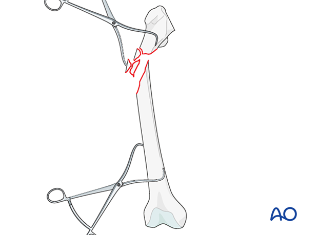
Provisional fixation of the fracture can be accomplished by using cables or wires, bone reduction clamps, or even bridging plate.
For additional techniques refer to the ORIF sections of femur shaft fracture management of the AO Surgery Reference.
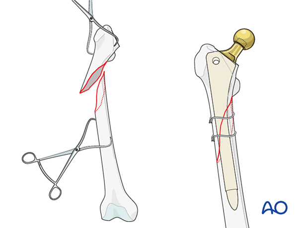
5. Femoral preparation
Implant selection
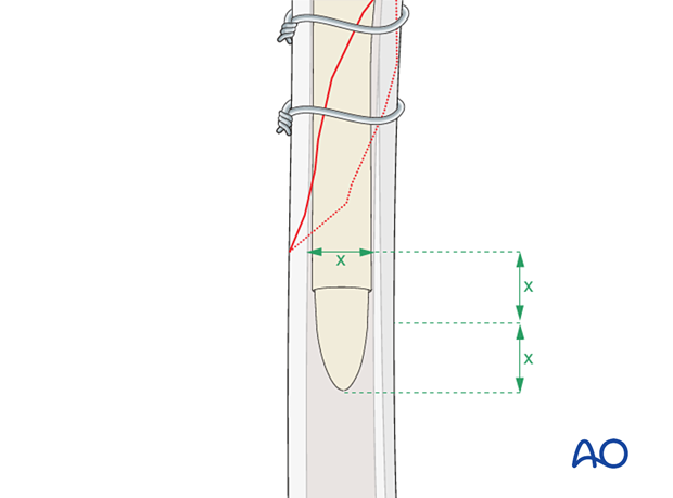
The new femoral stem must be of sufficient length to achieve rigid axial and rotational fixation into the distal diaphyseal femur fragment.
In general, the recommendation is a minimum length of two times the diameter of the femur below the fracture site. This length should be at least 4 to 6 cm of stem-to-cortex engagement in the distal fragment.
The diameter of the new stem is determined by the fit and rigidity of the reamer during the femoral preparation.
These are the general guidelines for femoral stem revision. Each implant manufacturer has specific recommendations and surgical technique for the specific device.
The surgeon has the option of using a modular or nonmodular stem.
Femoral canal reaming
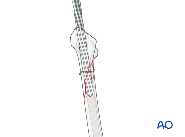
Femoral canal reaming is done using the reamer system from the specific device manufacturer.
The surgeon should ream in sequence, from the smallest diameter reamer to the largest diameter reamer that would achieve ideal stable fixation of the new stem. This is performed based upon both preoperative templating and intraoperative fit of the reamer.
Distal fragment protection
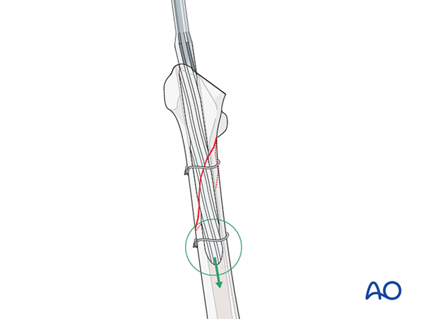
The distal fragment should be protected with clamps or provisional wires/cables during the reaming process to avoid further fractures or comminution.
If a curved stem is required, modified reamers should be used.
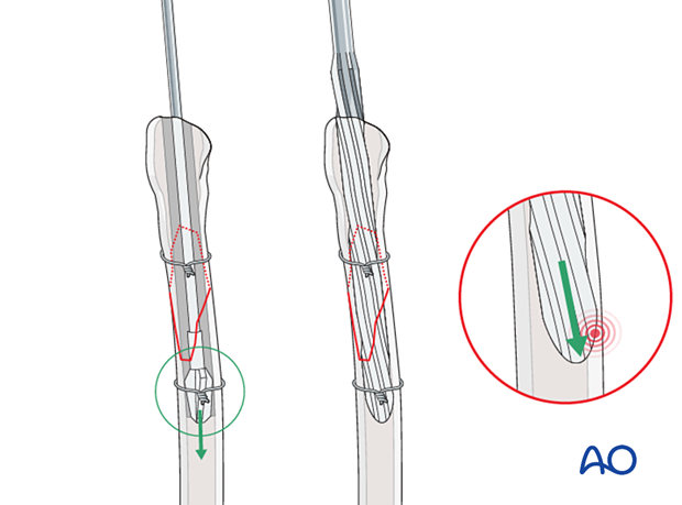
Proximal fragment protection
The proximal fragment may be protected with additional clamps, or provisional wires/cables during the broaching process to avoid further fractures or comminution.
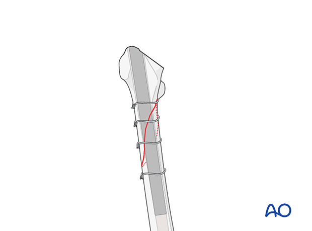
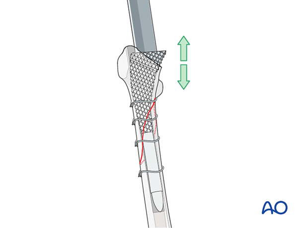
Metaphyseal segment preparation
Sequential broaching is performed to prepare the metaphyseal segment.
6. New implant insertion
Proper stem alignment must be achieved at this point.
A trial stem is inserted, and the hip is reduced to assess leg length, rotation, alignment of the limb, and hip joint stability. It is critical to mark the trial position prior to its removal.
The final stem is inserted guided by the previously made markings.
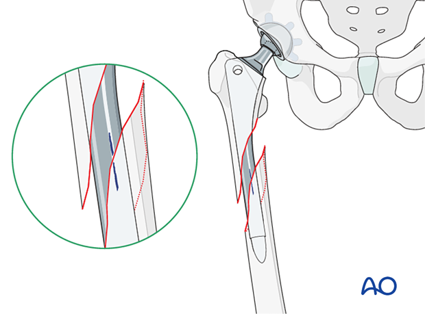
The selected femoral stem is inserted into the femur using specific tools from the device manufacturer. The surgeon must achieve proper stem rotation (anteversion) and leg length equalization.
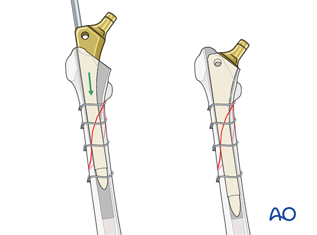
Hip reduction
The surgeon should do a trial reduction.
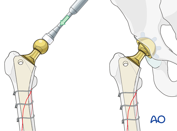
With the hip reduced, confirm the range of motion, leg length, and hip stability. Adjust the neck length if necessary.
Once satisfactory, attach the definitive femoral head to the stem, and reduce the hip. Confirm complete reduction, stability, and range of motion.
7. Final fracture reduction and fixation
Anatomic reduction verification
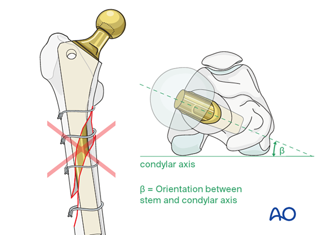
The surgeon must ensure anatomic limb alignment with regard to:
- Adequate contact of the fracture fragments
- Proper rotational alignment of the fracture fragments: the orientation of the distal femoral condylar axis should be aligned properly to the new femoral stem anteversion (recommended beta is equal to 5° to 15°)
- Appropriate leg length
Additional extramedullary fixation
The surgeon must determine if additional extra-medullary adjunct fixation is required. If necessary, additional wires/cables can be used.
If the initial provisional fixation prior to femoral canal preparation was performed using a plate, additional screws/cables can be added.

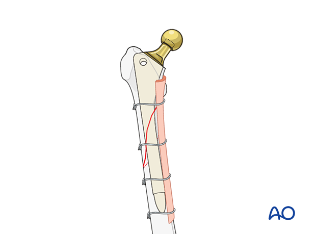
Adjunct strut graft
In selective cases of severe comminution, adjunct strut graft(s) may be considered to supplement the bone stock.
8. Aftercare following a femoral revision
Physiotherapy guidelines
Routine physiotherapy protocols for elective total hip arthroplasty is followed.
Early mobilization is recommended. Weight bearing status is individualized based upon fixation and implant stability.
Hip dislocation precautions are given to the patient.
Imaging
Postoperative radiographs can be made at 2 to 3 weeks to determine maintenance of satisfactory fracture reduction and fixation, no evidence of stem subsidence, or fracture displacement.
Follow-up radiographs can be made at 6 to 8 weeks after surgery to determine sufficient fracture fixation to increase the weight bearing status, and physical activities.
Future follow-up is similar to the routine and standard protocol for total hip arthroplasty.


