ORIF - Conventional plating
1. Principles
General considerations
These fractures are the most severe because there is a medial transarticular fracture that splits the joint in the coronal plane, resulting in anterior and posterior articular segments. They require a great deal of expertise and judgment and if possible, should be referred to someone who has the expertise and resources to cope with them. Anatomical reduction of the transarticular fracture component is absolutely mandatory. When dealing with the metaphyseal component of the fractures, it is more important to retain vascularization of the multiple fracture fragments than to reduce them anatomically.
In order to illustrate the difficulties that may be encountered, we have included a full discussion under the respective sections.
Main steps in treatment
Main steps in treatment include the following:
- Temporary reduction and immobilization until definitive surgery (eg, external fixator) typically dictated by resolution of soft-tissue injury
- Adequate preoperative planning (need for bone graft, approach(s), number and type of implants, sequence of reduction and fixation, additional equipment)
- Sub-meniscular arthrotomy for articular visualization and reduction, particularly laterally
- Anatomical reduction of the articular surface with disimpaction of fracture fragments
- Bone grafting of the metaphyseal defect once the fragments are disimpacted and the articular component elevated
- Direct and/or indirect reduction of the metadiaphysis to restore frontal and sagittal plane alignment
- Definitive stable fixation of the metaphysis using screws and plate(s) with preservation of the blood supply of the metaphyseal fragments
Timing of surgery
Surgery should be delayed until full recovery of soft tissues (swelling, blisters, abrasions, etc). This usually takes 3–10 days.
Until surgery, length of the leg should be maintained (eg, spanning external fixator, traction, etc).
Plate location
Buttress plating is fundamental to achieving stability. Plate locations are typically lateral, posteromedial, and/or anteromedial.
Choosing the proper reduction sequence
Commonly the medial column is generally addressed first. Based on a thorough understanding of the anterior and posterior medial segments, one reduces and fixes the fracture with the best read. Typically, the posteromedial fragment is fixed to the shaft first. Occasionally the anteromedial fragment is fixed to the shaft first. Sometimes there is no good metaphyseal read and the reduction is primarily driven by reducing the articular surface under direct vision. Key components should be articular reduction of the medial articular surface fracture fragments and restoration of frontal and sagittal plane alignment of the medial plateau. Stabilization of the medial column should be undertaken to avoid fixation of unreduced lateral column fragments. Also, medial-sided fixation should be placed to still allow fixation corridors for lateral-sided implants.
The lateral fracture is addressed next. Once a satisfactory reduction and fixation is reached on the lateral side, any remaining medial sided fixation is then completed, and the wounds are closed in a routine fashion.
Potential complications
The following complications should be checked for during surgery:
- Articular malreduction
- Alignment malreduction
- Intraarticular hardware
- Prominent fixation
- Vascular and nerve injury (peroneal nerve)
- Compartment syndrome
2. Patient preparation
Patient positioning
Depending on the approach, the patient may be placed in the following positions:
A tourniquet is helpful in most cases. Whether a tourniquet is used depends on the amount of bleeding. Exsanguinate the limb by elevating it.
To allow for intraoperative radiographic control of reduction and fixation, the use of a radiolucent table is mandatory.
The vast majority of subarticular and metaphyseal bone defects are currently filled with morcellized cancellous allograft and/or bone graft substitutes. These should be available during the procedure. Autologous bone graft is rarely utilized for void filling.
3. Approaches
Not all complete articular fractures can be perfectly reduced through a medial/posteromedial approach and/or an anterolateral/extended anterolateral approach. Sometimes fractures with a flexion injury mechanism need to be addressed through a prone posteromedial approach as well.
4. Preliminary reduction
General considerations
In situations treated with a temporizing spanning external fixator, the fixator is prepped and draped into the surgical field maintaining length, rotation, and alignment.
The typical sequence is to begin with the medial side first. Depending on the morphology of the fracture, a medial or posteromedial approach is performed.
In situations where metaphyseal fragmentation prevents accurate visual reduction of either segment, direct visualization is important to reduce and provisionally stabilize the joint. Applying ligamentotaxis to this restored medial condyle helps to achieve indirect reduction of the comminuted medial metaphyseal column.
All of these reduction maneuvers may require external fixator adjustment and/or placement of a medial distractor.
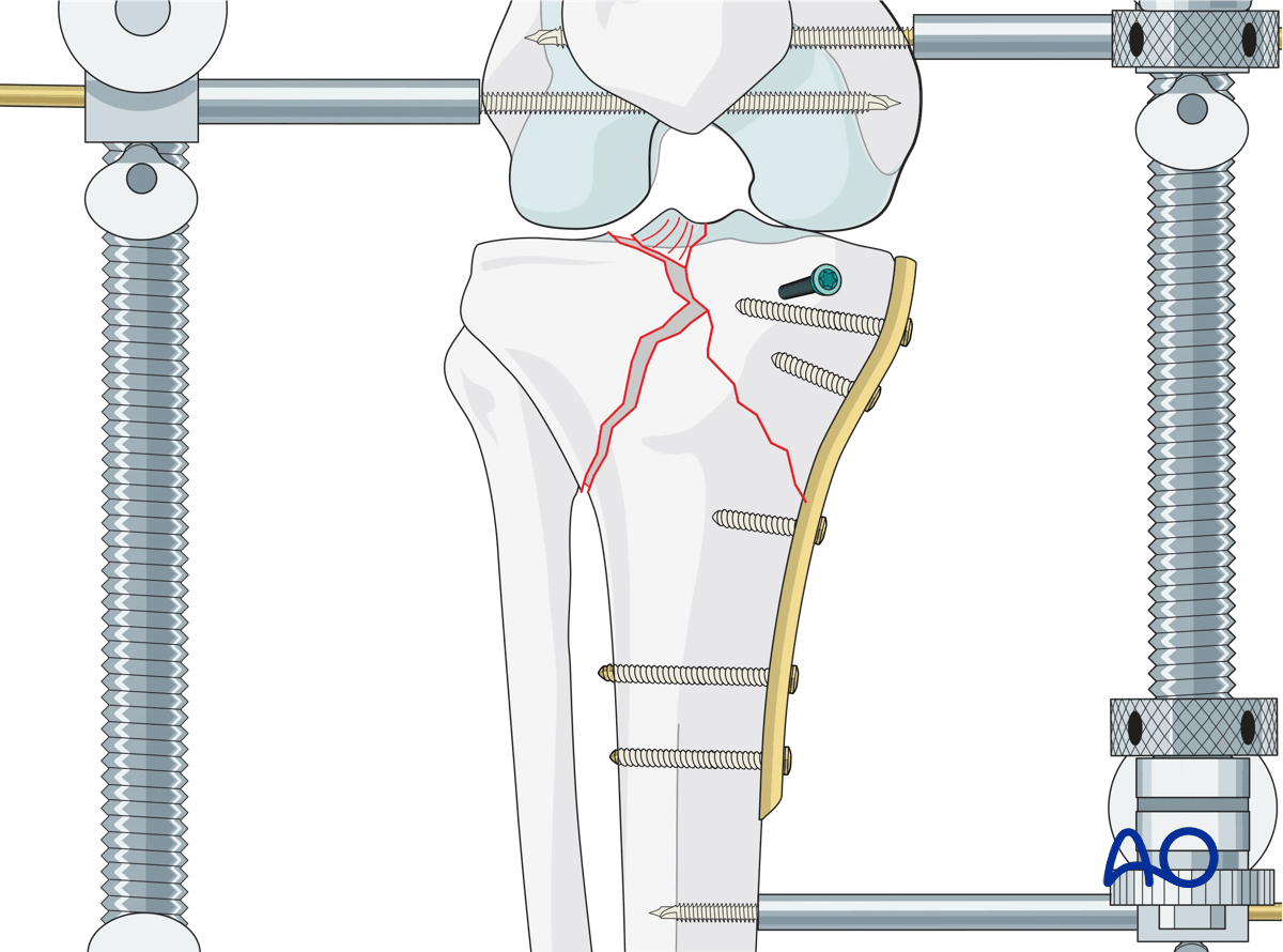
Transarticular coronal plane fracture
The fracture is roughly in the coronal plane, extending from the posteromedial portion of the lateral tibial plateau, and exiting along the medial cortex. The posteromedial fracture fragment often involves the posterior 50% of the articular surface. Invariably the fragment is displaced distally and often has a minimally comminuted exit point at the posteromedial metaphysis. Failure to identify, reduce, and stabilize this fragment often leads to posteromedial subluxation of the medial femoral condyle.
In situations without metaphyseal comminution, anatomic reduction of the posteromedial cortex and provisional fixation allows fixation of the anterior fragment. Dissection along the medial cortical fracture line to the joint surface allows visualization of the joint, corroborates the reduction, and, if needed, permits adjustment of the articular reduction.
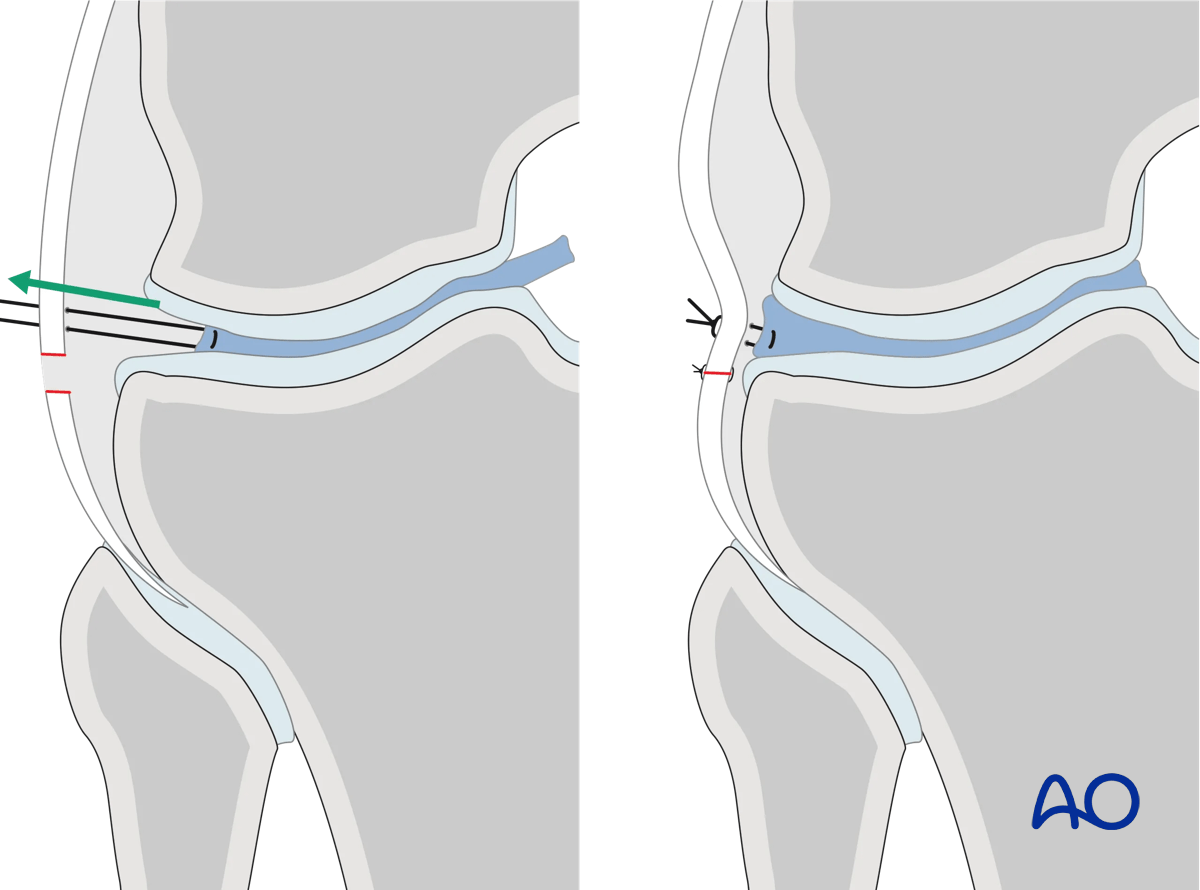
Pointed reduction clamps and anterior-posterior K-wires will provide provisional stabilization, as shown.
Plate application should ideally be directly overlying the apex of the fracture. Screws can then be positioned directly across the fracture plane from posterior to anterior, resulting in excellent stability of this fragment.
While conventional implants are acceptable for this, as they provide buttressing of the posteromedial fracture fragment, fixed-angle devices may be preferred given the fracture complexity.
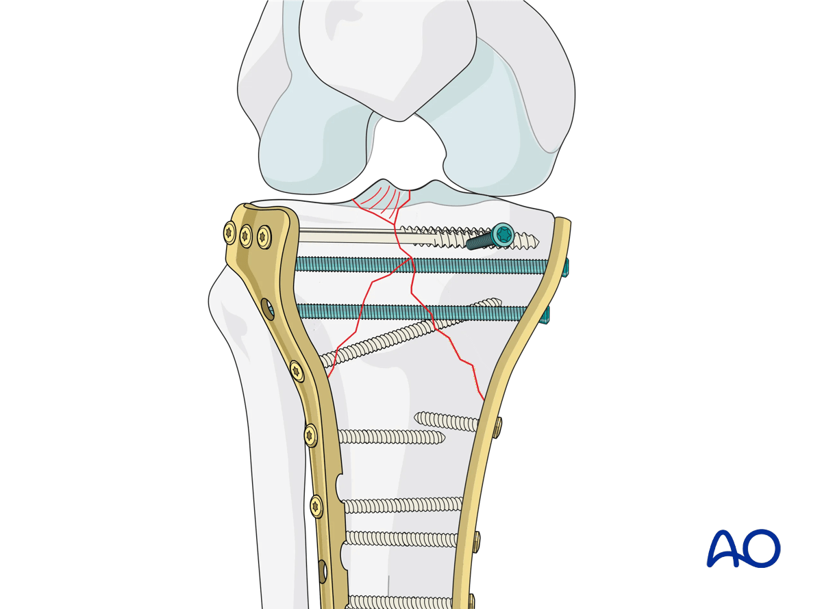
Application of medial plate
Typically, the medial plate is applied in the most mechanically advantageous position to support the medial column. This may be directly medial, posteromedial, or anteromedial, depending on the direction of displacement and fracture morphology. In situations with minimal comminution, lower profile, and malleable plates such as one third or one quarter tubular plates may be utilized. In situations with greater anticipated loads or inherent fracture instability, such as bone defects or comminution, stiffer 3.5 mm periarticular plates should be used. Screw fixation typically begins from distal to proximal, effectively buttressing the epiphysis. When using conventional implants, bicortical fixation is ideal. However, avoiding unreduced lateral-sided fracture fragments, or other fracture lines, can be very challenging. In these situations, unicortical conventional screws can be placed but their ability to create stability is suboptimal. An alternative is the use of low-profile or mini-fragment fixed angle or locking plates, which allow unicortical fixation with significant improvement in stability.
Reduction of lateral column
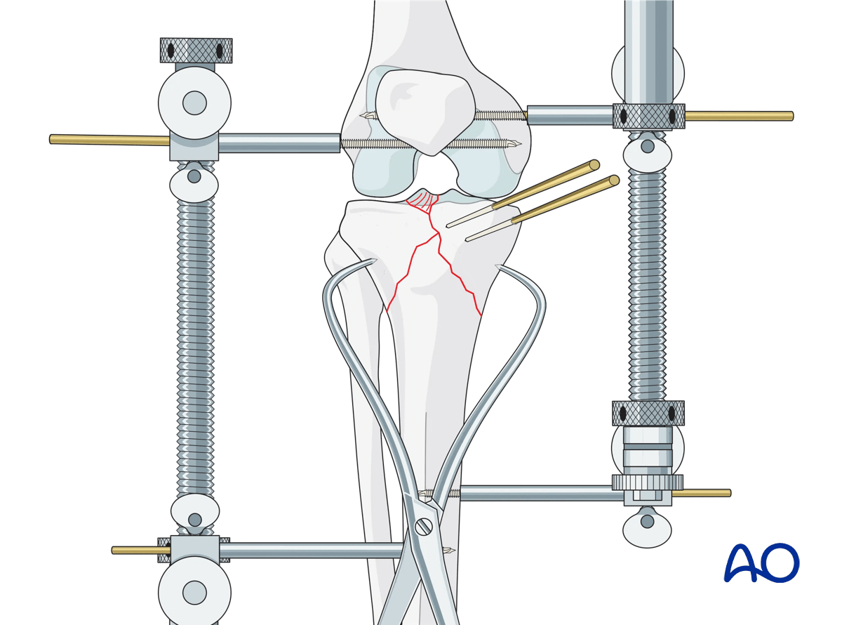
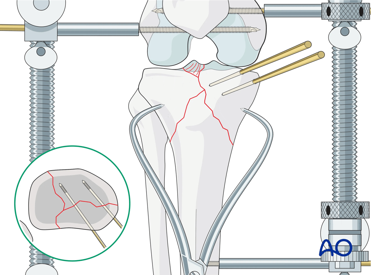
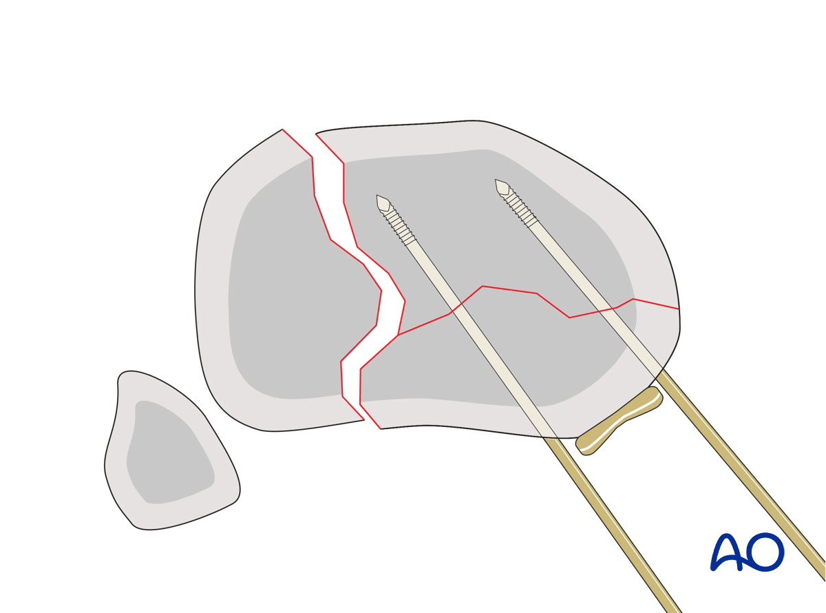
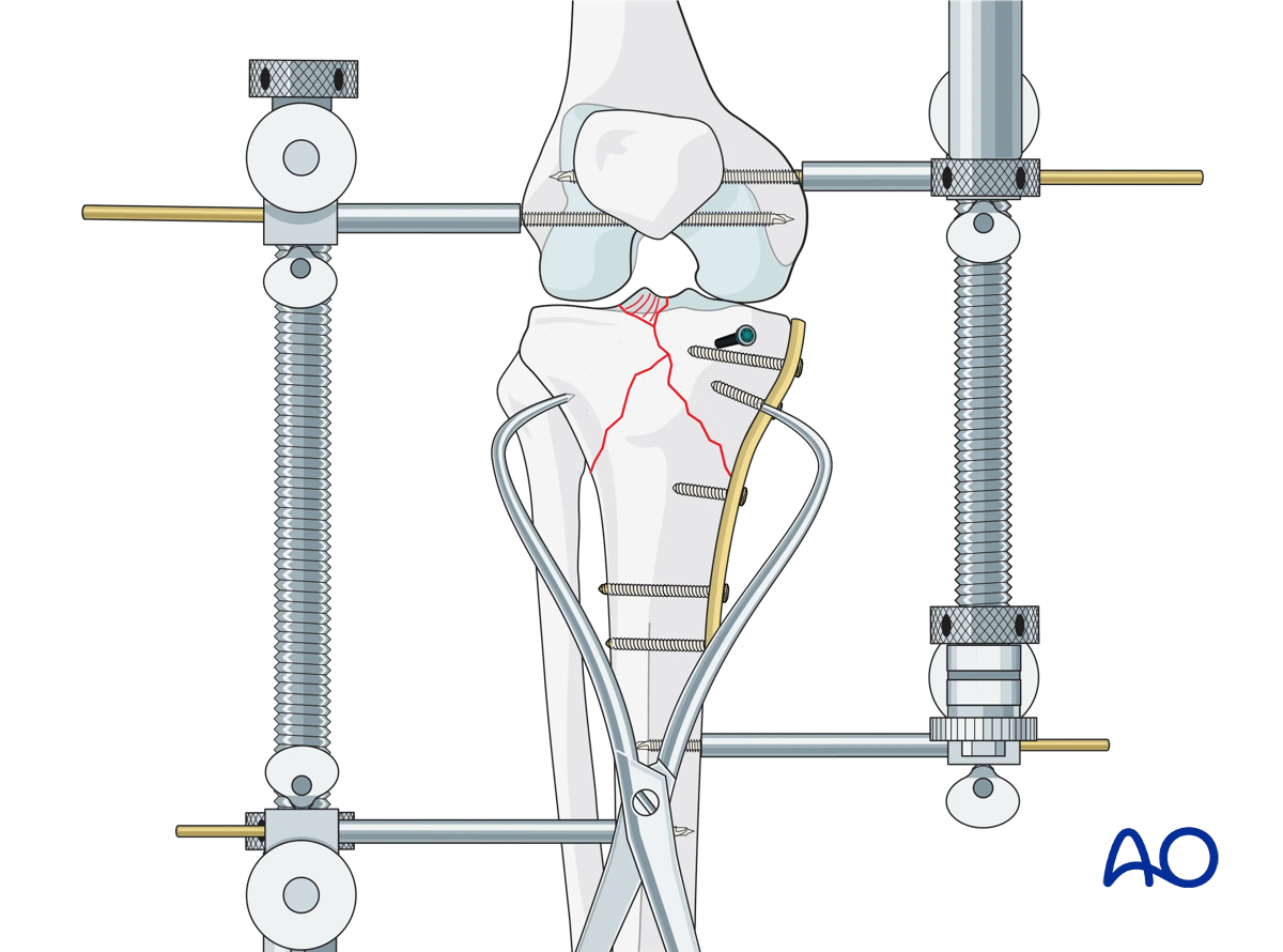
The lateral column is exposed using the anterolateral approach. Often, the fracture at this point is equivalent to a split depressed lateral plateau fracture. Reduction of any split component by reducing the cortical surfaces in the metaphysis is accomplished first. This often restores the length of the lateral column. Provisional fixation of this is performed with K-wires and clamps. Reduction of any depressed lateral articular fragments now requires joint visualization. A laterally-based femoral distractor is then used, and careful distraction is then applied.
The anterior external fixator may be retained to protect the medial-sided reduction and fixation. The femoral distractor will still provide sufficient distraction to enable joint visualization. If not, the external fixator can then be loosened. A lateral submeniscal arthrotomy is then performed and the joint is visualized. A cortical window is then performed through the metaphysis, and a bone clamp is used to elevate the depressed articular fragments. K-wires are used to maintain the reduction, and the bone defect is grafted. At this point, the articular surface should be reduced, and the frontal and sagittal plane alignment should be closely evaluated.
Fixation of lateral column
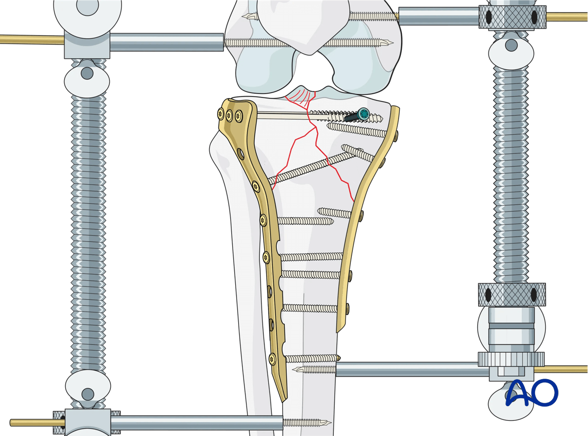
Lateral column fixation is typically the primary source of stability for the entire construct. Plate length should not be minimized and should travel distal to the medial column fixation. Periarticular plates contoured to the proximal lateral tibial plateau are the preferred implant. Similar to the medial side, fixation begins in the metadiaphyseal region and proceeds proximally, buttressing the metaphysis and the epiphysis. Conventional screw-plate devices will compress the tibial plateau metaphysis and minimize widening. A raft of screws from lateral to medial through the proximal portion of the plate will function to support the elevated lateral articular surface.
Occasionally a tibial tubercle fracture fragment is identified as part of this injury. Reduction and fixation of this is typically performed using the anterolateral approach. Fixation can be achieved with independent screw fixation, with or without the addition of a one third or one quarter tubular plate.
Final osteosynthesis
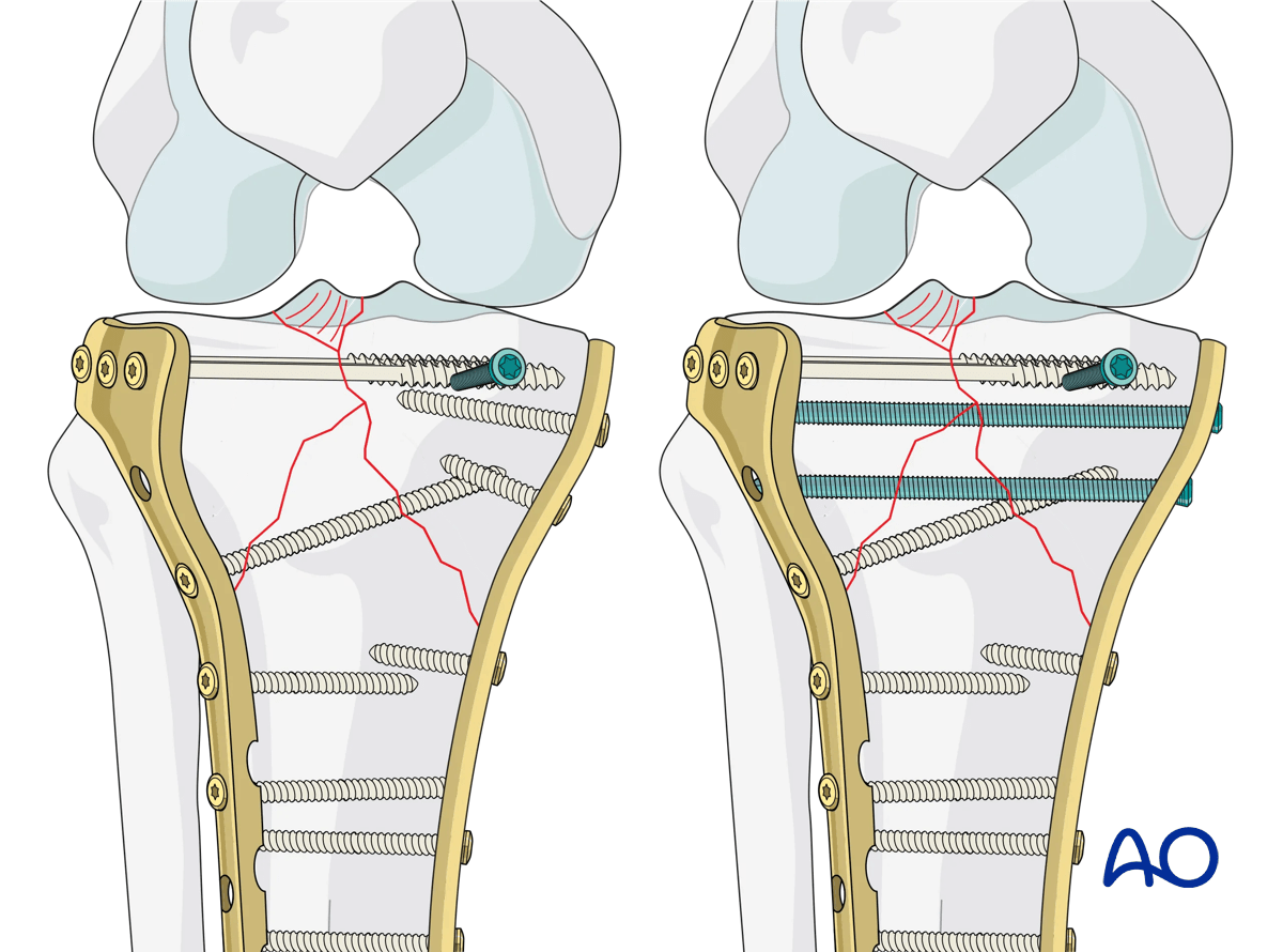
Once osteosynthesis is completed, make a final check with the image intensifier. If all is well, remove the external fixator and/or femoral distractor. At this point, short medial-column screws can be exchanged for screws with trajectories into the now reduced lateral column plateau. The knee should be ranged with a particular emphasis on full flexion to ensure fracture stability, and to disrupt quadriceps adhesions caused by the external fixator.
In situations with meniscal disruption from the capsule the meniscus can be repaired back to the capsule using absorbable mattress sutures. The submeniscal arthrotomy is then closed, ideally with a watertight closure. In situations where the capsule is deficient from the lateral proximal tibia the capsulotomy can be repaired to the proximal portion of the lateral plate.
5. Aftercare
Compartment syndrome and nerve injury
Close monitoring of the tibial compartments should be carried out, especially during the first 48 hours after injury and again after surgery to rule out compartment syndrome. More information is provided here:
The neurovascular status of the extremity must be carefully monitored. Impaired blood supply or developing neurological loss must be investigated as an emergency and dealt with expediently.
Consideration for DVT prophylaxis
Oral or subcutaneous administration of DVT prophylaxis for six weeks should be strongly considered.
Functional treatment
Optimal stability should be achieved at the time of surgery, in order to allow early range of motion exercises. Unless there are other injuries or complications, mobilization may be performed on post OP day 1. If available, continuous passive motion (CPM) splints can be very helpful in the early phase of rehabilitation. Static quadriceps exercises with passive range of motion of the knee should be encouraged. Afterwards special emphasis should be given to active knee and ankle movement.
The goal is to achieve as full range of motion as possible within the first 4–6 weeks.
Weight bearing
Weight-of-leg weight bearing is initiated depending on patient comfort. Depending on the severity of the articular displacement, weight bearing can begin as early as 6 weeks postoperatively. In situations where articular displacement was significant weight bearing should be delayed for 10–12 weeks.
Follow up
Wound healing should be assessed within the first two weeks. Subsequently, a 6- and 12-week follow-up with radiographic assessment is usually performed. If a delayed union is recognized, further surgical care may be necessary and should be carried out as soon as possible. Residual knee instability may require delayed reconstruction.
Implant removal
Implant removal is not mandatory and should be discussed with the patient.


