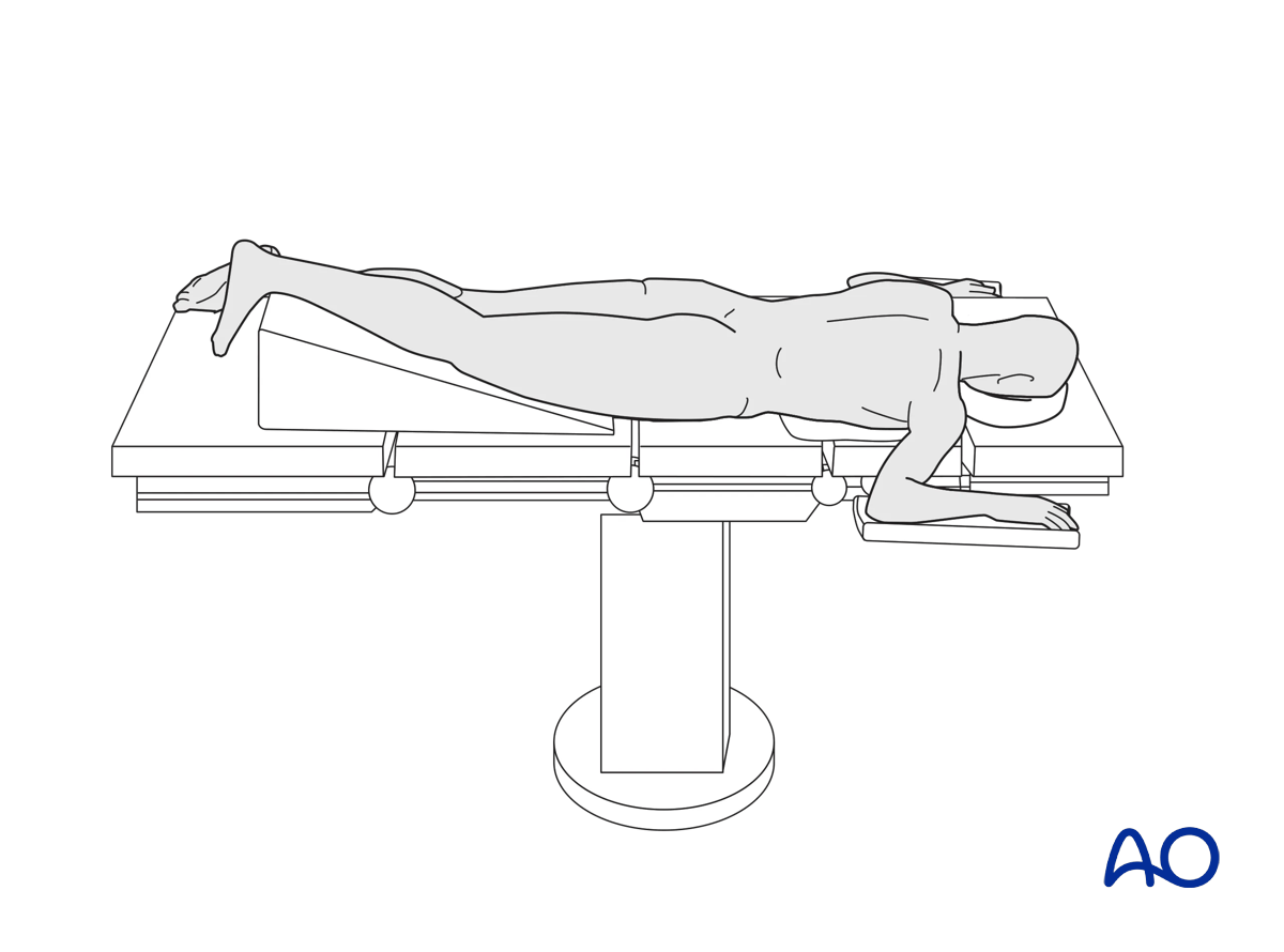
Prone position
1. Positioning
Place the patient prone. Prepare the limb from the ankle to the groin and drape the foot. This will allow full knee flexion.
The limb is placed on a raft elevating the injured knee above the level of the contralateral knee, to allow for unhindered lateral imaging.
2. Tourniquet usage
The use of a tourniquet may be necessary in open and direct reduction of articular fractures.
It is also helpful in open direct reduction and screw and plate fixation. It is not necessary whenever MIO techniques are employed. It is contraindicated for the insertion of external fixator pins, for intramedullary nailing, and under most circumstances when dealing with open fractures.

3. C-arm positioning
C-arm positioning must allow for perpendicular images of the plateau, especially accounting for the posterior slope of the proximal tibia. A true lateral image must be easily obtainable as well.


