ORIF - Plates without angular stability
1. Principles
Anatomical reduction
Anatomical reduction of the articular fracture component and fixation with lag screws (absolute stability) is mandatory.
After reduction of the articular fracture component, anatomical reduction of the metaphyseal component has to be achieved.

Double plating
In order to avoid varus collapse on the medial side double plating should be considered and if necessary carried out. In these fracture patterns the placement of the medial plate may be anteromedial rather than posteromedial. This depends of course on the fracture pattern and forces which have to be neutralized.

2. Patient preparation
Depending on the approach, the patient may be placed in the following positions:
3. Approaches
For this procedure the following approaches may be used:
4. Reduction
Indirect/open reduction
Indirect reduction may be achieved by external manipulation of the fracture fragment with clamps. In cases where adequate closed reduction is not achieved, the joint must be opened to carry out an open reduction.
If one is trying to carry out the procedure without opening the joint then reduction must be checked either with image intensification or with arthroscopy.
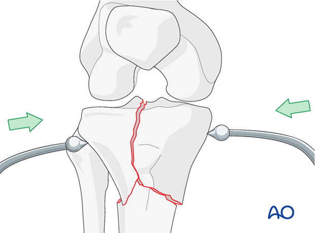
Pearl: clamp over plate
If you intend to keep the clamp on throughout the whole procedure it is best to slip the plate under the clamp prior to tightening the clamp to maintain reduction. Under these circumstances you determine which screw hole is best for the placement of the tip of the clamp from the pre-operative plan and intra-operative trial.
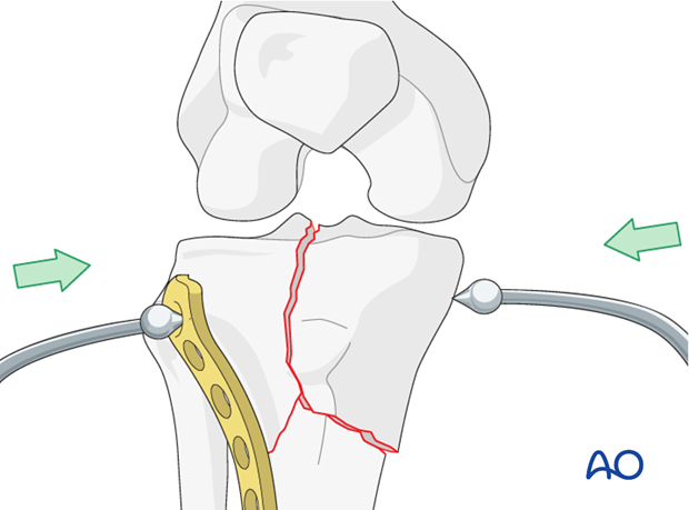
Secure reduction
Anatomical reduction of the articular surface is mandatory. Secure reduction and provisional fixation with K-wires.
Positioning of the knee is important for correct reduction and fixation. If the knee has a valgus injury, then the knee should be held with more varus positioning to ensure a good reduction. If the knee has a varus injury (medial condyle) then valgus positioning during reduction is important.
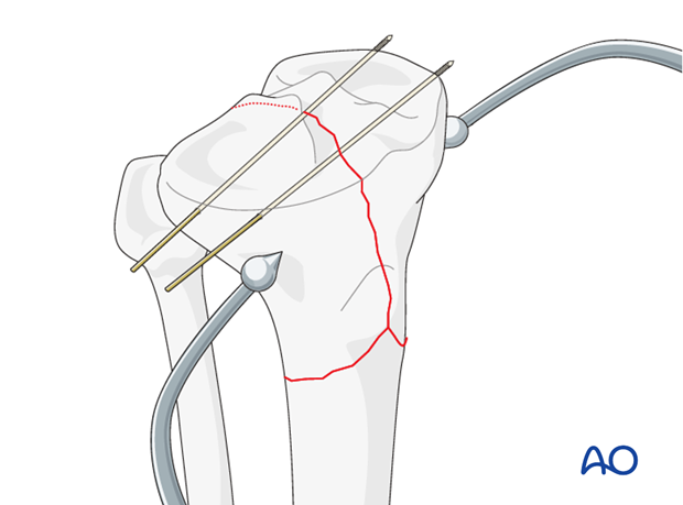
5. Fixation
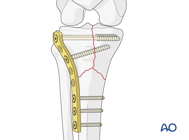
The positioning of the buttress plate is important as the ideal place is at the tip of the fragment and perpendicular to the fracture plane.
Lateral column fixation
The lateral plate is inserted in the space between muscle and periosteum. The plate must be contoured carefully to the bone. Fixation begins with the insertion of a lag screw to stabilize the joint. If this can be done through the plate the first screw inserted is then through the plate.
Click here for a detailed description of the lag screw technique.
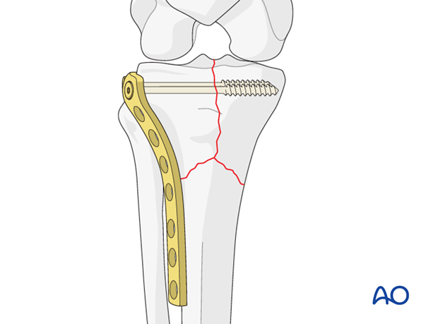
Compression of the metaphyseal fracture component
Using the dynamic compression principle screws are inserted eccentrically into one or two plate holes close the metaphyseal fracture line.
All remaining screws are placed in a neutral way.
Medial column fixation
The medial plate may be applied either posteromedially as we have shown in all our illustrations or anteromedially. The position of the plate is determined by a number of important factors such as the direction of the fracture lines, the forces to be neutralized, the exposure already made, and finally which application will allow for the least traumatic but biomechanically still sound application.
In this illustration we have used a cortex plate applied posteromedially. For fixation we have used only cortical screws.
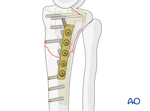
Illustration showing the final result.

Alternative: angular stable plate
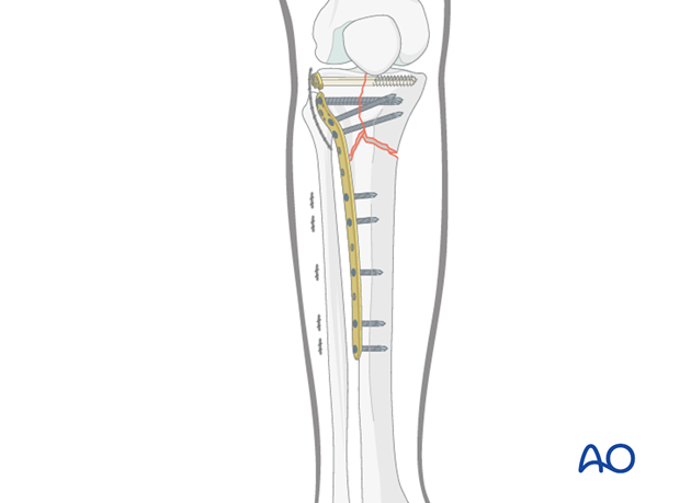
If one is using an angular stable implant (eg, the lateral tibial plate with locking screws) the application of a medial plate is not necessary.
6. Aftercare
Compartment syndrome and nerve injury
Close monitoring of the tibial compartments should be carried out especially during the first 48 hours after surgery to rule out compartment syndrome.
The neurovascular status of the extremity must be carefully monitored. Impaired blood supply or developing neurological loss must be investigated as an emergency and dealt with expediently.
Functional treatment
Unless there are other injuries or complications, mobilization may be performed on post OP day 1. Continuous passive motion (CPM) splints are very helpful in the early phase of rehabilitation. Static quadriceps exercises with passive range of motion of the knee should be encouraged. Afterwards special emphasis should be given to active knee and ankle movement.
Following any injury, and also after surgery, the neurovascular status of the extremity must be carefully monitored. Impaired blood supply or developing neurological loss must be investigated as an emergency and dealt with expediently. The goal of early active and passive range of motion is to achieve as full range of motion as possible within the first 4 - 6 weeks. Optimal stability should be achieved at the time of surgery, in order to allow early range of motion exercises.
Weight bearing
No weight bearing in the treatment of articular fractures for a minimum of 10 – 12 weeks.
Follow up
Wound healing should be assessed on a short term basis within the first two weeks. Subsequently a 6 and 12 week follow-up is usually performed. If a delayed union is recognized, further surgical care will be necessary and should be carried out as soon as possible.
Implant removal
Implant removal is not mandatory and should be discussed with the patient.


