Medial/posteromedial approach to the proximal tibia
1. Principles
If the patient’s hip is normal, position the patient supine, abduct and externally rotate the leg and put it in a figure of 4 position. If the hip is stiff position the patient in a lateral decubitus with the involved limb down.
2. Skin incision
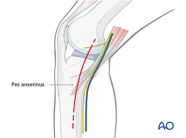
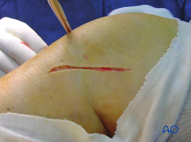
With the knee in slight flexion make a straight or slightly curved incision running from the medial epicondyle towards the posteromedial edge of the tibia. The incision can be extended as needed both proximally and distally as indicated by the dashed line.
Clinical image of the skin incision for the postero-medial approach.
3. Deep dissection
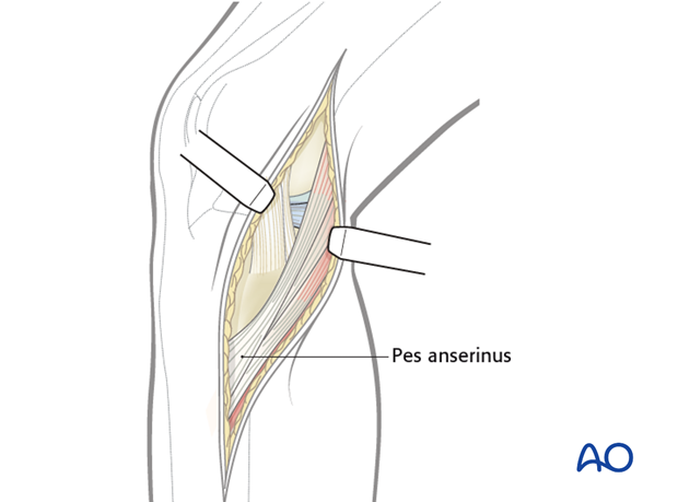
After opening of the fascia identity and expose the pes anserinus.
4. Access
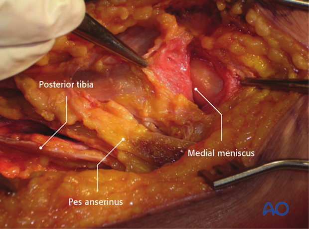
Retract the pes anteriorly and the gastrocnemius posteriorly and distally. Identify the medial edge of the tibial plateau.
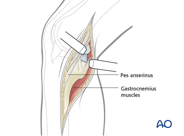
Identify the meniscus and incise the capsule between the meniscus and the edge of the tibial plateau thus gaining access to the knee joint.
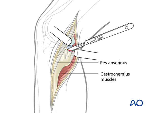
Exposure of the anteromedial part (medial column) of proximal tibia is possible by a subcutaneous dissecting anteriorly. The pes can be retracted posteriorly when dealing with the fracture in this part
Clinical picture showing medial plateau and medial meniscus, pes anserinus, and distally the gastrosoleus.
5. Wound closure
Close the capsule. If needed insert suction drains and close the soft tissues in a routine manner.


