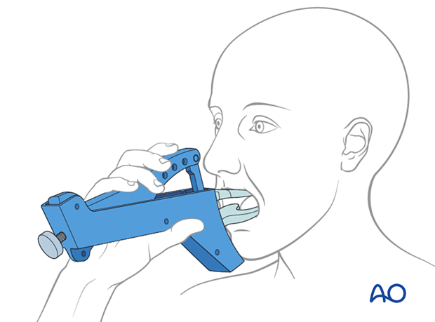
ORIF, lag screws
1. Principles
Lag screw fixation
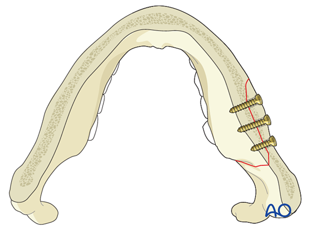
A fracture running obliquely from the buccal to the lingual side in the mandibular body is well suited for lag screw fixation due to the bony cortical thickness. At least two screws inserted perpendicular to the fracture surface is required for 3D stability and neutralization of the rotational forces.
Click here for a detailed description of the lag screw technique and its biomechanical principles.
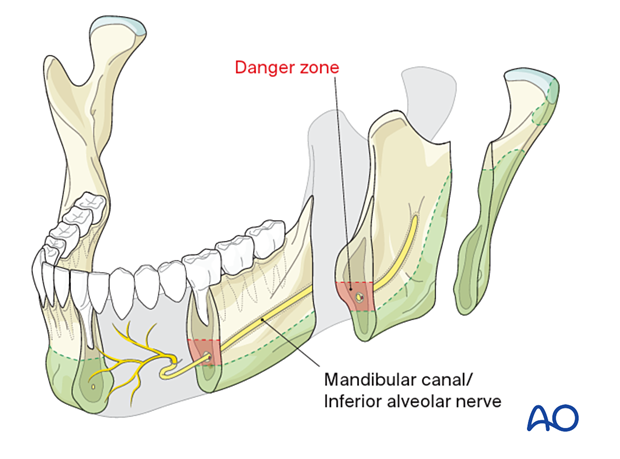
Zones for screw placement in the mandibular body
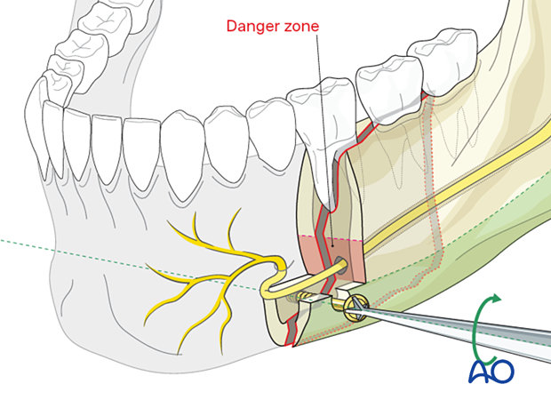
Two structures must not be harmed by screw insertion:
- the mandibular canal with the inferior alveolar nerve
- the tooth roots.
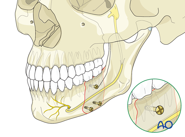
The classical description for screw placement in plate and screw osteosynthesis is depicted for orientation. Monocortical screws can be inserted above the level of the mandibular canal. The use of bicortical screws is restricted to the area below the nerve canal at the lower mandibular border.
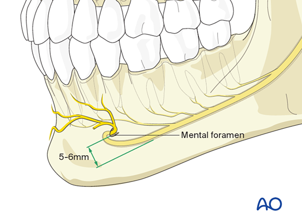
Bicortical screws should be inserted more than 5-6 mm caudal to the mental foramen because of the inferior alveolar nerve path in this area.
Screw insertion pattern
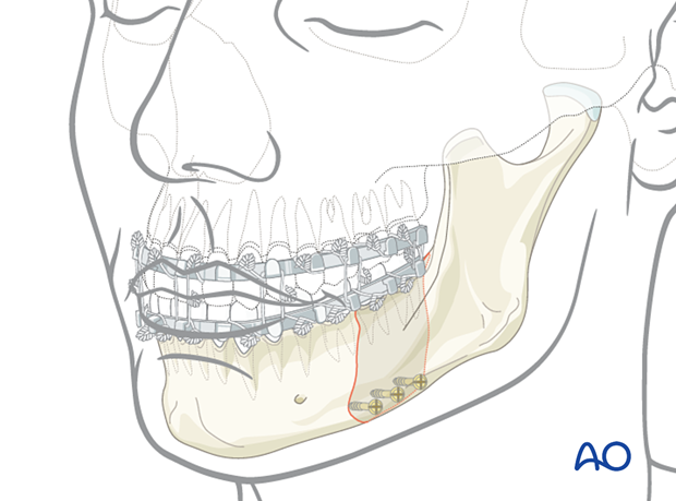
Screws can be inserted in a serial pattern perpendicular to the fracture line at the mandible's lower border.

Alternatively, screws can be placed in a tripod fashion using the lower and the upper insertion zone. In the upper zone, the anatomic relation between the nerve canal and the tooth apices varies.
Correct screw insertion could be dangerous and difficult to accomplish unless a tooth is missing.
Sometimes there is no space between the tooth apices and the nerve, which is a clear contraindication to follow this pattern.
Using the lag screws in a tripod fashion provides additional stability. Note how the superior screw avoids both the dental roots and the alveolar nerve in the illustration.
Number of screws
A minimum of two screws is required to withstand rotational forces. For additional stability, it is recommended to use additional screws.
Special considerations
The following special considerations may need to be considered:
2. Patient preparation
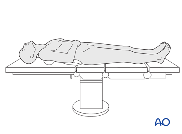
This procedure is typically performed with the patient placed in a supine position.
3. Selection of approach
These fractures can often be approached and treated through the transoral approach (combined with the transbuccal system when necessary).
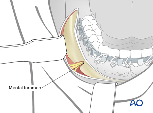
Depending on fracture severity, the difficulty of fixation, and/or the presence of a suitable laceration, a transcutaneous approach through the submandibular route may be indicated.
An advantage of a transcutaneous approach is that it offers an opportunity to place a bone clamp and visualize the lingual cortex reduction.
4. Reduction
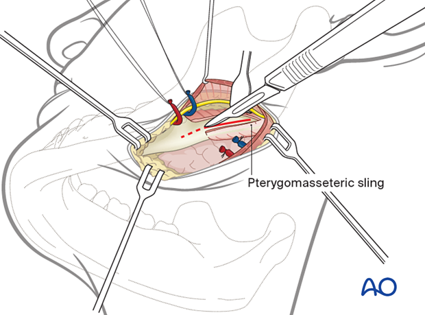
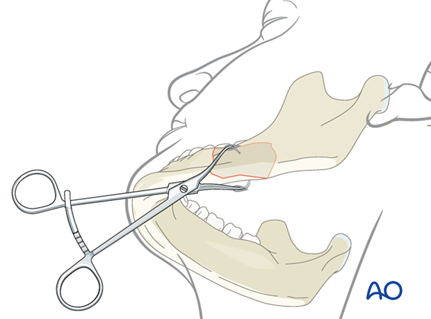
The patient is first placed into MMF. The bony fragments are then reduced manually.
A fixation clamp can be applied to maintain the reduction. Therefore, a towel clamp may be used transcutaneously to keep the reduction at the lower border with the prongs coming in from the medial side and laterally. Usually, there should be no risk of injuring the marginal mandibular branch of the facial nerve. The towel clamp may compromise the accessibility to the lower border.
5. Fixation
Alternative: transbuccal system
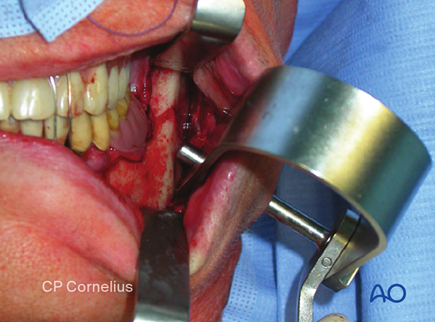
The transbuccal system's use may become necessary in the dorsal, caudal region of the mandibular body, which is inaccessible transorally with a screwdriver.
Click here for a detailed description of the transbuccal system.
Screw insertion
The screw should be angled perpendicular to the fracture line.
A crucial point is choosing the appropriate screw length so that the far tip of the screw fully engages the far cortex, with the screw tip exiting slightly beyond the bone's lingual cortex.
Click here for a detailed demonstration of the lag screw technique.
Completed osteosynthesis
The MMF is now released. It is critical to verify the occlusion.
Visual assessment of the lingual side reduction is not possible unless submandibular exposure has been performed. The anatomic reduction can be further assessed with intraoperative and postoperative CT imaging.
6. Aftercare following ORIF of mandibular symphysis, body, angle, and ramus fractures
Use of jaw bra
If significant degloving of the soft tissues of the mandible has occurred, there may be a consideration for using a jaw bra or similar support dressing.
Arch bars
If arch bars or MMF screws are used intraoperatively, they are usually removed at the conclusion of surgery if proper fracture reduction and fixation have been achieved. Arch bars may be maintained postoperatively if functional therapy is required or if required as part of the fixation.
X-Rays
Postoperative x-rays are taken within the first days after surgery. In an uneventful course, follow-up x-rays are taken after 4–6 weeks.
Follow up
The patient is examined approximately 1 week postoperatively and periodically thereafter to assess the stability of the occlusion and to check for infection of the surgical wound. During each visit, the surgeon must evaluate the patient's ability to perform adequate oral hygiene and wound care and provide additional instructions if necessary. Many patients need to be seen regularly for replacement of their intermaxillary elastics and to encourage range of motion in their TMJ in the later course of the treatment.
Follow-up appointments are at the discretion of the surgeon and depend on the stability of the occlusion on the first visit. If a malocclusion is noted and treatable with training elastics, weekly appointments are recommended.
The patient should be warned to continue routine follow up with their dentist. Fractures near the dental roots can often result in delayed loss of tooth viability, requiring periapical films and additional dental procedures.
Malocclusion
If a malocclusion is detected, the surgeon must ascertain its etiology (with appropriate imaging technique). If the malocclusion is secondary to surgical edema or muscle splinting, training elastics may be beneficial. The lightest elastics as possible are used for guidance, because active motion of the mandible is desirable. Patients should be shown how to place and remove the elastics using a hand mirror.
If the malocclusion is secondary to a bony problem due to inadequate reduction or hardware failure or displacement, elastic training will be of no benefit. The patient must return to the operating room for revision surgery.
Basic postoperative instructions
DietDepending upon the stability of the internal fixation, the diet can vary between liquid and semi-liquid to “as tolerated”, at the discretion of the surgeon. Any elastics are removed during eating.
Patients having only extraoral approaches are not compromised in their routine oral hygiene measures and should continue with their daily schedule.
Patients with intraoral wounds must be instructed in appropriate oral hygiene procedures. The presence of the arch-bars and any elastics makes this a more difficult procedure than normal. A soft toothbrush (dipping in warm water makes it softer) should be used to clean the surfaces of the teeth and arch-bars. Any elastics are removed for oral hygiene procedures. Chlorhexidine oral rinses should be prescribed and used at least three times each day to help sanitize the mouth. Chlorohexidine may cause staining of the teeth and should not be used longer than necessary. For larger debris, a 1:1 mixture of hydrogen peroxide (0.25%)/chlorhexidine (0.12%) can be used. The bubbling action of the hydrogen peroxide helps remove debris. A water flosser, providing a water jet, is a very useful tool to help remove debris from the wires. If a this is used, care should be taken not to direct the jet stream directly over intraoral incisions as this may lead to wound dehiscence.
Physiotherapy can be prescribed at the first visit and opening and excursive exercises begun as soon as possible. Goals should be set, and, typically, 40 mm of maximum interincisal jaw opening should be attained by 4 weeks postoperatively. If the patient cannot fully open his mouth, additional passive physical therapy may be required such as Therabite® or tongue-blade training.
