ORIF - Lag screw fixation
1. Principles
Anatomical reduction
Anatomical reduction of the articular fracture component and fixation with absolute stability is mandatory.
In theory, simple split fractures can be managed with lag screws only, and lateral plateau fracture treatment is shown here.
However, axial forces from joint reaction and even partial weight-bearing forces are significant and a buttress plate is usually preferred. This is particularly true for the medial plateau, where varus collapse is common due to the greater forces across the joint and reduced density of lateral cancellous bone to hold purchase of the screw thread.
2. Patient preparation and approach
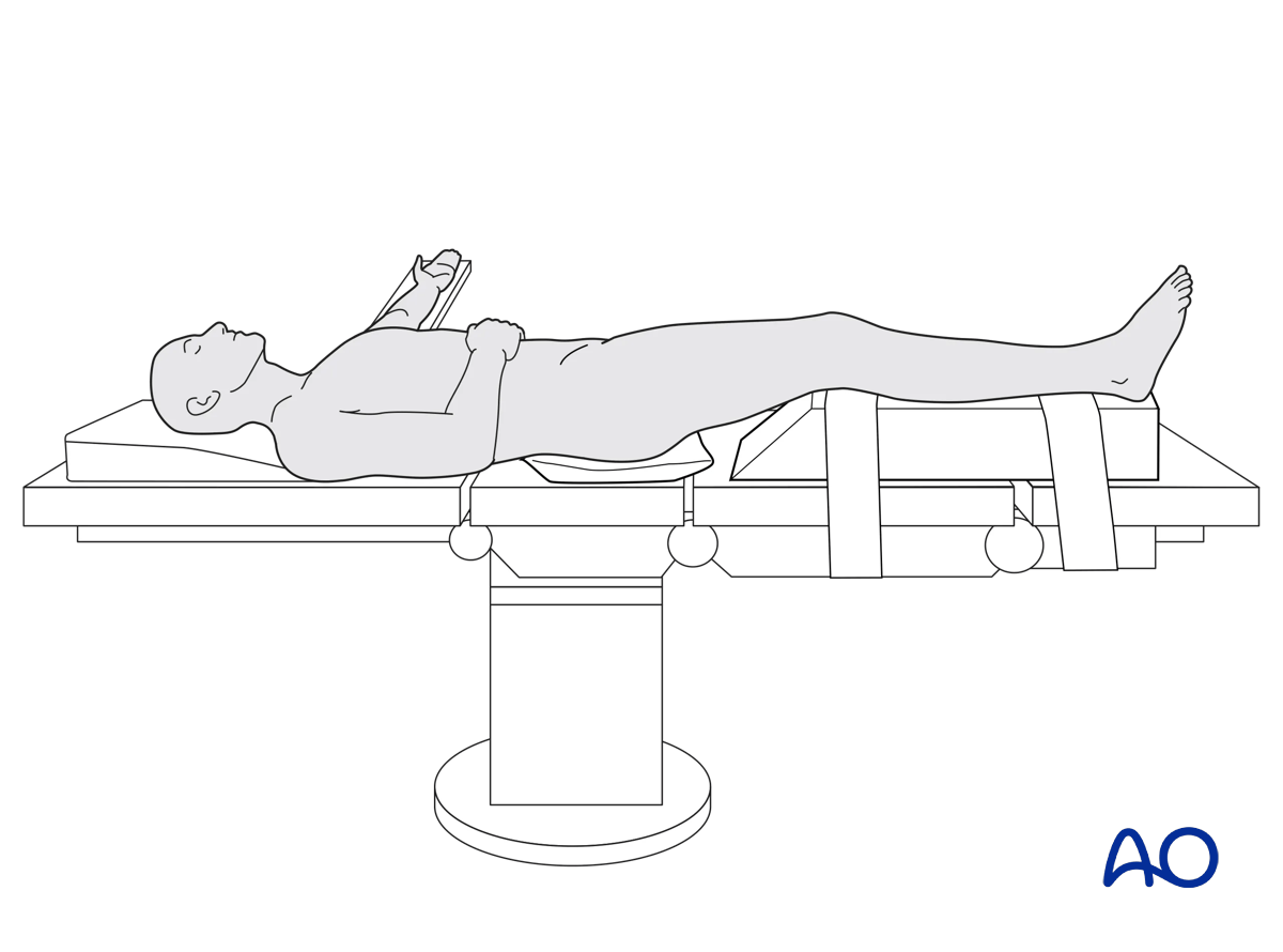
Patient preparation
This procedure is normally performed with the patient in a supine position.
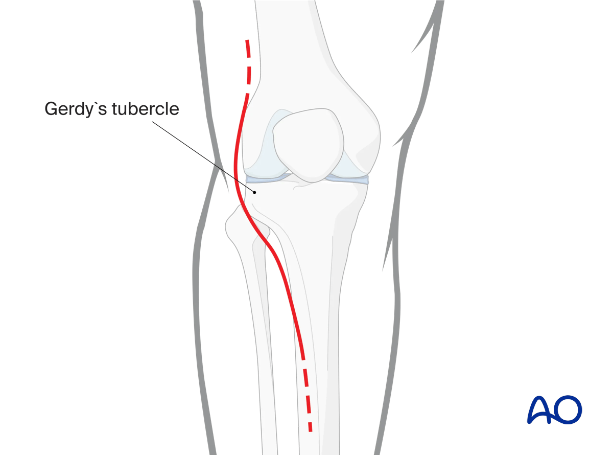
Approach
For this procedure an anterolateral approach is used.
3. Reduction
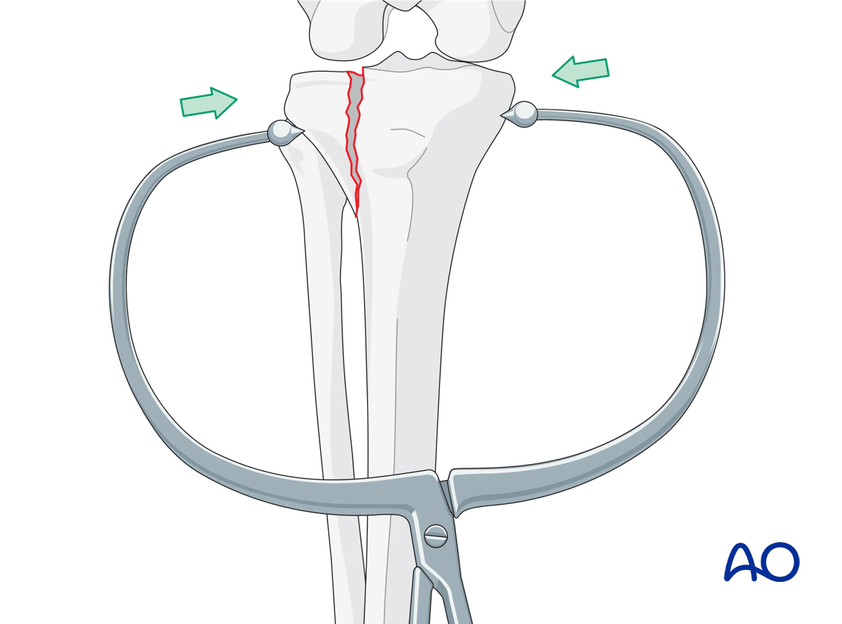
Clamps
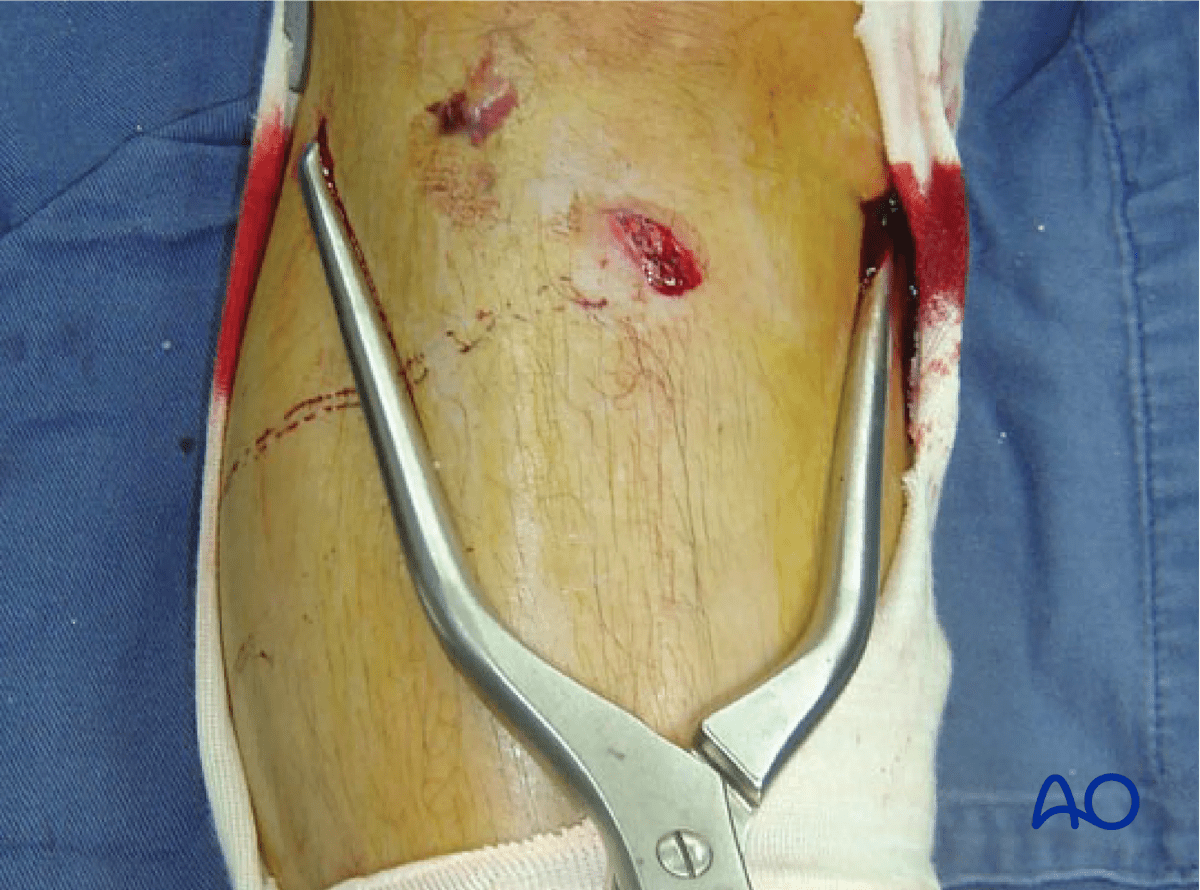
Indirect reduction may be attempted by external manipulation of the fractured fragment using clamps. The accuracy of the reduction should be checked with radiographic confirmation with or without arthroscopy. In cases where adequate closed reduction is not achieved the joint must be opened in order to carry out an anatomical reduction of the joint surface. This may also be necessary to repair the lateral meniscus which may be injured in association with these fractures.
Clinical image showing the clamp application.
4. Fixation
Lag screw application
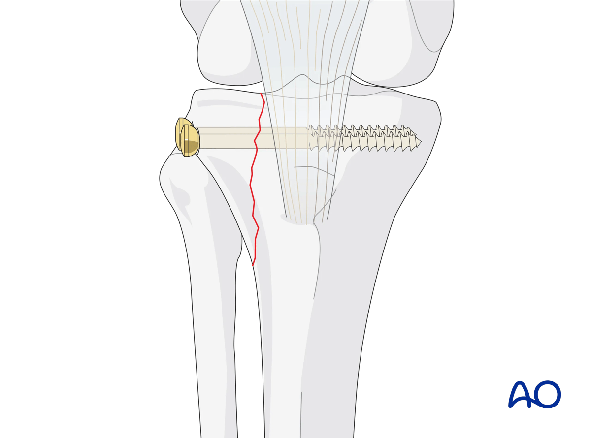
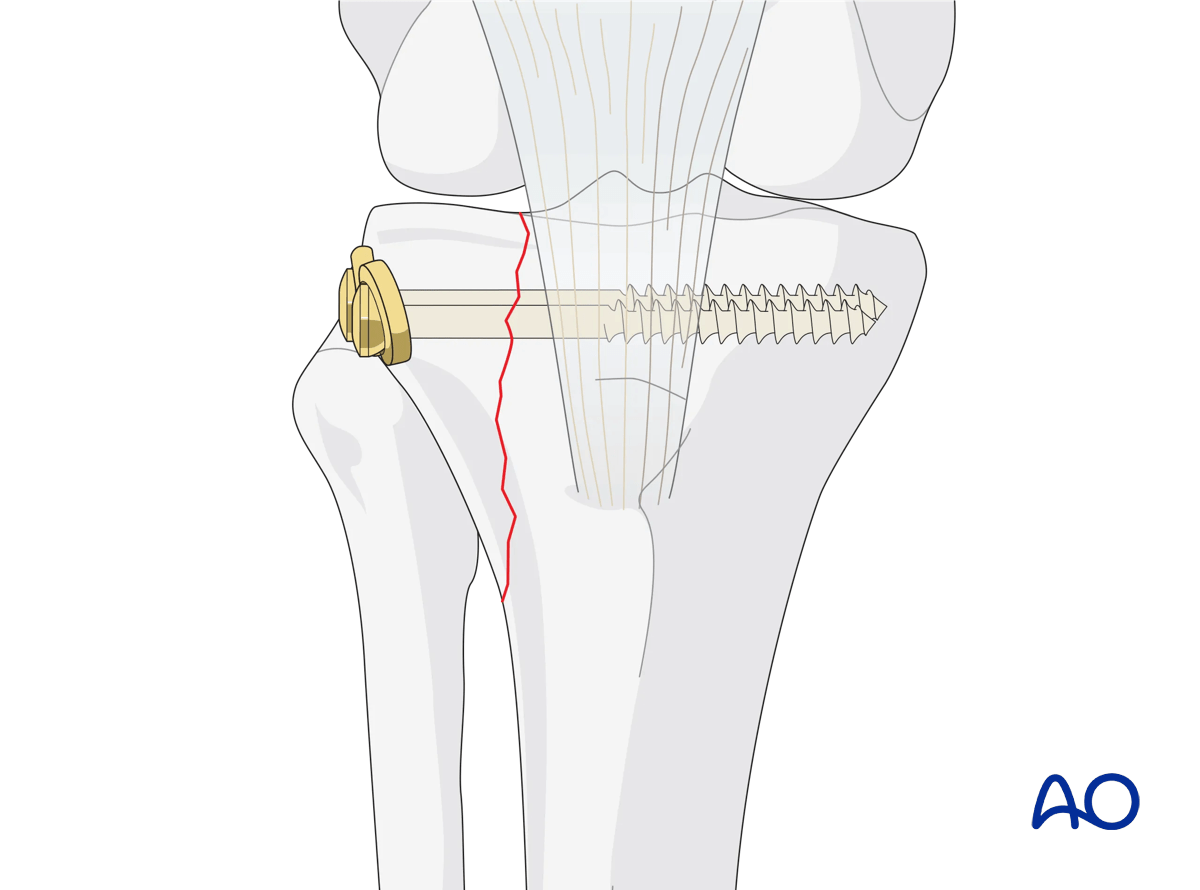
Once reduction of the joint fragment is obtained, two lag screws (by design or technique) can be employed to securely compress and stabilize the fracture, reducing the need for a buttress plate in compliant patients with good bone substance.
A detailed description of the lag screw technique is provided here:
Washers
The cortex in the lateral tibial head is thin and will not resist the strong force created when the screw threads advance into the subchondral bone of the medial plateau. Therefore, the use of washers and lag screws is advised.
5. Aftercare
Compartment syndrome and nerve injury
Close monitoring of the tibial compartments should be carried out, especially during the first 48 hours after injury and again after surgery to rule out compartment syndrome. More information is provided here:
The neurovascular status of the extremity must be carefully monitored. Impaired blood supply or developing neurological loss must be investigated as an emergency and dealt with expediently.
Consideration for DVT prophylaxis
Oral or subcutaneous administration of DVT prophylaxis for six weeks should be strongly considered.
Functional treatment
Optimal stability should be achieved at the time of surgery, in order to allow early range of motion exercises. Unless there are other injuries or complications, mobilization may be performed on post OP day 1. If available, continuous passive motion (CPM) splints can be very helpful in the early phase of rehabilitation. Static quadriceps exercises with passive range of motion of the knee should be encouraged. Afterwards special emphasis should be given to active knee and ankle movement.
The goal is to achieve as full range of motion as possible within the first 4–6 weeks.
Weight bearing
Weight-of-leg weight bearing is initiated depending on patient comfort. Depending on the severity of the articular displacement, weight bearing can begin as early as 6 weeks postoperatively. In situations where articular displacement was significant weight bearing should be delayed for 10–12 weeks.
Follow up
Wound healing should be assessed within the first two weeks. Subsequently, a 6- and 12-week follow-up with radiographic assessment is usually performed. If a delayed union is recognized, further surgical care may be necessary and should be carried out as soon as possible. Residual knee instability may require delayed reconstruction.
Implant removal
Implant removal is not mandatory and should be discussed with the patient.


