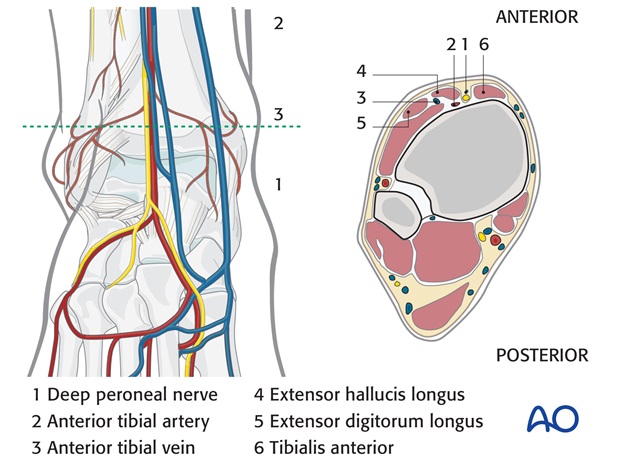
ORIF
1. Principles
General considerations
If the Volkmann's fracture has a significant proportion of the articular surface, it should be fixed anatomically.
It is possible to obtain good access to the posterolateral fracture and fibular fracture through a single posterolateral approach. Direct reduction of the Volkmann's fragment is possible, facilitating anatomic reduction and fixation. This requires the patient to be positioned prone.
Alternatively, the fibular fracture can be fixed through a standard lateral approach and the Volkmann's fragment can be indirectly reduced by dorsiflexing the ankle and then secured with screws inserted through anterior stab incisions.

Order of fixation
In these factures the talus is often unstable within the ankle mortise. Stability is restored when the fibular fracture is stabilized. The Volkmann's fragment can then be reduced and fixed.
Choice of implant – Lateral fixation
As this is a simple fracture a lag screw and neutralization plate is the most appropriate method of fixation.
Anatomic plates are available, and their lower profile may reduce postoperative discomfort due to prominent hardware. As these plates use locking screws, they may provide more secure fixation in osteoporotic bone.
Medial injury
There is a rupture of the deltoid ligament. Once the talus is stabilized within the ankle mortise, the ruptured deltoid ligament usually heals without the need for surgical repair.
Occasionally the ligament may lie between the talus and the medial malleolus blocking reduction, in which case the medial side will need to be opened to remove ligament. Once the medial side is open the surgeon may choose to repair the ligament.
2. Patient preparation
Depending on the approach, the patient may be placed in the following positions:
- Supine position
- Lateral position
- Supine position, figure-of-four
- Prone position (preferred for posterior fractures)
3. Approaches
If the direct reduction and fixation of the Volkmann's fragment is chosen, a posterolateral approach is used, both for this fragment and the fibular fracture.
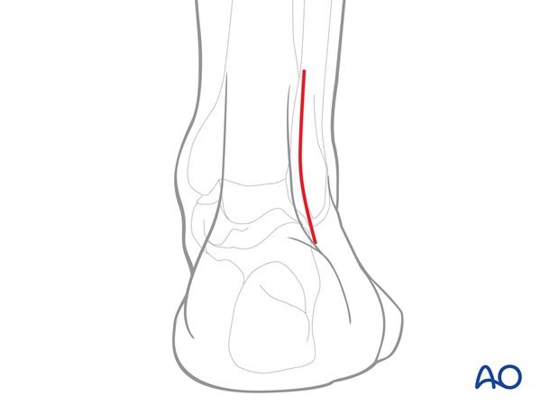
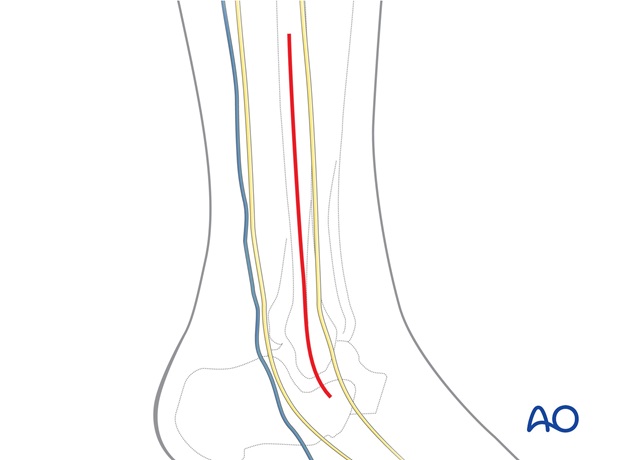
Alternatively, the fibular fracture may be approached directly through a standard lateral approach with additional anterior stab incisions.
4. Fixation
Lateral fixation
The lateral side is usually a simple oblique fracture, and is fixed with a lag screw and neutralization plate.
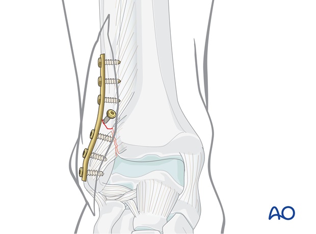
Alternatively, and in particular if there is poor bone or a small fragment, an Antiglide plate may be used.
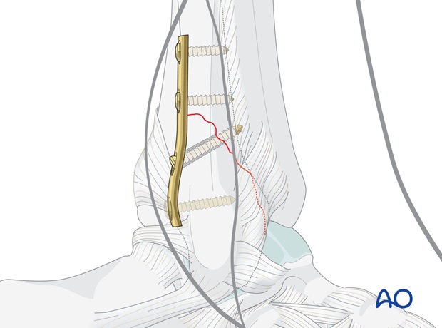
Fixation of Volkmann's fragment
This may be fixed through direct reduction via a posterolateral approach with lag screws.
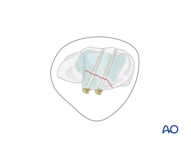
If indirect reduction is chosen, sagittal lag screws are inserted through separate stab incisions.
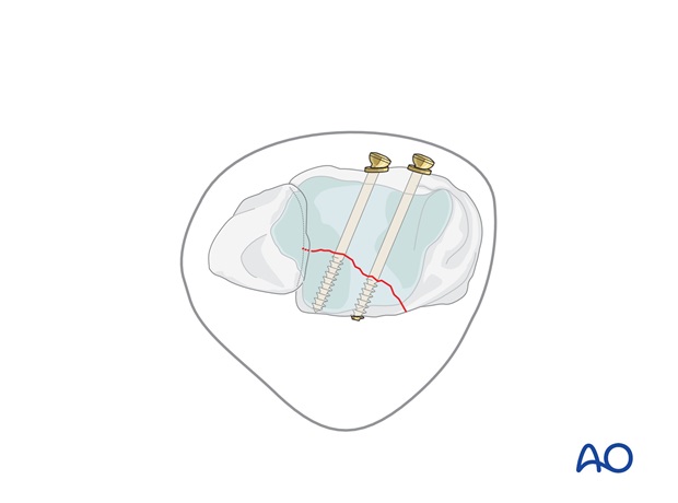
5. Check of osteosynthesis
Check the completed osteosynthesis by image intensification.
Make sure the intra articular components of the fracture have been anatomically reduced.
Make sure none of the screws are entering the joint. This needs to be confirmed in multiple planes.
6. Postoperative treatment of infra- and trans-syndesmotic malleolar fractures
A bulky compression dressing and a lower leg backslab, or a splint, are applied, and the limb is kept elevated for the first 24 hours or so, in order to avoid swelling and to decrease pain.
In anatomically reconstructed, stable malleolar fractures, early active exercises and light partial weight bearing are encouraged after day one. In osteoporotic bone, weight bearing should be postponed.
X-ray evaluation is made after 1 week and then monthly until full healing has occurred. Progressive weight bearing is recommended as tolerated.
