ORIF
1. General considerations
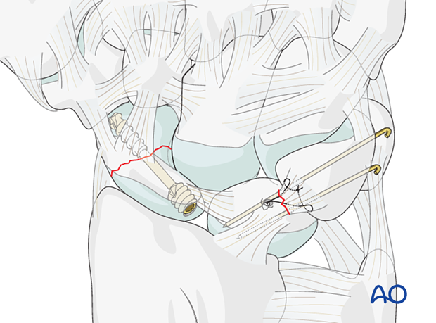
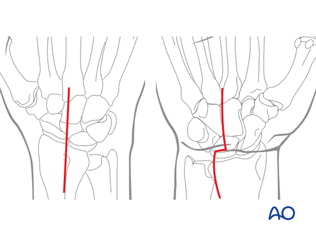
Greater arc injuries are a combination of fractures and ligament injuries.
A scaphoid fracture is most frequently involved and needs ORIF. A combination with other fractures and ligament injuries depends mainly on the wrist position during the fall.
Here, the most frequent injury patterns and their surgical management sequence are presented.
Imaging
Conventional radiographs do not adequately demonstrate the complete extension of the injury. A CT or MRI scans are recommended to reveal the degree of displacement and extent of soft tissue injury.
2. Preliminary treatment
Urgent closed reduction is indicated. This is particularly important if surgery is likely be delayed, eg, if transfer to a specialized hand and wrist unit is planned.
Closed reduction is a preliminary to operative treatment and has the following benefits:
- Reducing the risk of median nerve injury
- Restoration of carpal alignment
- Pain relief
- Facilitating surgical repair
If closed reduction is not successful, open reduction is necessary as soon as possible (due to the risk of median nerve compromise).
Splint immobilization
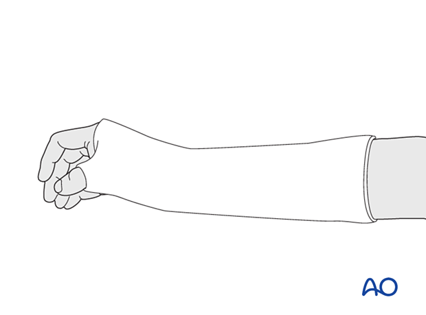
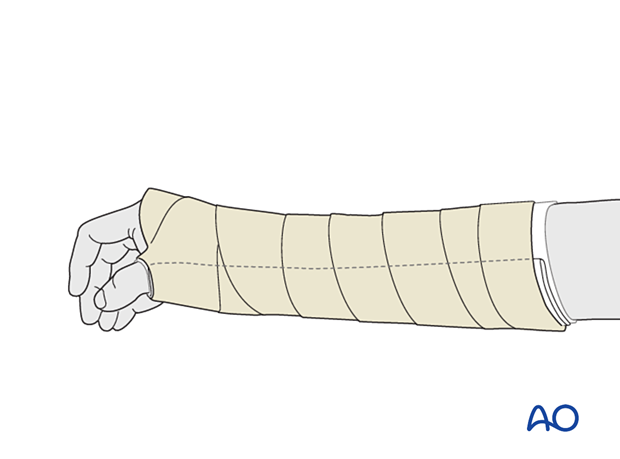
After emergency reduction, the wrist is immobilized in a palmar plaster splint in the neutral position.
3. Patient preparation
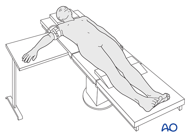
The patient is usually supine with the arm on a radiolucent side table.
4. Approaches
For this procedure, a dorsal approach may be used.
If the reduction is not anatomical or the median nerve needs to be explored (carpal tunnel decompression), a combination with a palmar approach is necessary.
5. Scaphoid fractures
For surgical treatments of isolated scaphoid fractures, see the section on the corresponding scaphoid fracture:
6. Scaphoid fracture and lunotriquetral ligament injury
The treatment sequence for this injury is:

7. Scaphoid and capitate fracture
The treatment sequence for this injury is:
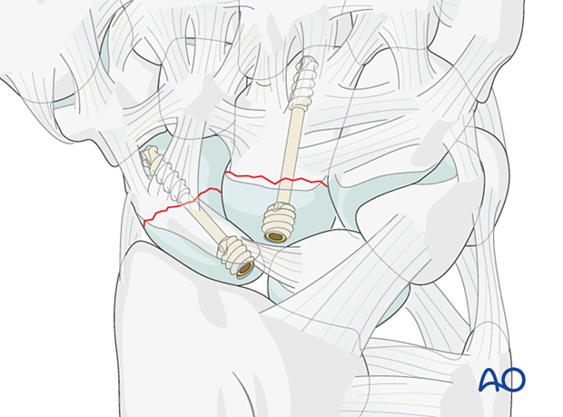
8. Scaphoid, capitate, and triquetral fracture +/- ulnar styloid fracture
The treatment sequence for this injury is:
- Screw fixation of a capitate fracture
- Screw or K-wire fixation of a triquetrum fracture
- Antegrade screw fixation of a scaphoid fracture
A small triquetral fragment may be stabilized with K-wires.
Associated ulnar styloid fractures often do not need separate fixation.
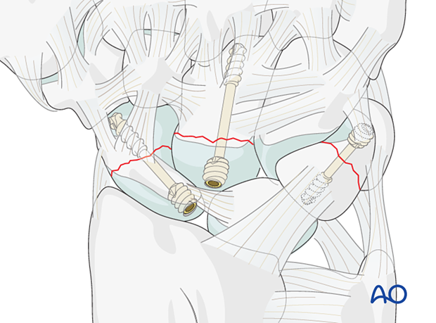
9. Final check
After final confirmation, using image intensification, cut and bend over any K-wires, so they do not protrude through the skin.
10. Aftercare
The aftercare can be divided into four phases of healing:
- Inflammatory phase (week 1–3)
- Early repair phase (week 4–6)
- Late repair and early tissue remodeling phase (week 7–12)
- Remodeling and reintegration phase (week 13 onwards)
Full details on each phase can be found here.
Pain control
To facilitate rehabilitation, it is important to control the postoperative pain adequately. To do so, the following should be considered:
- Management of swelling
- Appropriate splintage
- Appropriate oral analgesia
- Careful consideration of peripheral nerve blockade
Immediate postoperative treatment
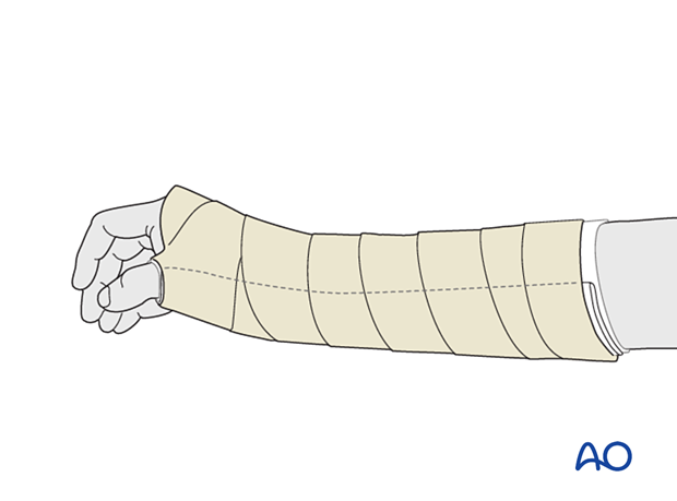
Immobilize the wrist with a well-padded below-elbow splint for 2 weeks.
Splinting helps with soft-tissue healing, especially of the ligaments cut during a palmar approach.
Follow-up
After 2 weeks, remove the sutures, check the skin over the K-wires, and apply a below-elbow scaphoid cast with slight dorsiflexion at the wrist for 6–8 weeks. Then the K-wires are also removed with appropriate pain control.
Check the skin regularly every 2 weeks in the outpatient clinic.
