Posterior C2–C3 fusion
1. Introduction
Posterior instrumentation is either combined with open reduction or is performed after closed reduction has been successfully achieved.
2. Reduction
Reduction may be performed by gentle traction, especially in acute cases. If not achieved or in more delayed cases, surgical reduction is indicated.
This involves soft tissue release (ligaments, capsules, and scar tissues found in delayed presentation) followed by gentle manipulation.
The final reduction must be confirmed using a C-arm.
3. Approach and positioning
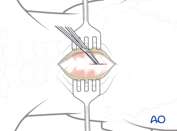
This procedure is performed using a posterior approach with the patient placed in the prone position.

4. C2–C3 posterior fusion
C2 Screws
Polyaxial pedicle screws are inserted into C2 following the standard technique.

C3 Screws
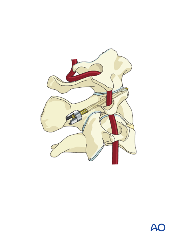
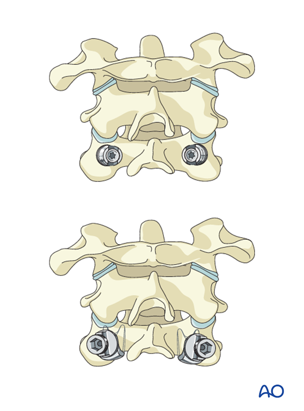
For fixation of C3, one of the following techniques can be applied:

Rod insertion
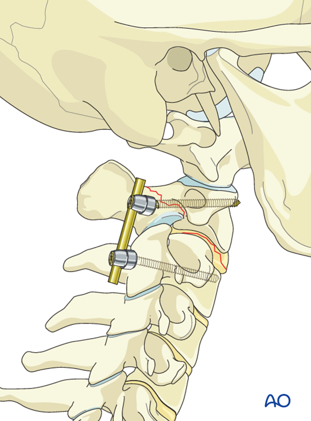
The rod is placed, and screws are closed with slight compression to enhance the stability of the construct.

5. Aftercare
Patients are made to sit up in bed in the evening after the operation.
A collar is commonly used following surgical stabilization to moderate patient activity.
The purpose of a collar is to prevent ranges of motion outside of limits deemed favorable for fracture healing. The collar is optional.
Patients with intact neurological status are made to stand and walk on the first day after surgery. Patients can be discharged when medically stable or sent to a rehabilitation center if further care is necessary. This depends on the comfort levels and presence of other associated injuries.
Patients are generally followed with periodical x-rays at 6 weeks, 3 months, 6 months, and 1 year.


