Prone position
1. Positioning
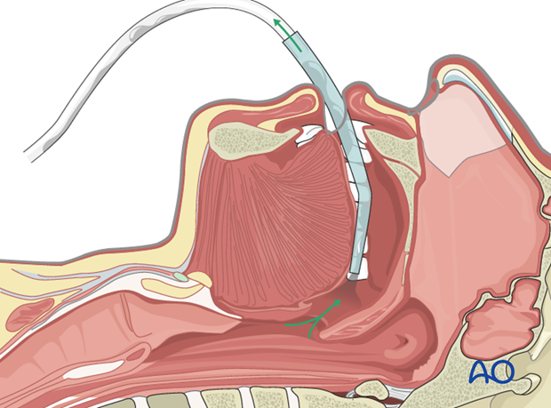
The patient is anesthetized and intubated while taking care not to mobilize the unstable cervical spine.
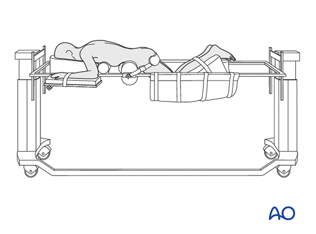
The head is attached to Gardner-Wells tongs or a halo, and suspended with two ropes. One rope (extension rope) goes over a pulley while the other one pulls in line with the spine in a neutral position. Note that this setup requires a Jackson spine frame. A 10 kg weight attached to either rope will suspend the head and keep it stable. The arms are tucked at the side and taped in place.
The entire table is placed in a reversed Trendelenburg position to minimize bleeding as well as intraoccular pressures.
Make sure that there are adequate personnel to receive and turn the patient from supine to prone position on a radiolucent operating table.
Because rotation or flexion movements at the level of injury can result in worsening of neurological status, a turning frame can be used, if available, to minimize spinal displacement during positioning.
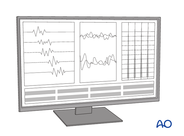
In patients with unstable injuries, who are at risk for neurological deterioration during positioning, base line electrodiagnostic signals can be obtained to allow for comparison after positioning. Other options in high-risk patients include awake positioning or a wakeup test after positioning.
Lateral fluoroscopic views should be obtained immediately after positioning to confirm acceptable alignment.
The patient is placed on two horizontal padded bolsters (one at the level of sternum and one at the level of anterior iliac spine) or a frame. Designated radiolucent spine surgery frames with similar supports are ideal.
When lower cervical levels need to be seen on lateral images, adhesive straps are used to pull the shoulders downwards.

2. Anesthesia
With unstable fractures, either an awake or fiberoptic intubation should be performed. In patients with spinal cord injury, it is essential to avoid hypotensive anesthesia and the mean arterial blood pressure should be maintained above 80 mmHg.

3. Preoperative antibiotics
Antibiotics should be administered 30–60 minutes prior to the incision.
A cephalosporin antibiotic with good Gram-positive coverage is generally recommended.
A second-generation cephalosporin and clindamycin is typically used. Another alternative is ampicillin, clindamycin, and gentamycin.
4. Spinal cord monitoring
Spinal cord monitoring is optional.

5. Fluoroscopy/x-ray control
Preoperative fluoroscopy is mandatory. Before draping, ensure that a good lateral fluoroscopy view can be obtained throughout all levels being instrumented. However, this can be challenging or even impossible at the cervicothoracic junction.
The incision can be planned based on the lateral fluoroscopic view.


