ORIF of transverse fractures
1. Principles
Successful treatment of acetabular fractures noted intraoperatively is critical to achieve component ingrowth, pain control, and long-term survivorship of the arthroplasty.
For further information about standard techniques for ORIF in native acetabular fractures, please refer to the acetabulum section of the AO Surgery Reference.
2. Approach
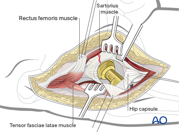
Intraoperative unstable fractures already have a significant surgical approach that can be extended to address the fracture. New minimally invasive techniques make it more challenging to diagnose these fractures.
Surgeons that routinely utilize fluoroscopy during total hip replacement have the advantage of identifying fractures fluoroscopically. Without fluoroscopy, diagnosis depends on the surgeon experience noting abnormal cup mechanics during insertion.
The surgical approach may predict the fracture location:
- Direct anterior and anterolateral approaches are more commonly associated with anteriorly based fractures.
- Direct lateral and posterior approaches to the hip are more commonly associated with posterior type fractures.
Medial wall fractures can be associated with any approach.
If the fracture extends across the superior aspect of the acetabulum towards the posterior wall, the anterior approach to the acetabulum can be extended to a Smith-Petersen approach which will allow for adequate visualization of the outer table, with the patient in the supine position.
3. Cup removal
In the setting of infection, loosening, or osteolysis, cup removal may lead to acute bone loss and/or the creation of periprosthetic fractures. If this destabilizes the acetabular support of the arthroplasty, fracture specific reduction and fixation techniques are necessary. Depending on the fracture pattern, ORIF with plate and screw and/or column fixation may be indicated.
It is critical to remove the cup without causing additional fractures or bone defects.
Liner removal
The cup liner is removed prior to cup removal.
Cup screw removal
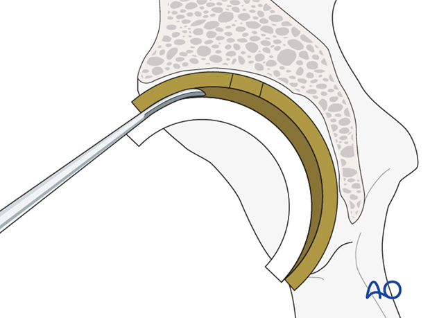
Ensure that all the cup screws are removed prior to attempting to remove the cup itself.
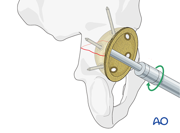
Acetabular component removal
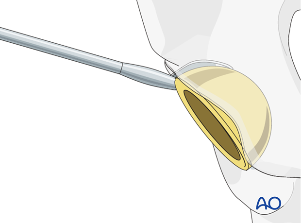
4. Reduction and fixation of transverse fractures
Transverse fracture reduction
In these fractures, anatomic reduction of the articular surface is not relevant. Columnar stability and wall containment are needed.
Reduction of a transverse fracture requires two main steps: lateralization, and derotation of the ischiopubic fragment. It is important that the reduction is correct across the entire fracture, as the posterior surface may appear to be reduced satisfactorily while anterior rotation or displacement persists.
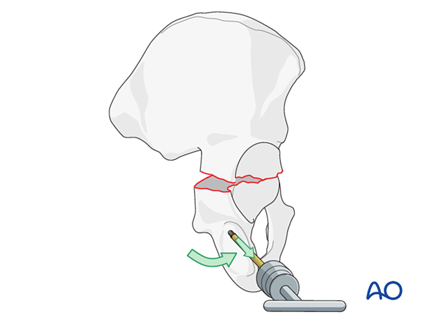
The reduction is temporarily fixed with two clamps.
Transverse fracture stabilization
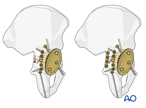
Transverse fractures can be stabilized with lag screws across the posterior aspect of the fracture with neutralization plating. The anterior aspect of the fracture should be further stabilized with the anterior column screws utilizing standard techniques.
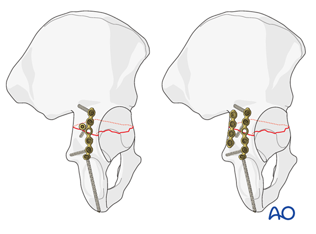
Radiographic confirmation
Reduction and stabilization of the fracture should be confirmed using fluoroscopy.
5. New acetabular cup implantation
With the anatomy of the acetabulum restored, a standard multi-hole cup can be utilized.
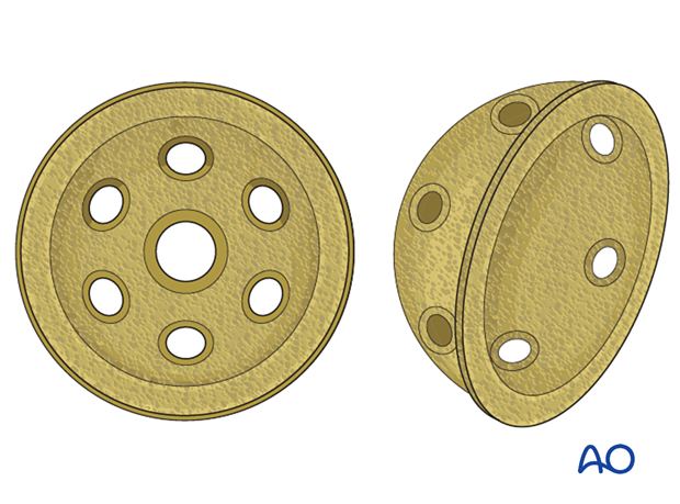
Acetabular reaming
The acetabulum should be reamed gently as not to compromise the reduction. The new cup can be implanted.
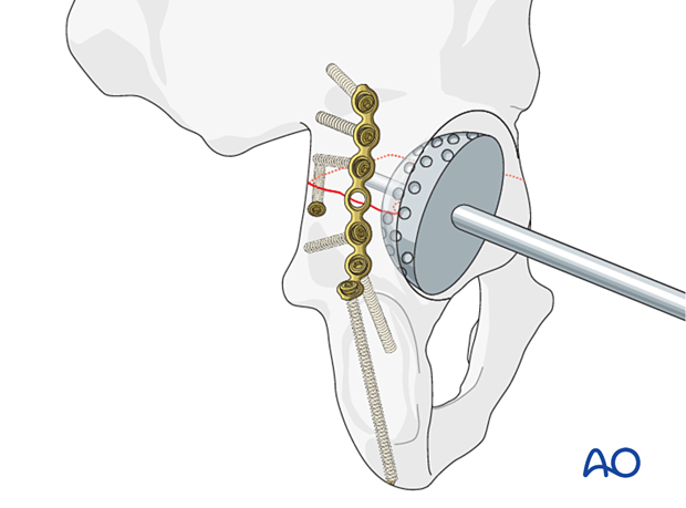
Cup screw fixation
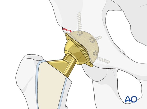
The cup should achieve rim fit. Insert multiple screws in different planes to achieve stabilization to allow bony ingrowth.
Residual bony defects should undergo bone grafting with autograft or allograft, per surgeon's preference.

For further details about the multihole press-fit cup implantation please refer to the treatment: Revision of cup to multihole press-fit cup.
6. Aftercare following ORIF
Postoperative management
Postoperative management should include careful monitoring of hematocrit and electrolytes particularly in the elderly patients.
Postoperative IV antibiotics should be administered up to 24 hours.
Consideration should be given to anticoagulation for a minimal course of 35 days. If there are thromboembolic complication this treatment is extended.
Drains can be discontinued when output is less than 30 to 50 cc per 12 hours.
Patient mobilization
Immediate mobilization of the patient should commence. If fracture stability will allow, the patient should be made weight bearing as tolerated as soon as possible. Long periods of limited weight bearing are extremely detrimental to patient recovery.
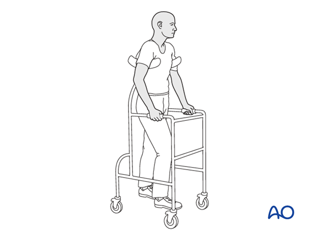
Precautions against hip dislocation
Hip precautions can be extremely important in patients who have suffered intraoperative acetabular fracture. Much work has been done to minimize the surgical exposure during hip arthroplasty to decrease the risk of dislocation. These advantages are typically removed when acetabular stabilization need to be performed. A dislocation in the postoperative course of such a patient can be disastrous.
Patients are instructed to follow standard hip precautions against dislocation based upon the surgical approaches for hip arthroplasty.
Wound healing
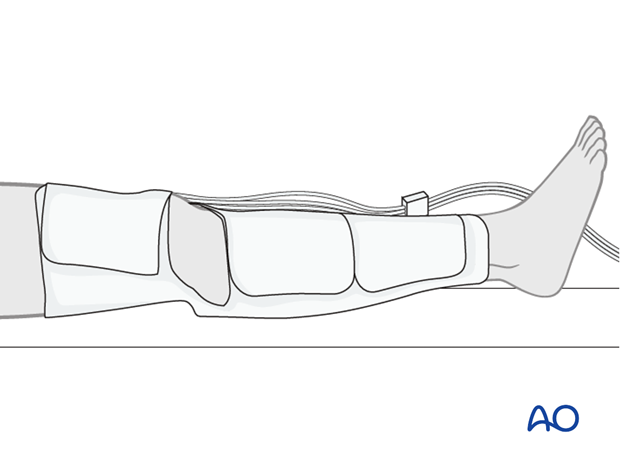
Avoidance of edema postoperatively is critical for both wound healing and patient mobilization. This can be aided by pneumatic compression devices. If negative pressure wound therapy is utilized, it can be discontinued after 5 to 7 days. Staples or sutures are typically removed at 14 to 21 days.
