Lag-screw fixation
1. General considerations
Partial articular fractures require anatomical reduction and may be fixed with lag screws if the size of the fragment allows.
The reduction can be assisted with arthroscopy if skill and equipment are available.
The joint may collapse if there is impaction or comminution while the lag screw is tightened. In this case, plate fixation should be considered for chondral support.
This fracture type may be associated with interphalangeal (IP) joint dislocation. In this case, the dislocation must be manipulated, and any interposed soft-tissue structures removed.
Fracture configuration
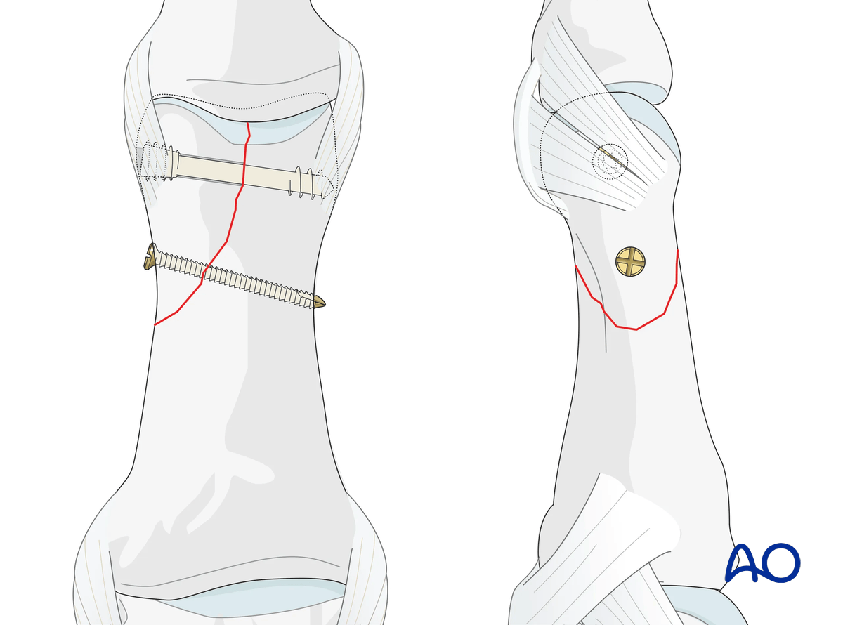
Fractures in the sagittal plane as well as in the dorsal coronal plane are accessible to direct screw fixation.
Fractures in the palmar coronal plane are particularly difficult to treat as direct fragment stabilization is not possible because direct screw fixation would damage the bearing zone of the articular surface.
Anatomical reduction
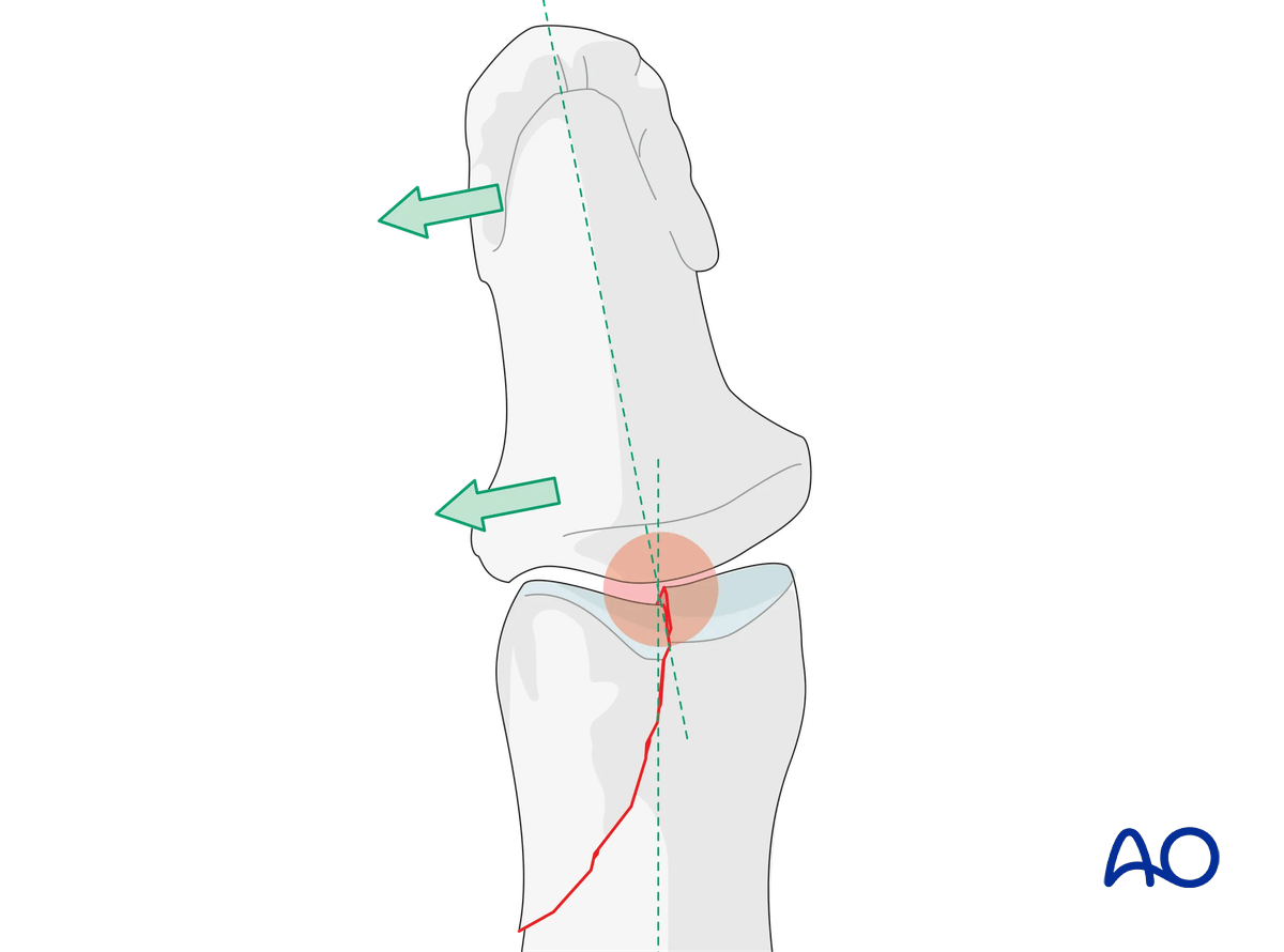
Articular fractures must be reduced anatomically. Otherwise, the articular cartilage may be damaged, leading to painful degenerative joint disease and digital deformity.
This illustration shows how even slight unicondylar depression with malreduction may lead to angulation of the finger.
Percutaneous vs open reduction and fixation
Percutaneous reduction and fixation may be performed.
The advantage of a percutaneous reduction and fixation is:
- Shorter operation time
- Less soft-tissue damage
- Faster mobilization
This treatment option needs some skills and experience and special reduction forceps to avoid impingement of swollen soft tissue (atraumatic technique).
These reduction forceps allow to hold the reduction at the planned screw placement, and drilling and screw insertion. There is no need for additional K-wire fixation or reduction forceps placement.
If a percutaneous reduction is not achievable, the treatment can be changed to open surgery.
AO teaching video
Thumb, head of the proximal phalanx—Unicondylar fracture—Fracture fixation using 2.0 mm Lag Screw
Renato M Fricker, Regula Steiger, Klaus Lowka
2. Patient preparation
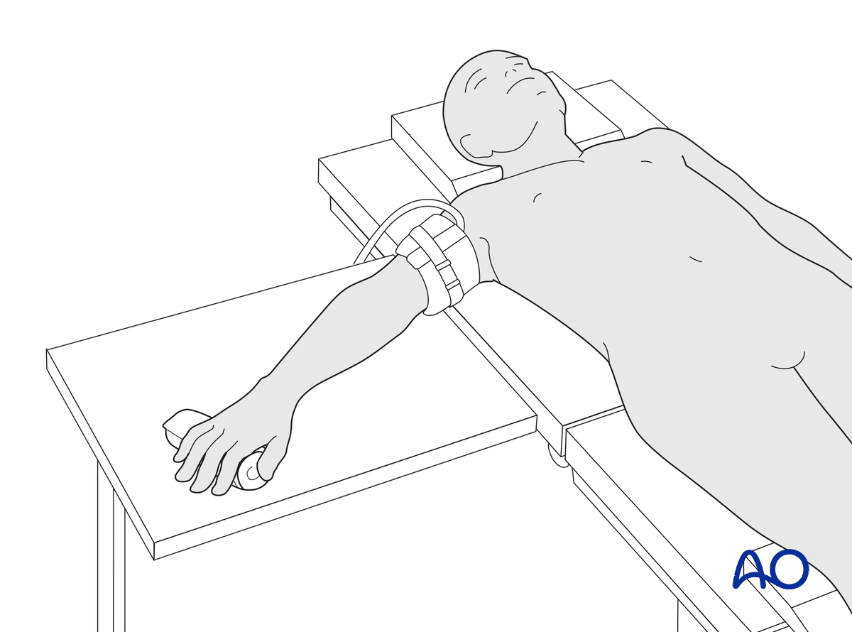
This procedure is usually performed with the patient in a supine position with the arm on a radiolucent side table.
3. Approach
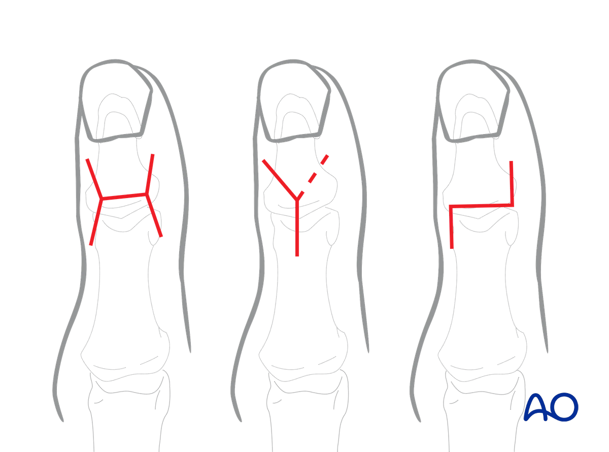
For this procedure, a dorsal approach to the IP joint with proximal extension can be used.
4. Reduction
Closed reduction
Reduction starts with traction to restore length.
Exert lateral pressure with your thumb and index finger or with dedicated percutaneous reduction forceps to reduce the fracture.
Confirm reduction with an image intensifier.
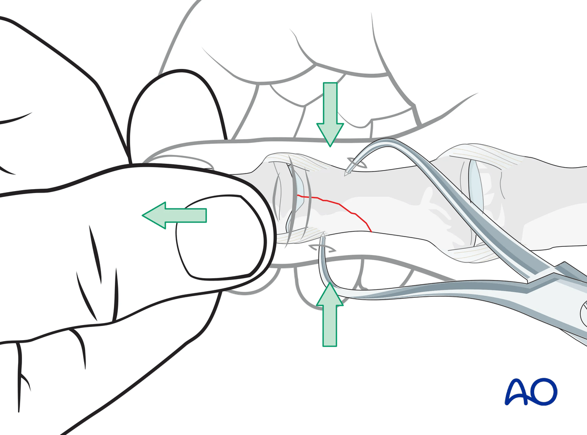
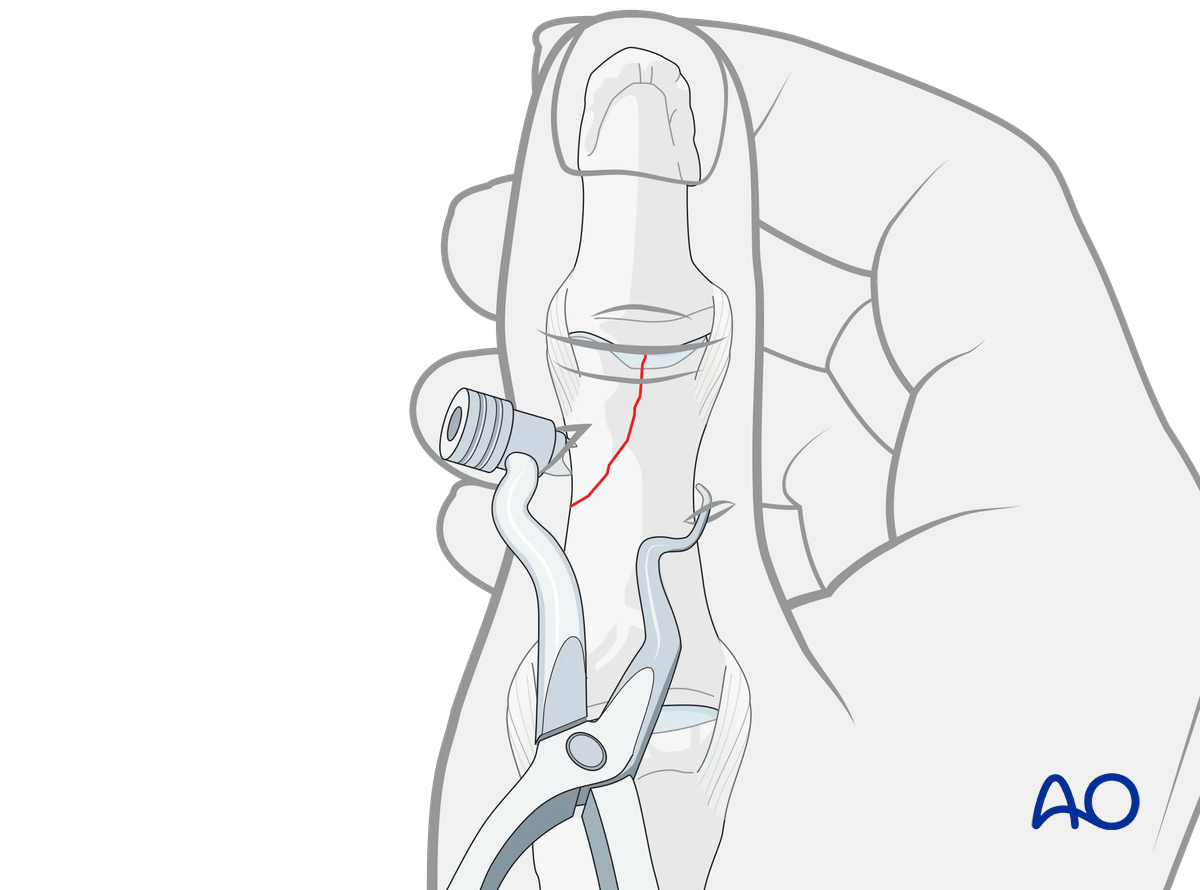
Special reduction forceps designed for percutaneous fixation may be used to hold the reduction.
Open reduction
Manipulate the articular fragment manually or with a dental pick, small K-wire, or a small periosteal elevator.
Small K-wires can also be used for preliminary fixation. Depending on fracture configuration, pointed forceps may be useful for reduction.
Application of excessive force can result in fragmentation.
Confirm reduction with an image intensifier.
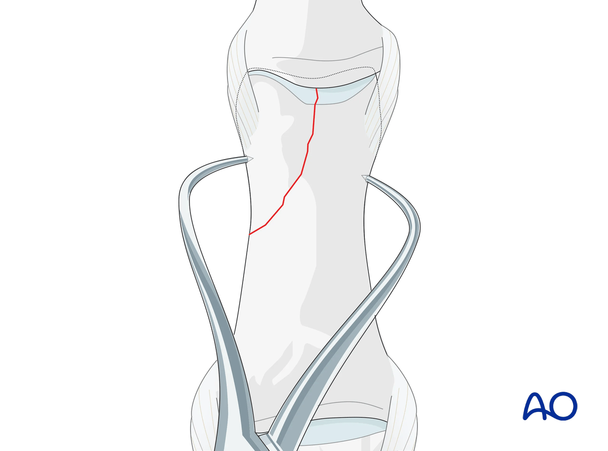
Preliminarily fix the fragments by inserting a K-wire. Be careful to place it so it will not conflict with later screw placement.
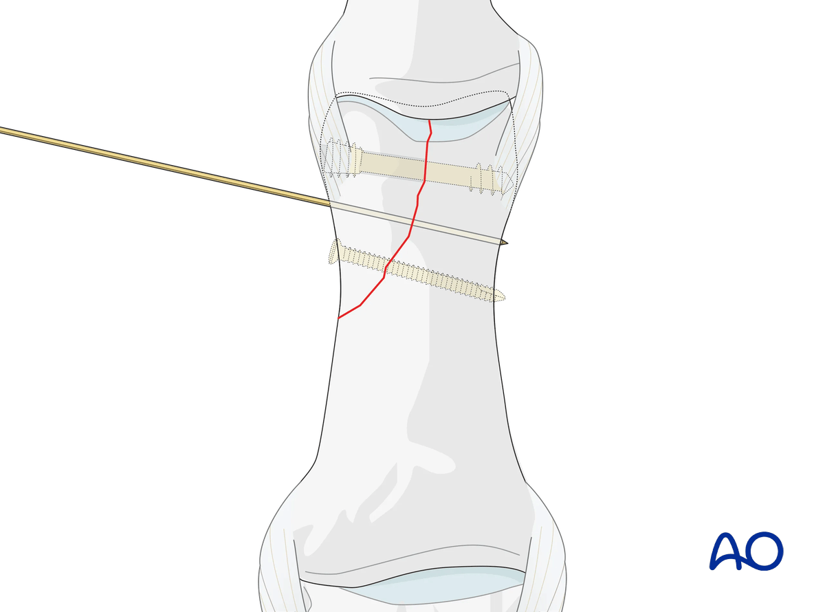
Stability evaluation
Confirm reduction with an image intensifier and check the joint stability by flexion and extension. This should show congruent movement.
5. Checking alignment
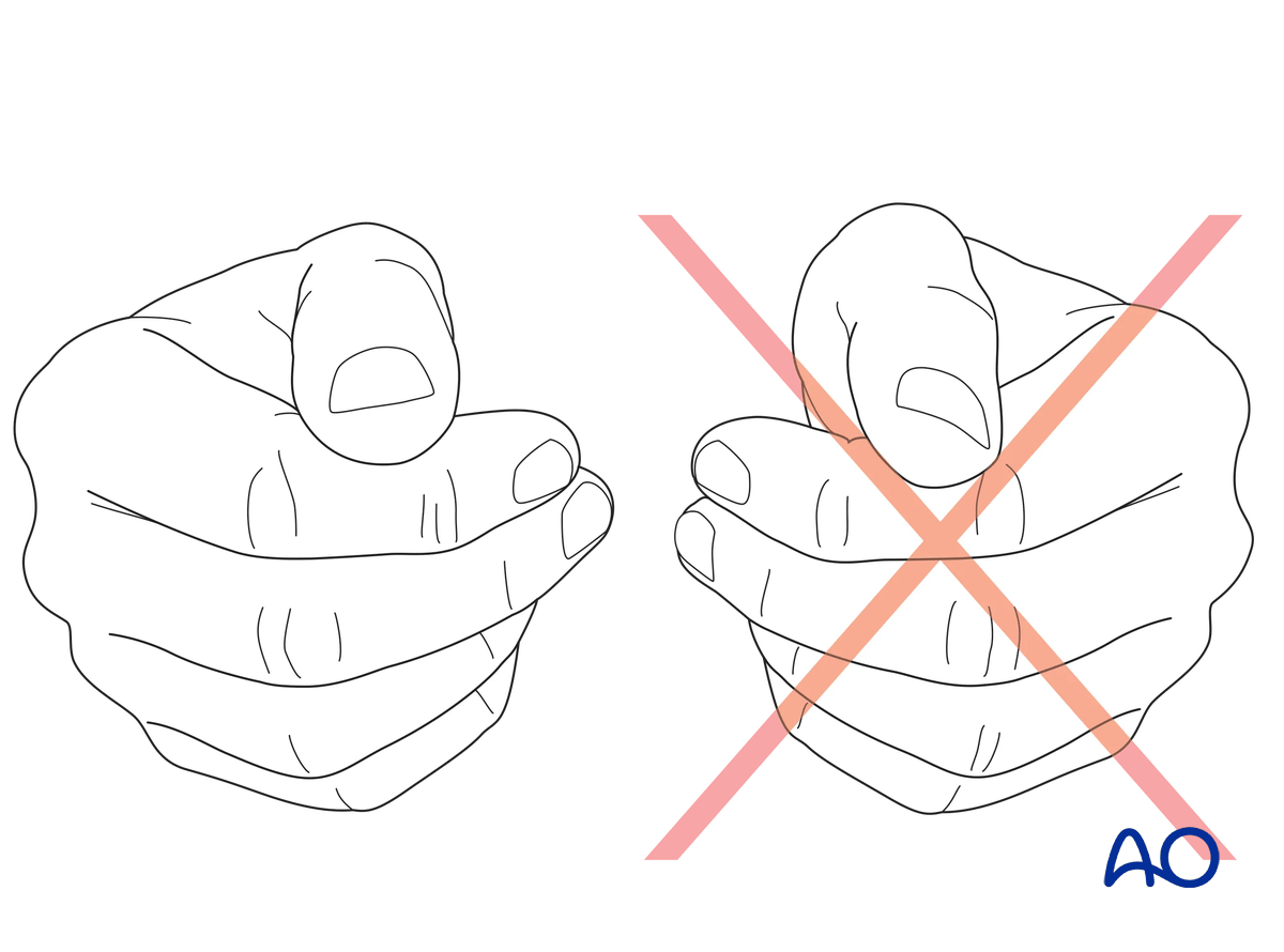
Confirm correct rotational reduction by inspecting the thumb nail plate in relation to the index finger. Compare it to the contralateral side if necessary.
6. Fixation of a large fragment
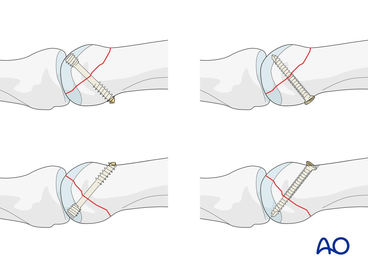
Planning for screw insertion
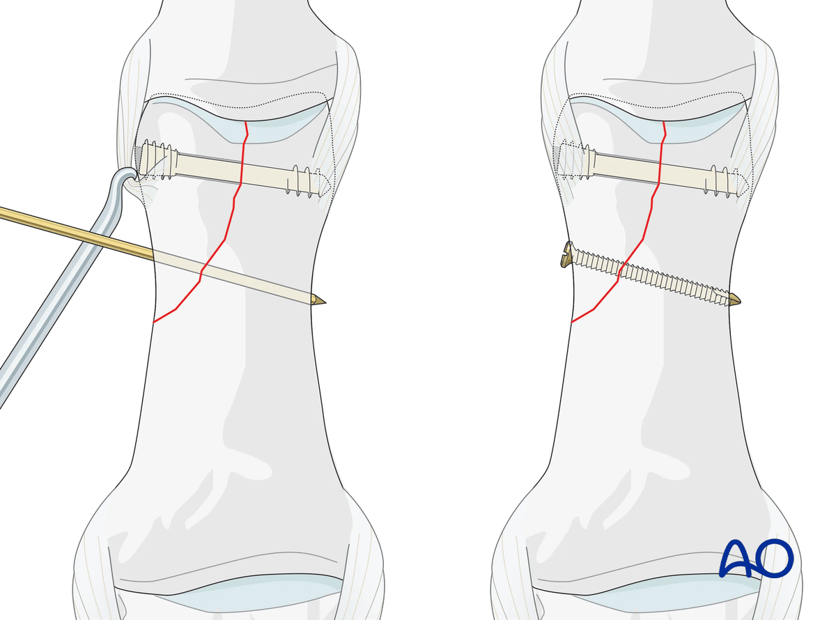
Each lag screw must be inserted perpendicularly to the fracture plane.
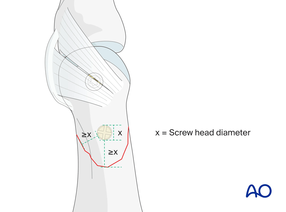
Do not insert screws too close to the fracture apex or the subchondral bone. A minimal distance from the fracture line, equal to the screw head diameter, must be observed.
The screw insertion should avoid conflicts with the MCP ligaments.
The screw length needs to be adequate for the screw to penetrate and purchase in the opposite (trans) cortex.
Screw-size selection
The exact size of the diameter of the screws used will be determined by the fragment size and the fracture configuration.
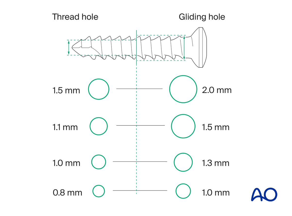
The various gliding and thread hole drill sizes for different screws are illustrated here.
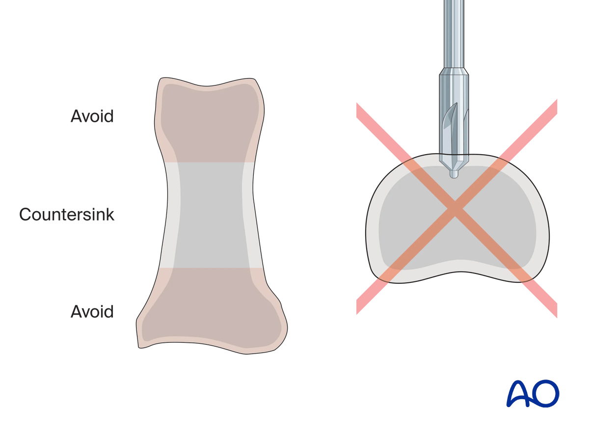
Pitfall: countersinking in the metaphysis
Screw insertion
Insert the screw closest to the articular surface first.
Alternate tightening of the two lag screws helps to avoid tilting the fragment and applies even compression forces across the fracture surface.
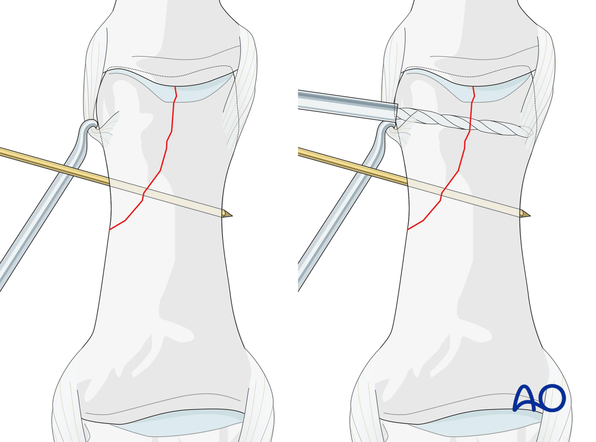
In the next step, it is replaced with a screw.
7. Fixation of a small fragment
Screw positioning in a small fragment
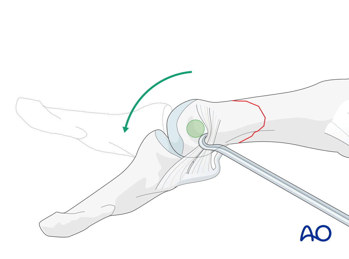
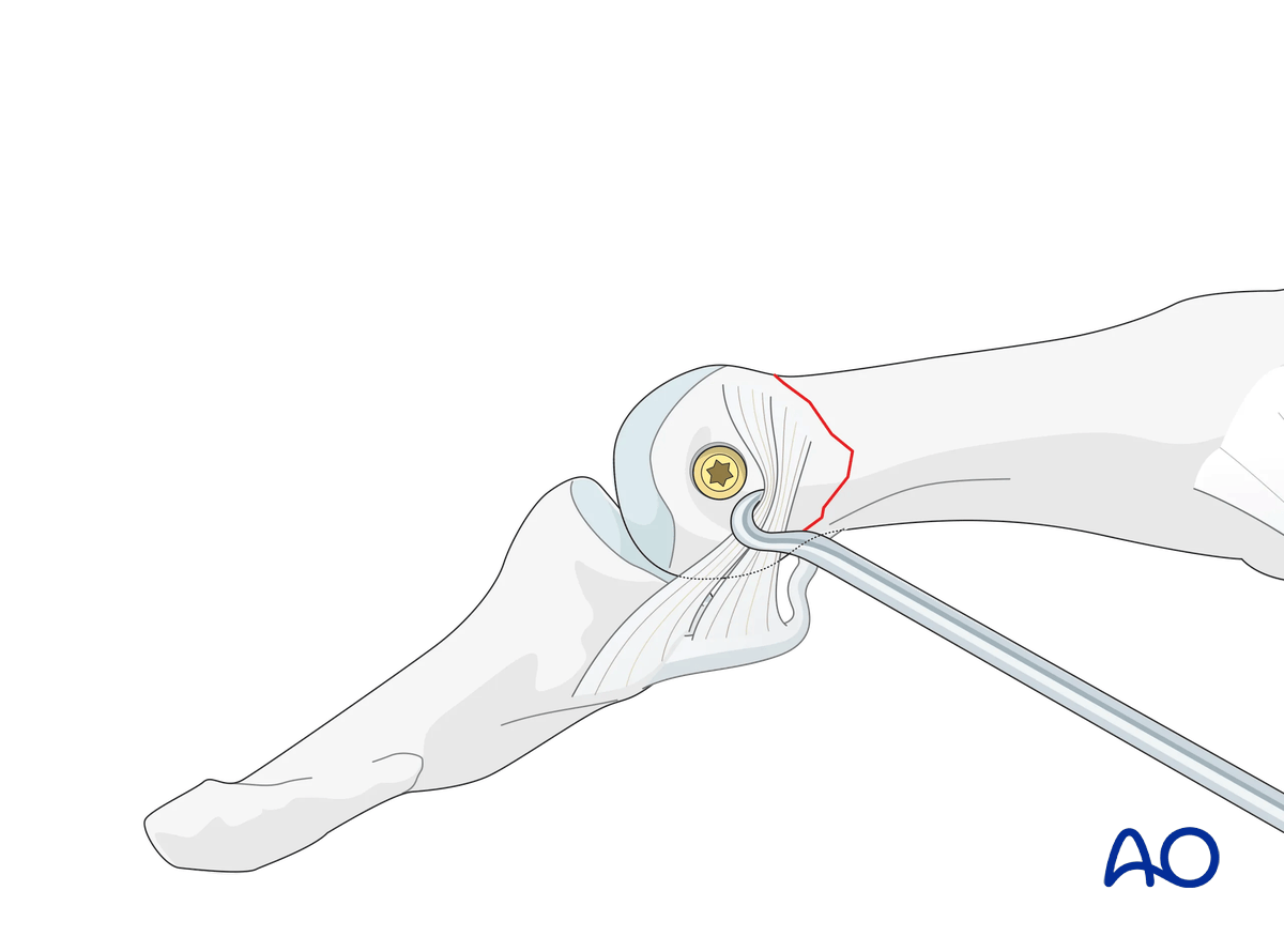
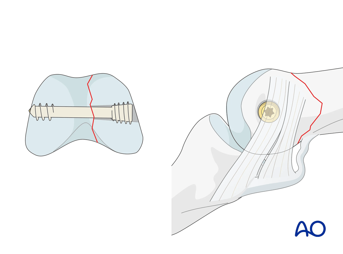
If only one screw can be inserted into a small fragment, it will have to be placed within the joint cavity but through the nonarticular face of the condyle, distal to the collateral ligament.
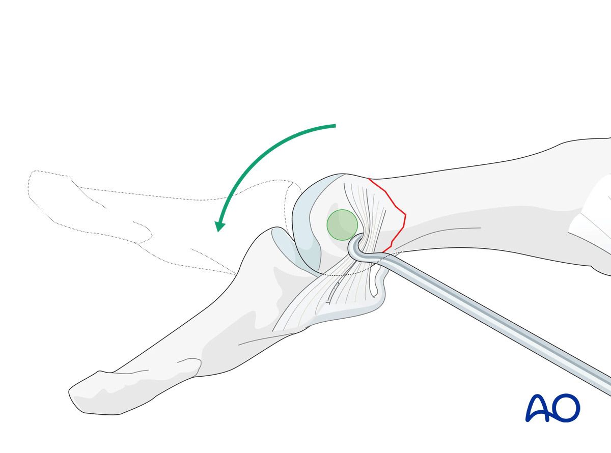
The lateral aspect of the phalangeal head, which is safe for screw placement, can be approached by flexing the interphalangeal (IP) joint.
A headless cannulated screw is recommended to avoid ligament irritation due to a protruding screw head and eventual joint stiffness.
Determining screw size
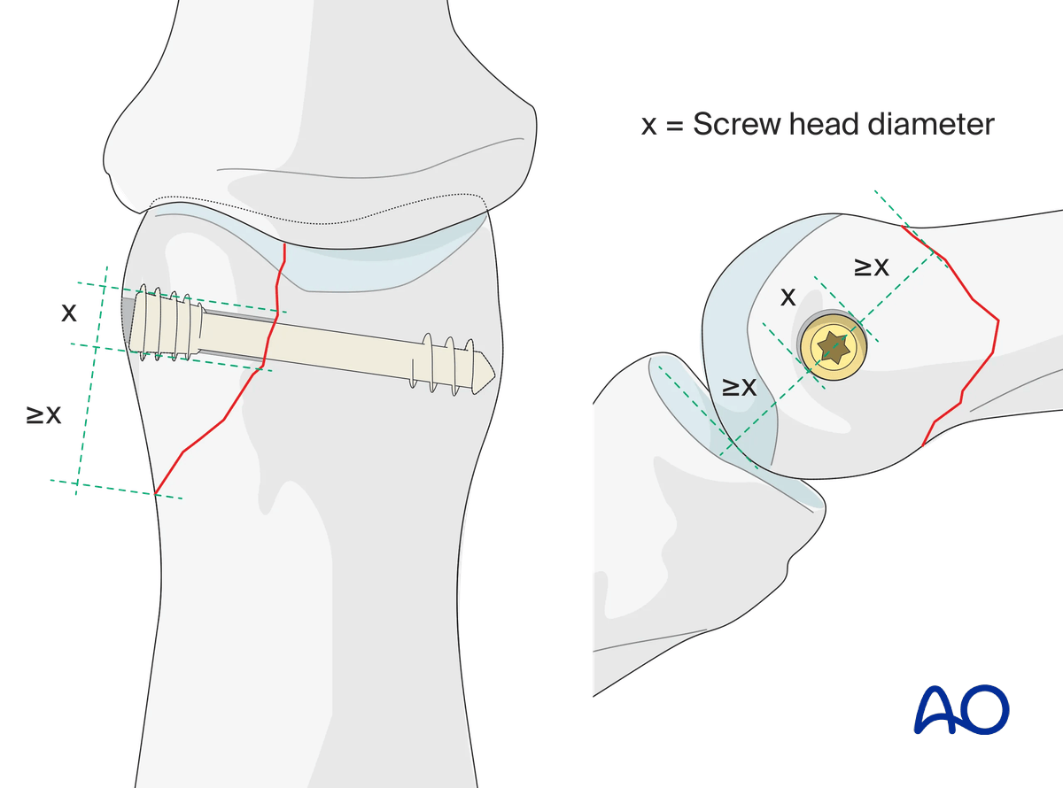
The screw length needs to be adequate for the screw just to penetrate the opposite cortex.
Keep in mind that at the apex of the fragment, the minimal distance between the screw head and the fracture line must be at least equal to the diameter of the screw head. If necessary, a screw of a smaller diameter will have to be chosen.
Preparation for drilling
The optimal way to approach the outer surface of the phalangeal head is by flexing the PIP joint and retracting the collateral ligament in a volar direction.
Lag-screw insertion
Insert the headless screw and gently tighten it to compress the fracture.
8. Final assessment
Confirm anatomical reduction and fixation with an image intensifier.
9. Aftercare
Healing phases
The aftercare can be divided into four phases of healing:
- Inflammatory phase (week 1–3)
- Early repair phase (week 4–6)
- Late repair and early tissue remodeling phase (week 7–12)
- Remodeling and reintegration phase (week 13 onwards)
Full details on each healing phase can be found in the chapter Postoperative treatment of thumb injuries.
Postoperative care
Early range of motion, without load bearing, in the hand is important for optimal outcome.
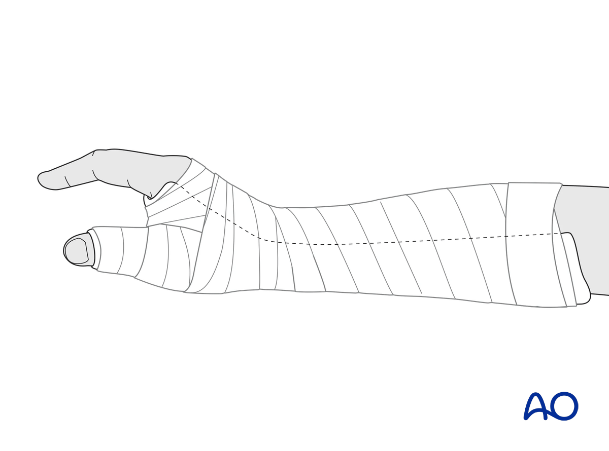
Immediately postoperatively, a temporary plaster splint is applied. This also helps with swelling and pain control.
The plaster splint can be switched to a custom-made thermoplastic splint after a week. The splint should be worn until fracture healing has been confirmed.
Hand therapy should be initiated as soon as possible.
The arm should be actively elevated to help reduce any swelling.
Mobilization
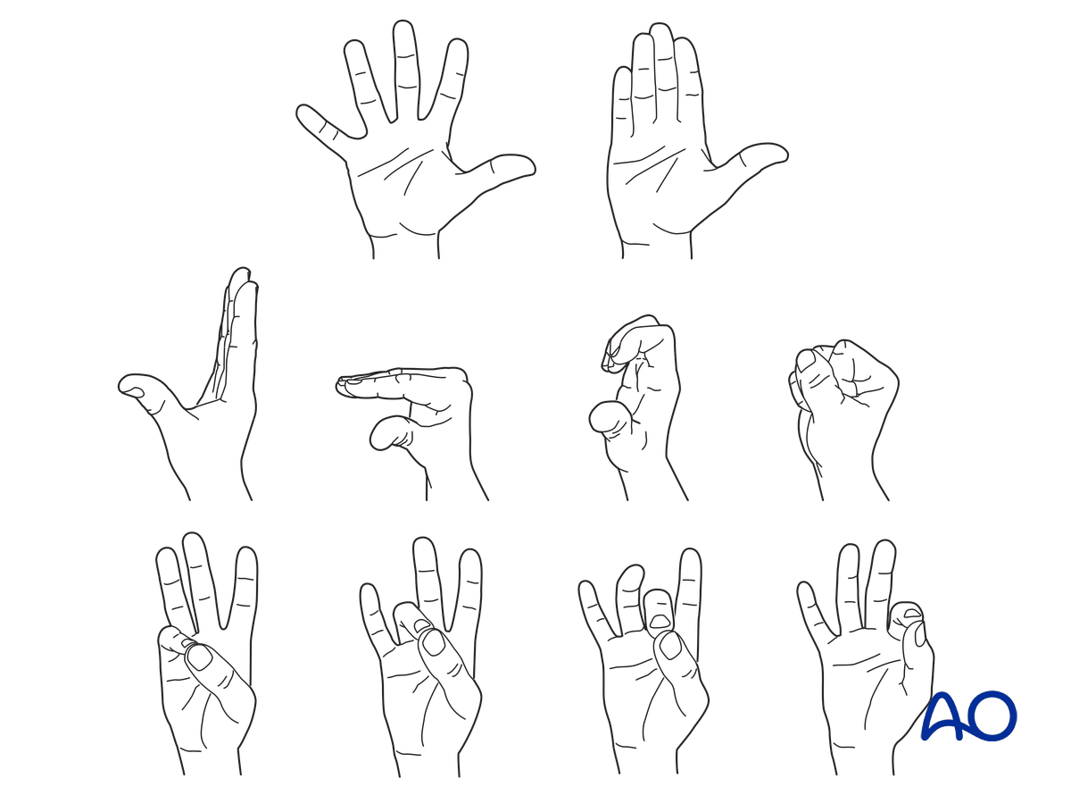
To prevent joint stiffness, the patient should be instructed to begin active motion (flexion and extension) of all nonimmobilized joints of the hand immediately after surgery.
In compliant patients with stable fixation, the splint can be removed under the supervision of a hand therapist, and early active motion exercises started after a few days.
Weight bearing should be avoided until fracture healing is confirmed.
Follow-up
The surgical wound is checked as necessary depending on soft tissue status. The wound is monitored for evidence of infection.
To allow for early joint mobilization and to prevent wound dehiscence, leave the sutures for 2 weeks.
Confirm fracture healing clinically and radiologically after about 5–6 weeks (depending on the fracture configuration).
10. Case
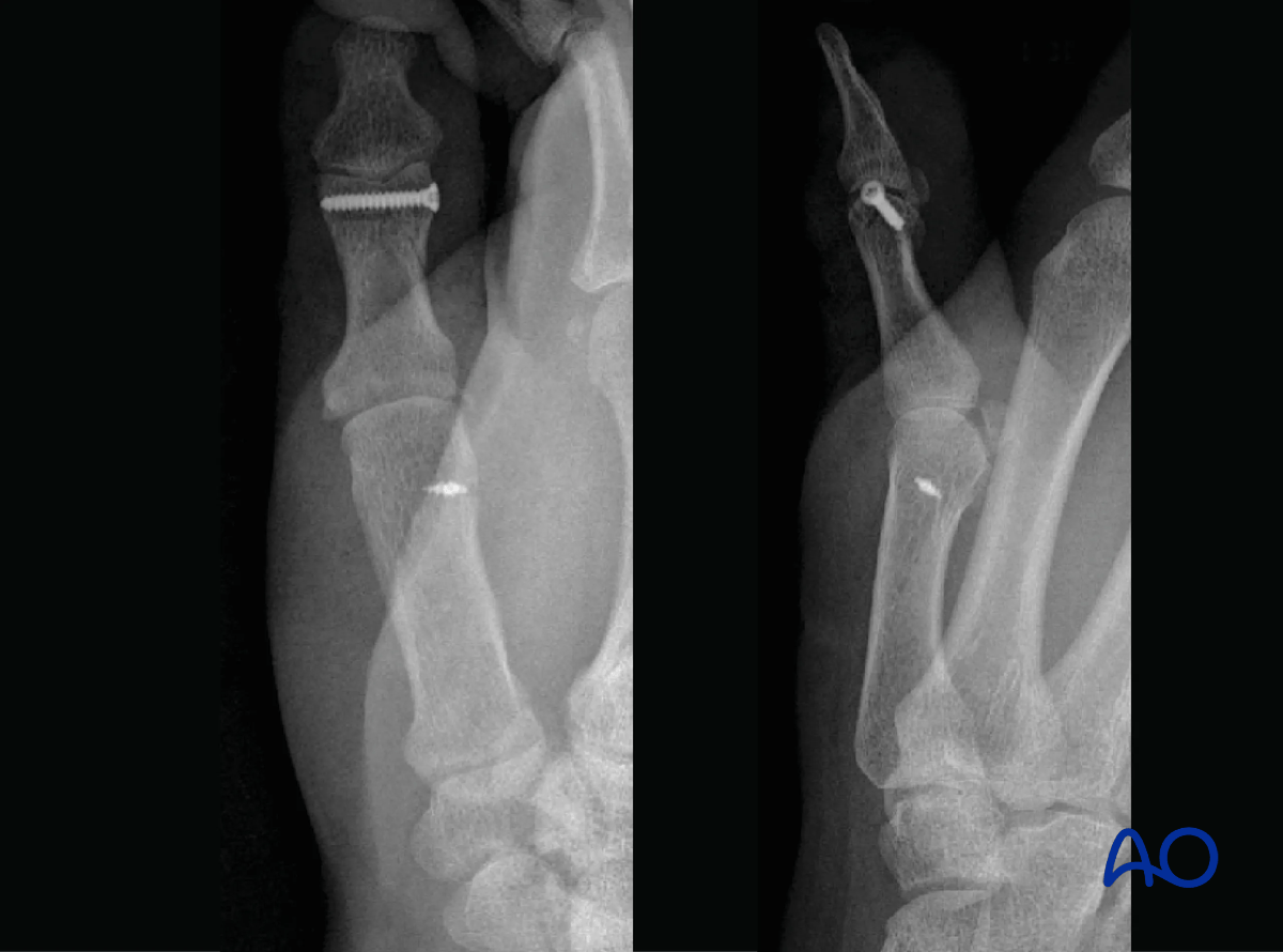
X-rays of a partial articular fracture of the proximal phalangeal head (distal end segment)
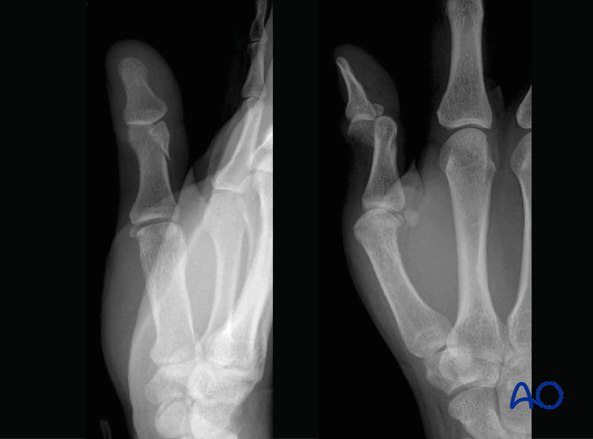
X-rays of the final construct with screw fixation