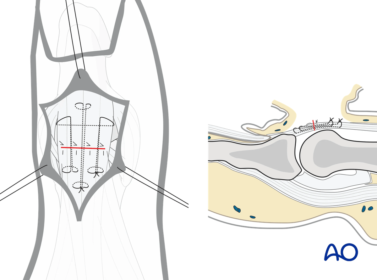
Dorsal approach to the interphalangeal joint of the thumb
1. Introduction
Indications
The dorsal approach to the interphalangeal (IP) joint of the thumb is indicated for:
- Interphalangeal arthrodesis
- Intraarticular fractures of the interphalangeal joint
- Dorsal avulsion fractures of the base of the distal phalanx by the extensor pollicis longus (EPL) tendon
- Fracture avulsions of collateral ligament of the IP joint
If the underlying tendons are retracted dorsally, a Y-shaped incision may allow for management of collateral ligament avulsions.
AO teaching video
Thumb—Interphalangeal joint—Dorsal approach
Renato M Fricker, Klaus Lowka, Regula Steiger
2. Surgical anatomy
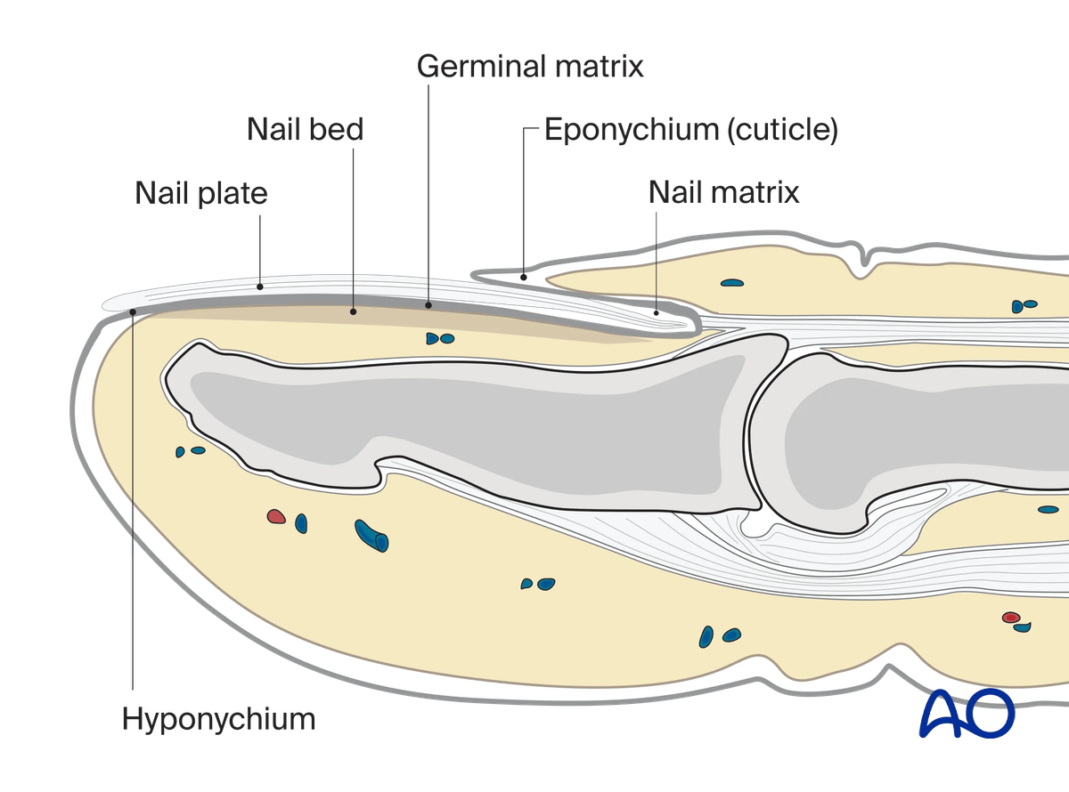
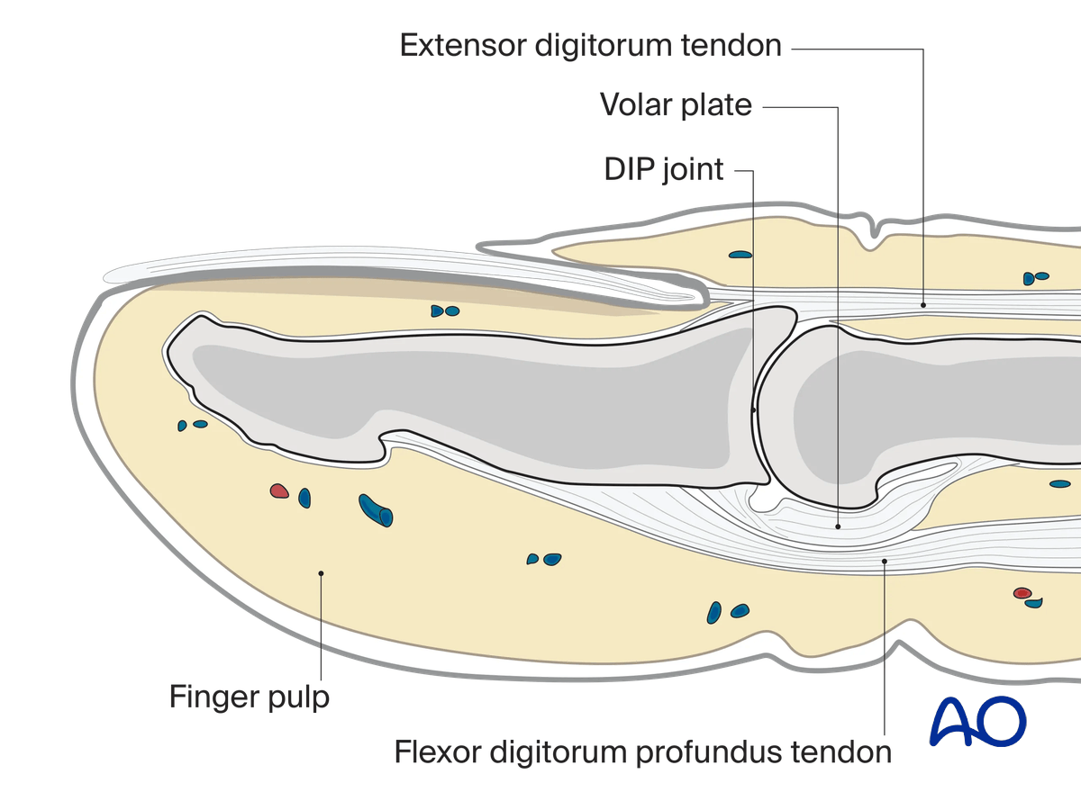
The base of the distal phalanx has a prominent dorsal crest at the insertion of the extensor tendon. The tendon is also adherent to the interphalangeal (IP) joint capsule.
On the palmar surface is the insertion of the flexor pollicis longus tendon.
The flexor tendon occupies the whole width of the base of the distal phalanx. It is made up of two different fibers. The superficial fibers attach to the lateral aspects of the phalanx. The deep fibers run centrally and attach more distal in the palmar aspect of the phalanx.
The volar plate is very flexible, allowing hyperextension of the IP joint and pulp-to-pulp pinch.
The vascularity of the extensor tendons is more precarious than that of the flexor tendons. This prolongs extensor tendon healing time.
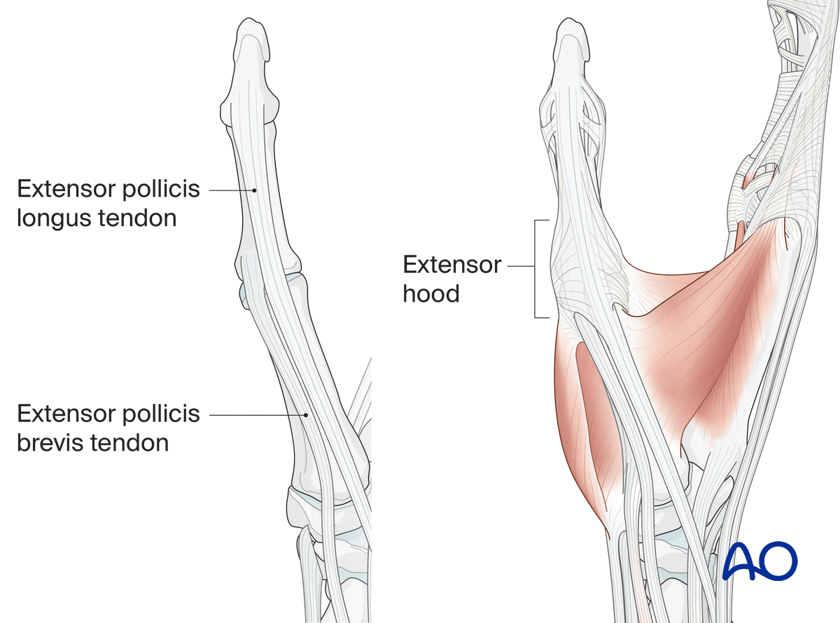
The extensor hood of the thumb consists of the radially located extensor pollicis brevis tendon (insertion: base of the proximal phalanx) and the ulnarly located extensor pollicis longus tendon (insertion at the base of the distal phalanx).
Both tendons are held in position by the sagittal bands at the level of the MCP joint and the extensor hood at the level of the proximal phalanx.
At the level of the MCP joint the abductor pollicis aponeurosis radiates into the extensor hood.
3. Skin incision
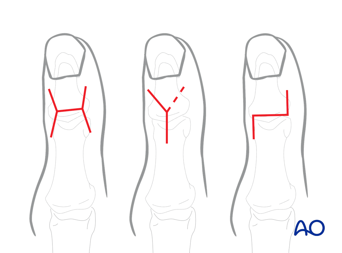
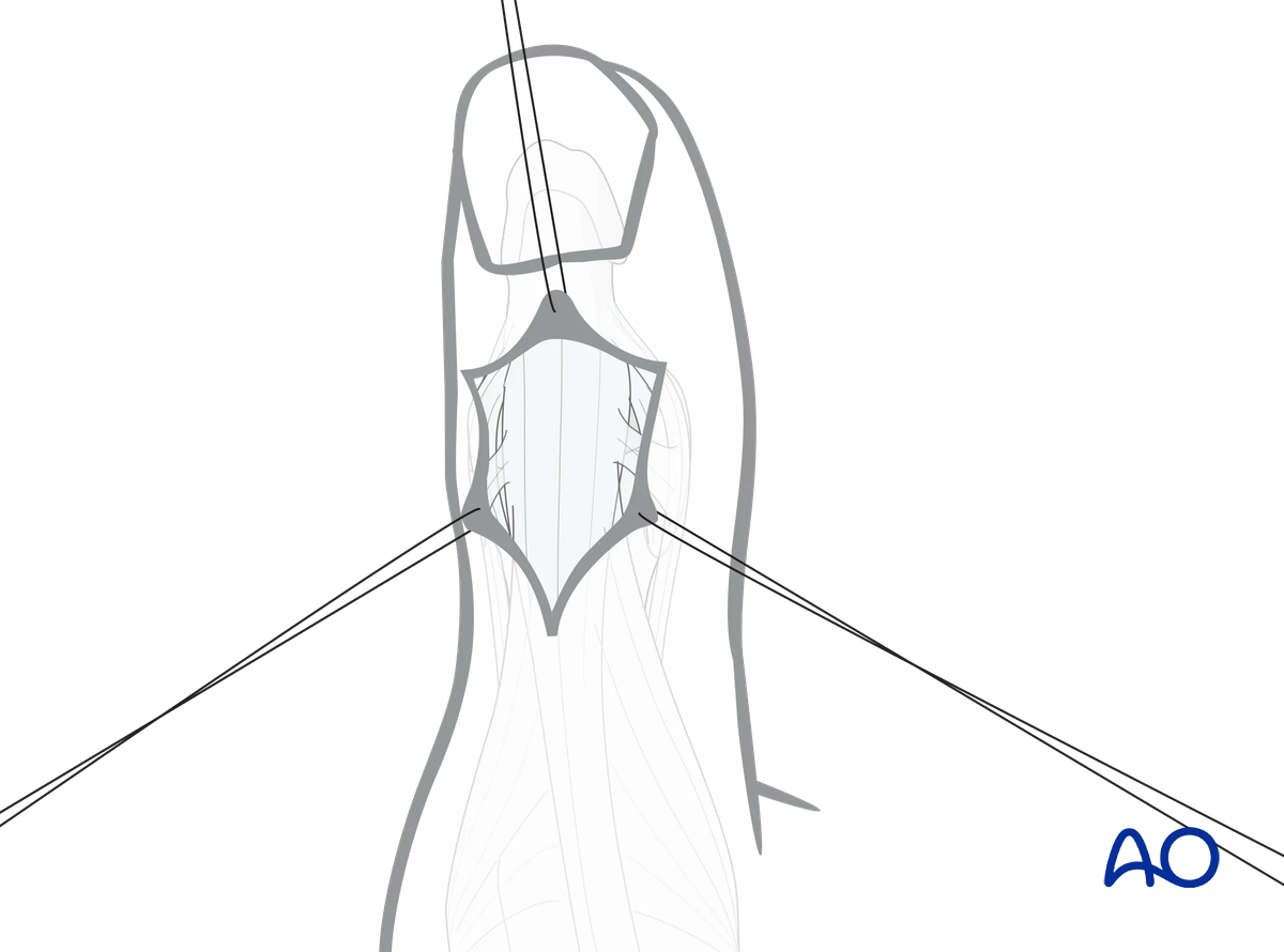
There are three common skin incisions: The H-shaped, the Y-shaped, and the Z-shaped incisions.
The H-shaped incision is usually modified by diverging the sides slightly, to minimize vascular insult.
A Y-shaped incision may be limited to a hockey-stick incision and extended if necessary.
Using these incisions helps to preserve the blood supply to the eponychium.
4. Elevation of the skin flaps
Depending on the shape of the skin incision, flaps should be elevated and held with fine sutures to minimize soft-tissue trauma.
Tiny veins will appear and should be coagulated with the bipolar forceps, as necessary, to gain exposure to the joint.
5. Division of the extensor tendon
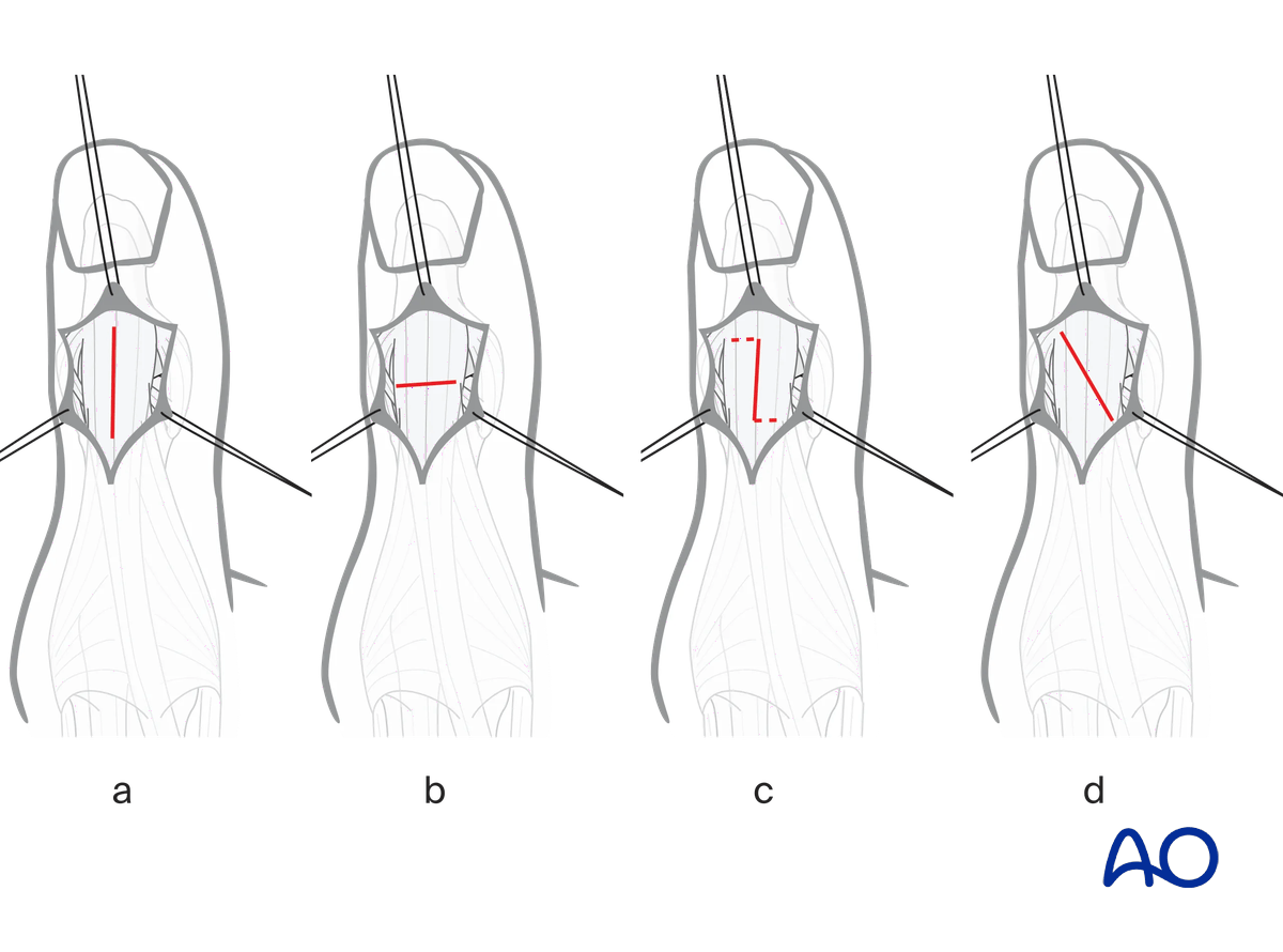
Divide the distal tendon of the EPL longitudinally (a).
For arthrodesis or full articular exposure, a transverse tenotomy is used (b).
A step cut (c) or an oblique cut (d) facilitates subsequent repair.
Alternative: retraction of tendon
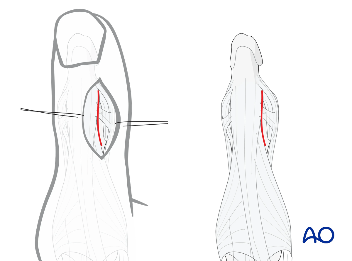
A straight dorsoradial, or dorsoulnar, skin incision will allow access to the interval between the EPL tendon and the collateral ligaments of the IP joint. This exposes either the radial or ulnar aspect of the IP joint, eg, for management of condylar fractures or collateral ligament repair.
This alternative only allows access to one condyle, but never to both.
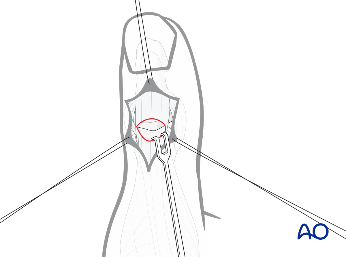
6. Joint exposure
In case of a transverse tenotomy, retract the EPL tendon proximally to expose the IP joint.
7. Wound closure
Repair the tenotomy using fine interrupted mattress sutures.
After transverse tenotomy, it is recommended to perform a core suture (eg, with a 3-0 or 4-0 PDS) followed by a superficial, uninterrupted adaptation suture (eg, with 6-0 PDS).
Postoperatively, the IP joint must be consequently immobilized in (hyper-)extension like a tendon repair (6–8 weeks in an IP splint).