

Plate fixation
1. General considerations
Open anatomical reduction and internal fixation is the treatment of choice for Rolando fractures.
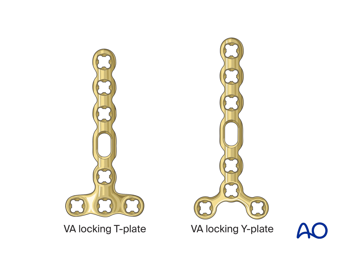
They may be fixed with a dorsal T- or Y-plate.
The articular fragments are reduced and fixed using the transverse part of the plate. In the second step, the extraarticular fracture component is reduced and fixed.
Noncomminuted fracture components, eg, between the articular fragments or between the articular block and the diaphysis, may be fixed with compression.
If the articular fragment cannot be reduced or are too small to be fitted with a plate, conversion to arthrodiastasis should be considered.
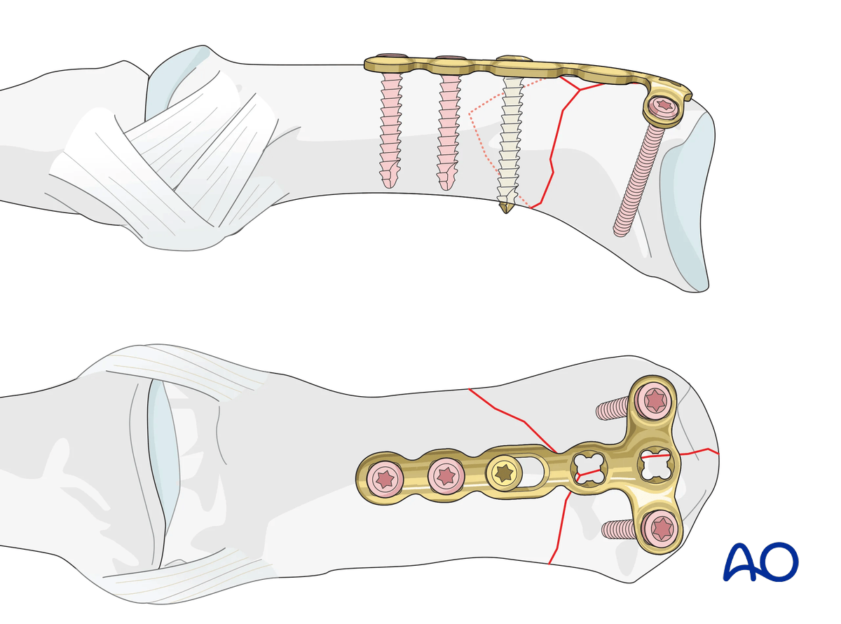
Plate selection
A T-shaped plate is best suited for these fractures.
A 2.0 mm locking compression plate (LCP) is the implant of choice in cases of additional metaphyseal comminution.
A plate with variable-angle (VA) locking-head screws has the advantage to allow insertion of two or three screws in variable angles into the articular block. The rounded plate edges avoid soft-tissue irritation and adhesion.
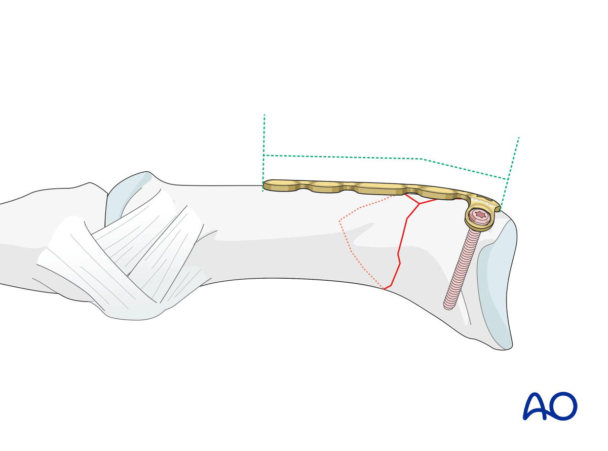
The plate needs to be contoured to fit the anatomy of the end segment.
AO teaching video
Metacarpal I, Base—Rolando Fracture—Plate Fixation Using Various Types of Treatment
Doug Campbell, Ladislav Nagy, Renato Fricker, Sudhir Warrier
2. Patient preparation
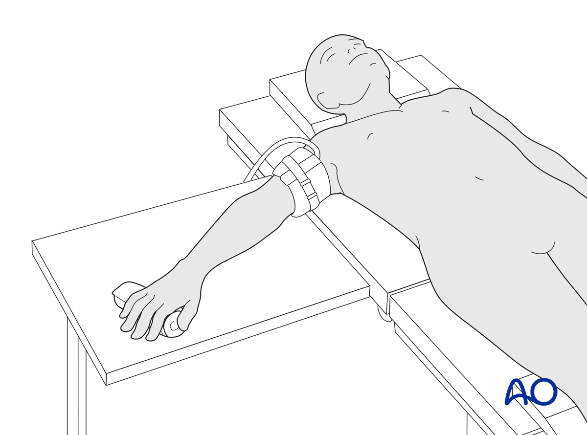
This procedure is usually performed with the patient in a supine position with the arm on a radiolucent side table.
3. Approaches
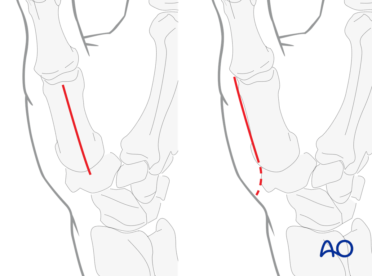
For Y- or T-shaped patterns in the frontal plane, a straight dorsal approach is preferred.
For a Y- or T-shaped fracture pattern in the sagittal plane, the radiopalmar approach is preferred.
4. Reduction
Any axial displacement should be reduced to gain full length of the metacarpal.
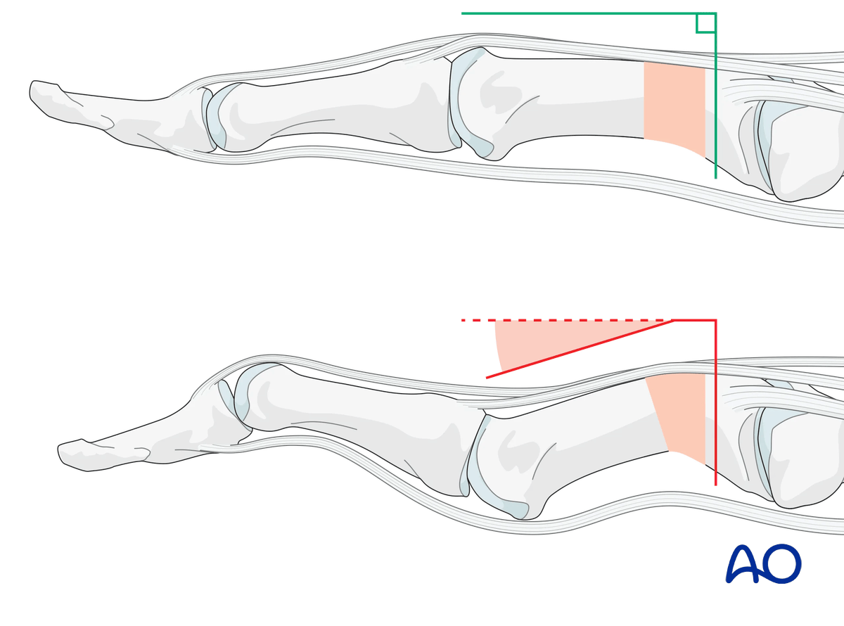
Preliminarily fix the articular fragments of the base of the thumb with a pointed reduction forceps.
Confirm accurate reduction under direct vision and using image intensification.
A preliminary K-wire may be inserted perpendicularly to the vertical fracture plane to stabilize the construct additionally. The K-wire can be useful when the reduction forceps will have to be removed for correct plate placement.
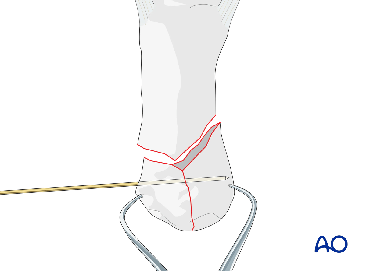
After stabilization of the articular fragments with forceps or a position screw, reduction of the diaphyseal fragment to the articular fragments can be performed with axial traction on the thumb and simultaneous pressure on the dorsal aspect of the diaphysis near the fracture.
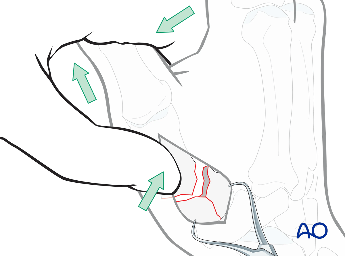
Preliminary fixation of diaphysis
The diaphysis is preliminarily fixed to the base with a K-wire.
Confirm articular congruency and anatomical reduction using image intensification. A limited capsulotomy will permit visual inspection of the joint surface.
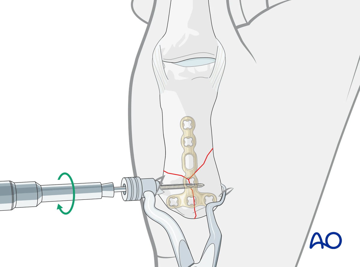
5. Screw fixation of the articular block
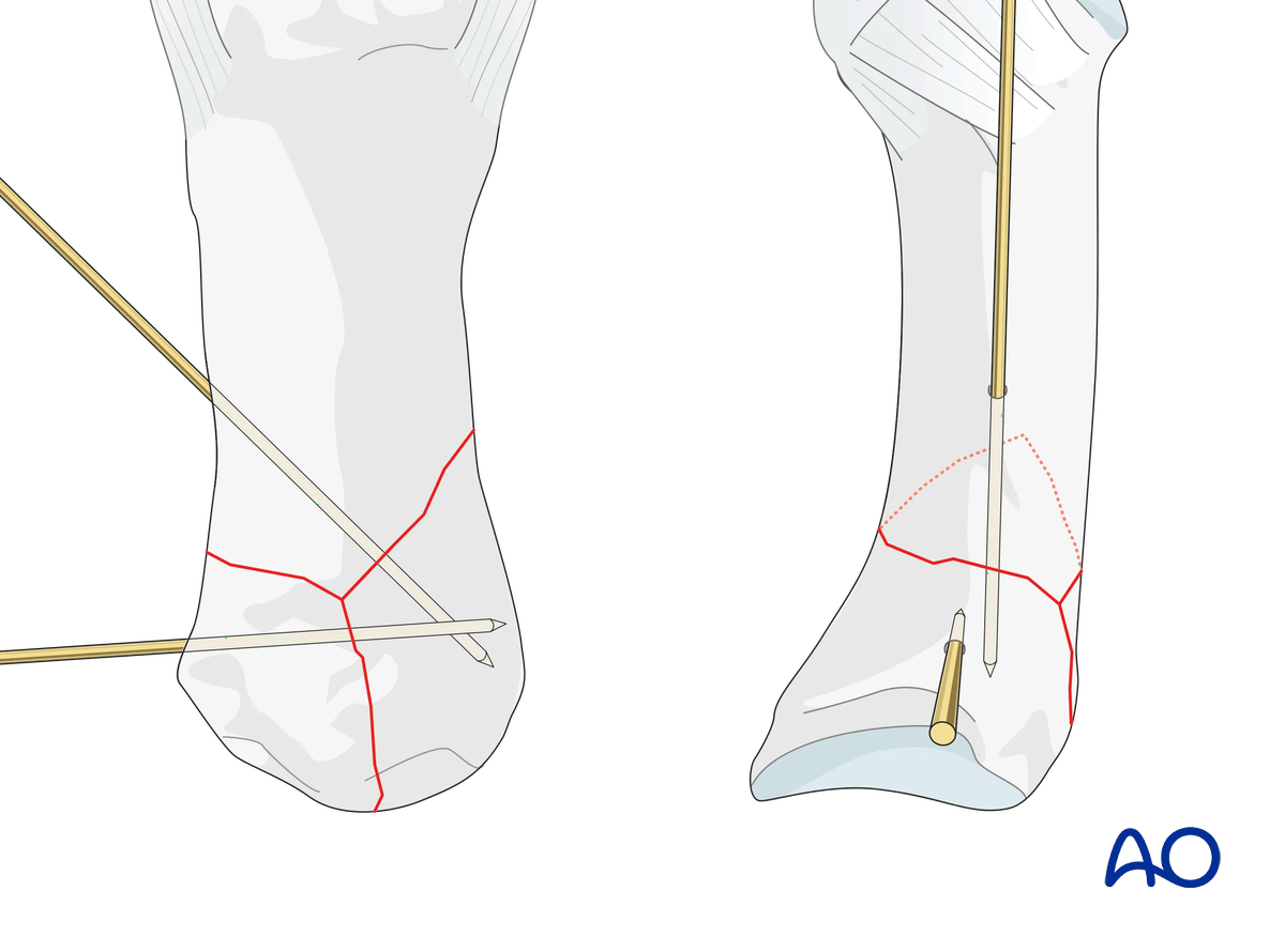
If the fracture line allows, eg, in a T- or Y-shaped fracture with the articular fracture line running from palmar to dorsal, a lag screw may be inserted to stabilize the articular fracture. This converts a three-part fracture to a two-part fracture.
Make sure the screw track is not interfering with later plate application.
6. Plate preparation
Plate trimming
Adapt the plate length to the length of the metacarpal. Avoid sharp edges, which may be injurious to the tendons.
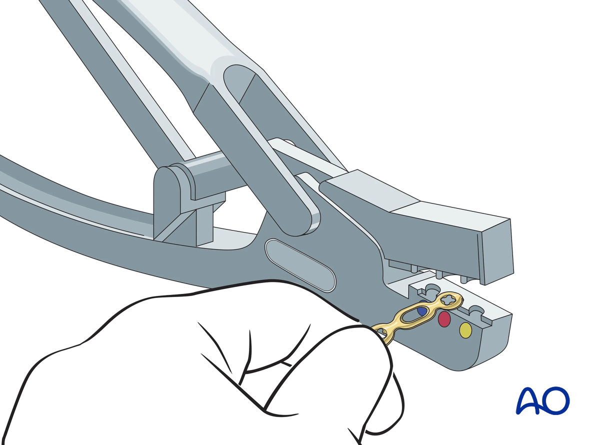
There should be at least 3 plate holes distal to the fracture available for fixation in the diaphysis. At least two screws need to be inserted into the diaphysis.
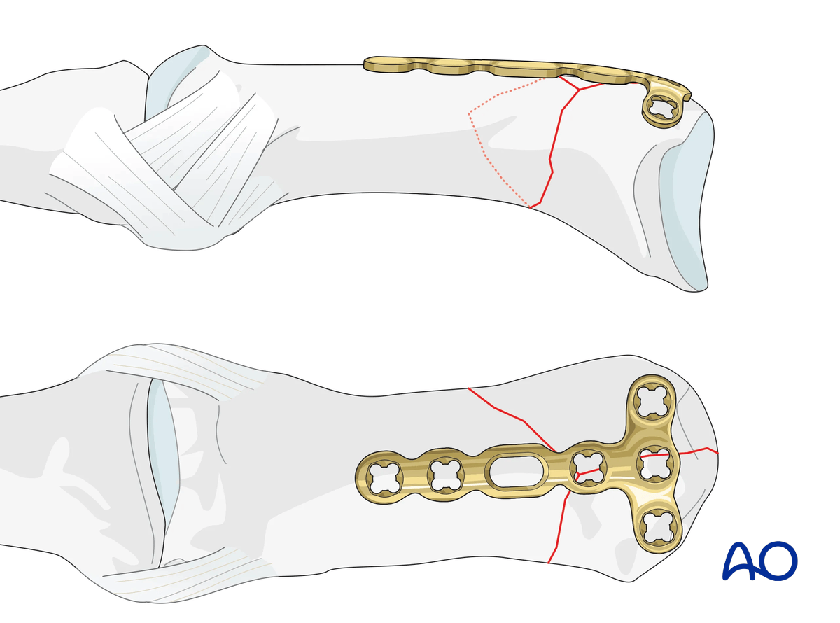
Contouring
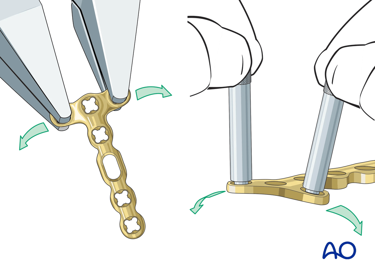
The plate must be adapted accurately to the contour of the proximal fragment. Use bending pliers to contour the plate. Be careful to bend in between the holes by positioning the pliers over the plate holes. Alternatively, insert the threaded bolt into the plate holes to bend the plate.
The bend should never go through a hole as this may damage the locking mechanism.
Adding a slight bend
To prevent the screw from penetrating the joint, a slight bend may be introduced to the articular part of the plate. Be careful not to overbend, as there is a comminuted zone at the opposite cortex, which cannot withstand compression.
Alternatively, if the fracture allows, the plate may be positioned more distally.
7. Plate fixation
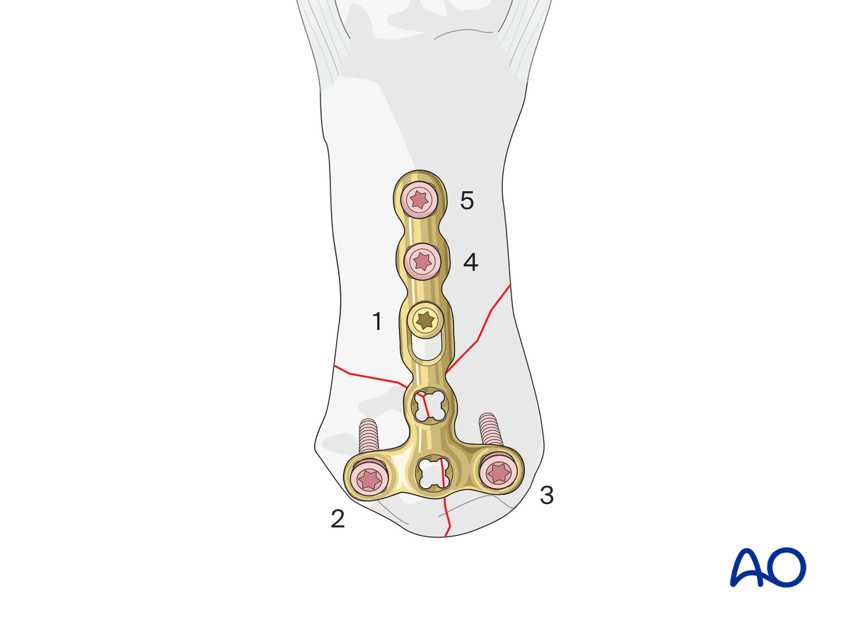
Position the plate and hold it preliminarily with a cortical screw, not fully tightened, in the oblong hole. This allows for later adjustment of the plate position.
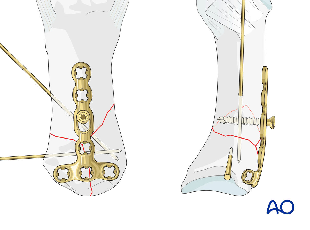
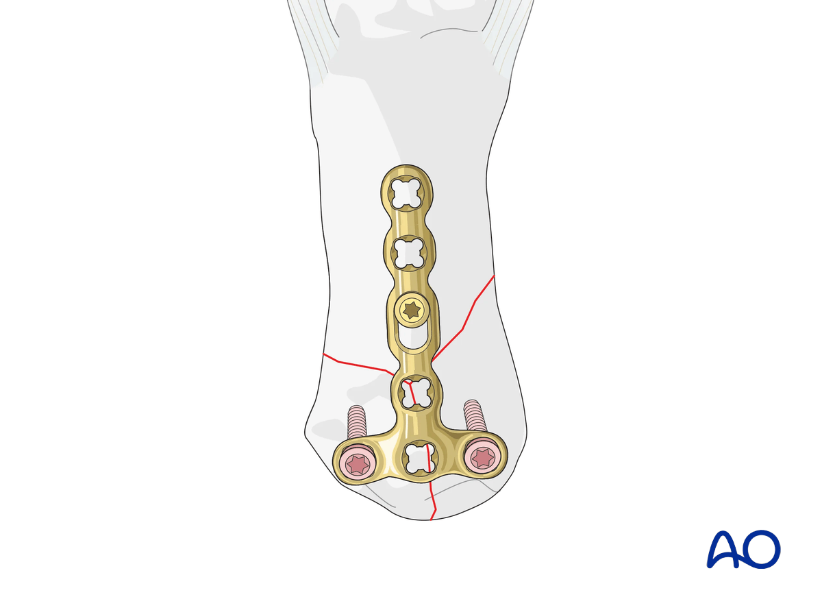
Insert two screws into the articular block:
Tighten the cortical screw to pull the distal fragment onto the plate. This avoids angular deformity.
If a preliminary position screw has not been used, insert a cortical screw in the most distal plate hole to pull the bone onto the plate.
This screw can also be used to apply compression through the plate if the metadiaphyseal fracture allows.
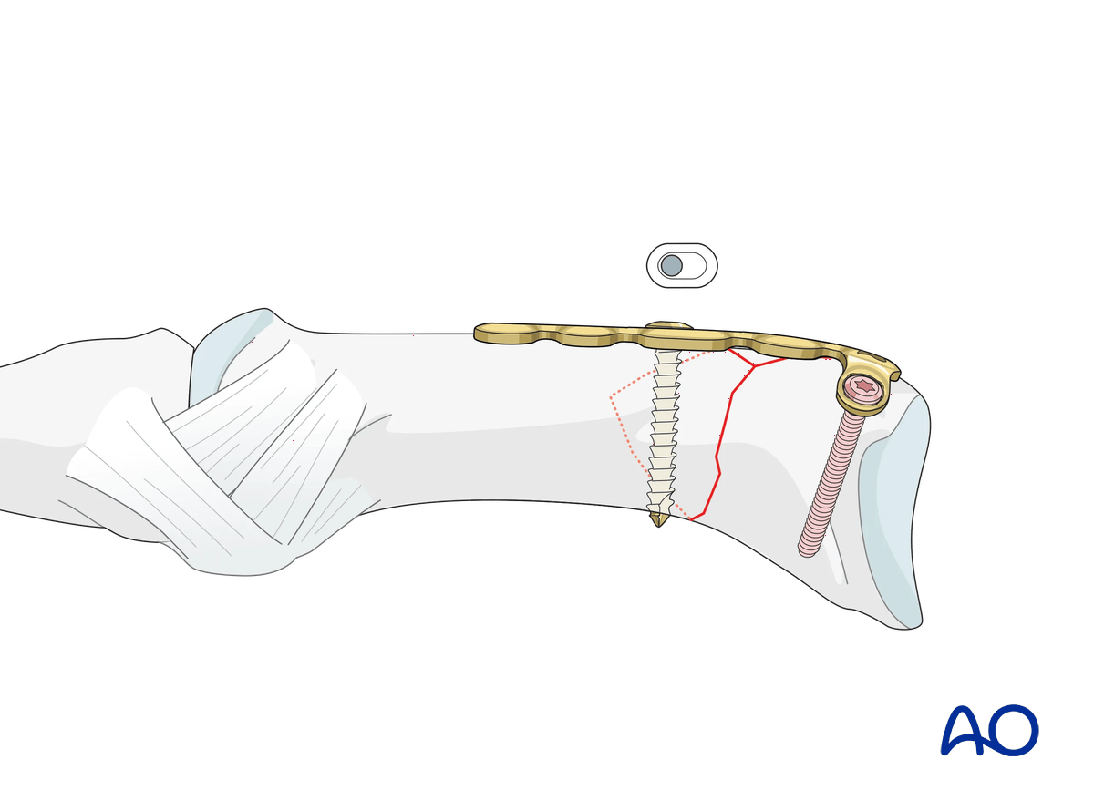
Insertion of distal screws
Insert at least one locking head screw in the diaphysis to provide additional angular stability.
8. Final assessment
Confirm correct rotational alignment by clinical examination.
Image intensification should be used to confirm anatomical reduction and correct placement of implants in two views.
9. Aftercare
Healing phases
The aftercare can be divided into four phases of healing:
- Inflammatory phase (week 1–3)
- Early repair phase (week 4–6)
- Late repair and early tissue remodeling phase (week 7–12)
- Remodeling and reintegration phase (week 13 onwards)
Full details on each healing phase can be found in the chapter Postoperative treatment of thumb injuries.
Postoperative care
Early range of motion, without load bearing, in the hand is important for optimal outcome.
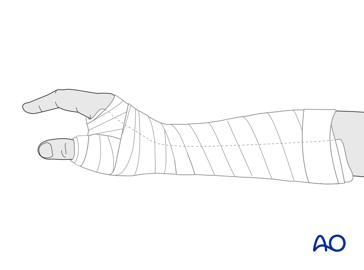
Immediately postoperatively, a temporary plaster splint is applied. This also helps with swelling and pain control.
The plaster splint can be switched to a custom-made thermoplastic splint after a week. The splint should be worn until fracture healing has been confirmed.
Hand therapy should be initiated as soon as possible.
The arm should be actively elevated to help reduce any swelling.
Mobilization
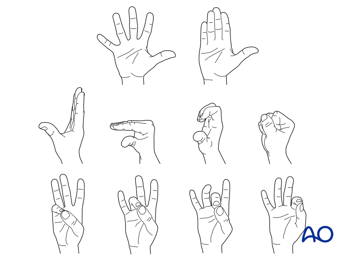
To prevent joint stiffness, the patient should be instructed to begin active motion (flexion and extension) of all nonimmobilized joints of the hand immediately after surgery.
In compliant patients with stable fixation, the splint can be removed under the supervision of a hand therapist, and early active motion exercises started after a few days.
Weight bearing should be avoided until fracture healing is confirmed.
Follow-up
The surgical wound is checked as necessary depending on soft tissue status. The wound is monitored for evidence of infection.
To allow for early joint mobilization and to prevent wound dehiscence, leave the sutures for 2 weeks.
Confirm fracture healing clinically and radiologically after about 5–6 weeks (depending on the fracture configuration).
Implant removal
The implants may need to be removed in cases of soft-tissue irritation.
In case of joint stiffness or tendon adhesion restricting finger movement, arthrolysis or tenolysis may become necessary. In these circumstances, the implants can be removed at the same time.


