Dorsal approach to the metacarpal of the thumb
1. Introduction
Indications
This approach is indicated for extraarticular fractures of the first metacarpal, and for periarticular fractures of the first carpometacarpal joint.
It can also be extended proximally for first carpometacarpal joint arthrotomy for arthrodesis, arthroplasty, or trapeziectomy.
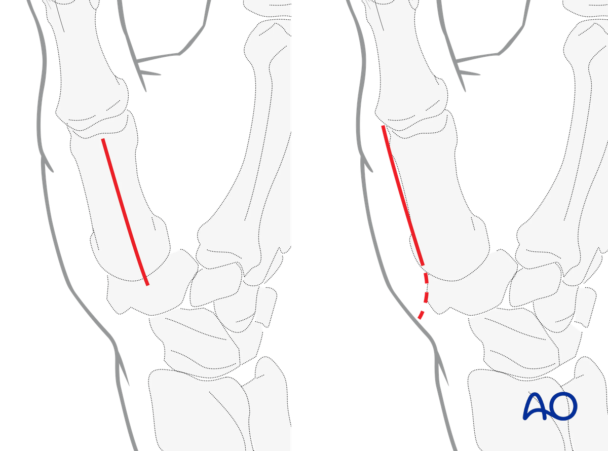
The radial-sided approach is made along the line between the palmar (glabrous) and dorsal skin of the thenar. If the incision is extended proximally in the ulnar direction as depicted, it is referred to as the Wagner approach.
AO teaching video
Thumb—1st metacarpal—Dorsal approach
Renato M Fricker, Klaus Lowka, Regula Steiger
2. Surgical anatomy
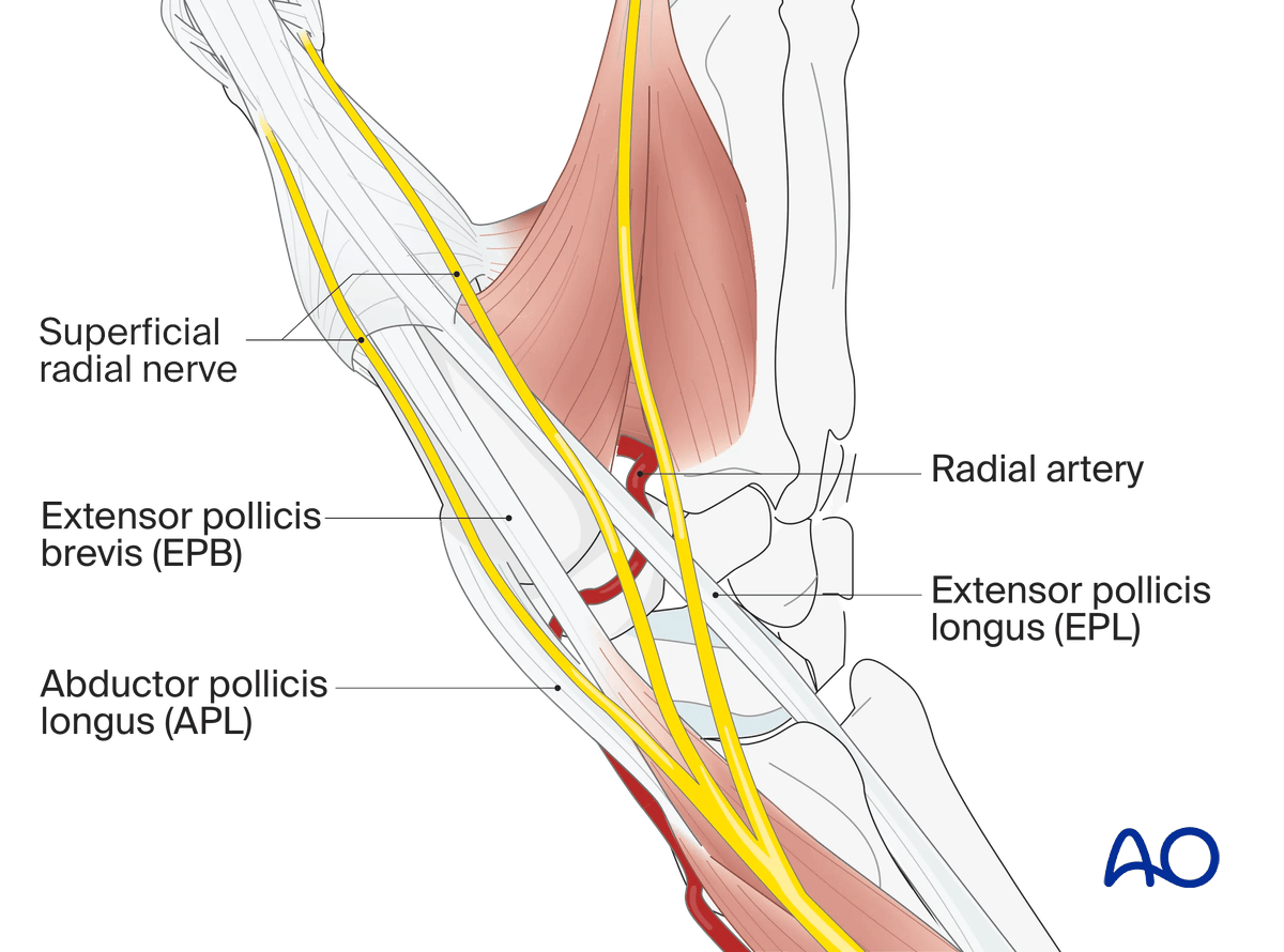
In the subcutaneous tissue, the dorsoulnar and dorsoradial sensory branches of the superficial radial nerve must be protected.
The radial artery crosses over the trapezium underneath the APL, EPB and EPL tendons.
The surgical approach is made between the tendons of extensor pollicis longus (EPL) and extensor pollicis brevis (EPB).
3. Skin incision
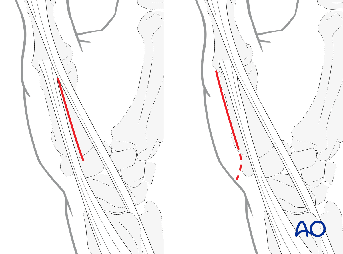
Perform a straight skin incision over the interval between EPL and EPB throughout the length of the first metacarpal.
Alternatively, the incision can be performed radial to the EPB. This incision can be extended proximally, curving in a radiopalmar direction (Wagner approach). For optimal aesthetic results, incise at the boundary of the glabrous (non-hairy) skin.
Identify and protect the sensory branches of the superficial radial nerve, the distal branches of the lateral antebrachial cutaneous nerve (Wagner approach) and the longitudinal veins.
4. Separation of the tendons
Separate the EPL from the EPB tendon and retract them to either side.
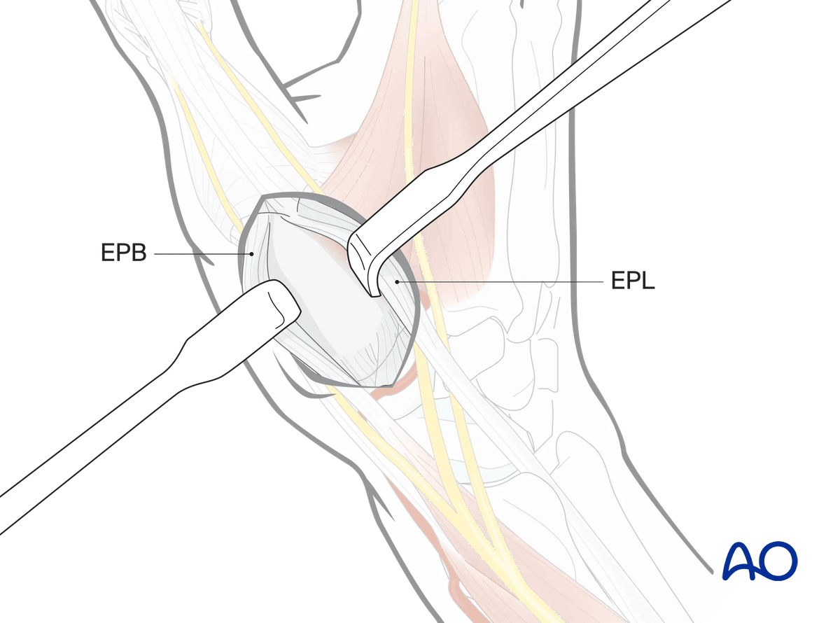
Alternative incision
If the more radial skin incision has been chosen, retract both tendons in a dorsoulnar direction and release the radial attachment of the thenar muscles to expose the metacarpal bone.
This incision may be used for the management of:
- Fractures of the metacarpal shaft
- Intraarticular fractures and fracture-dislocations involving the radial basal condyle of the metacarpal (Bennett fracture)
- CMC joint dislocations
- Trapezial fractures
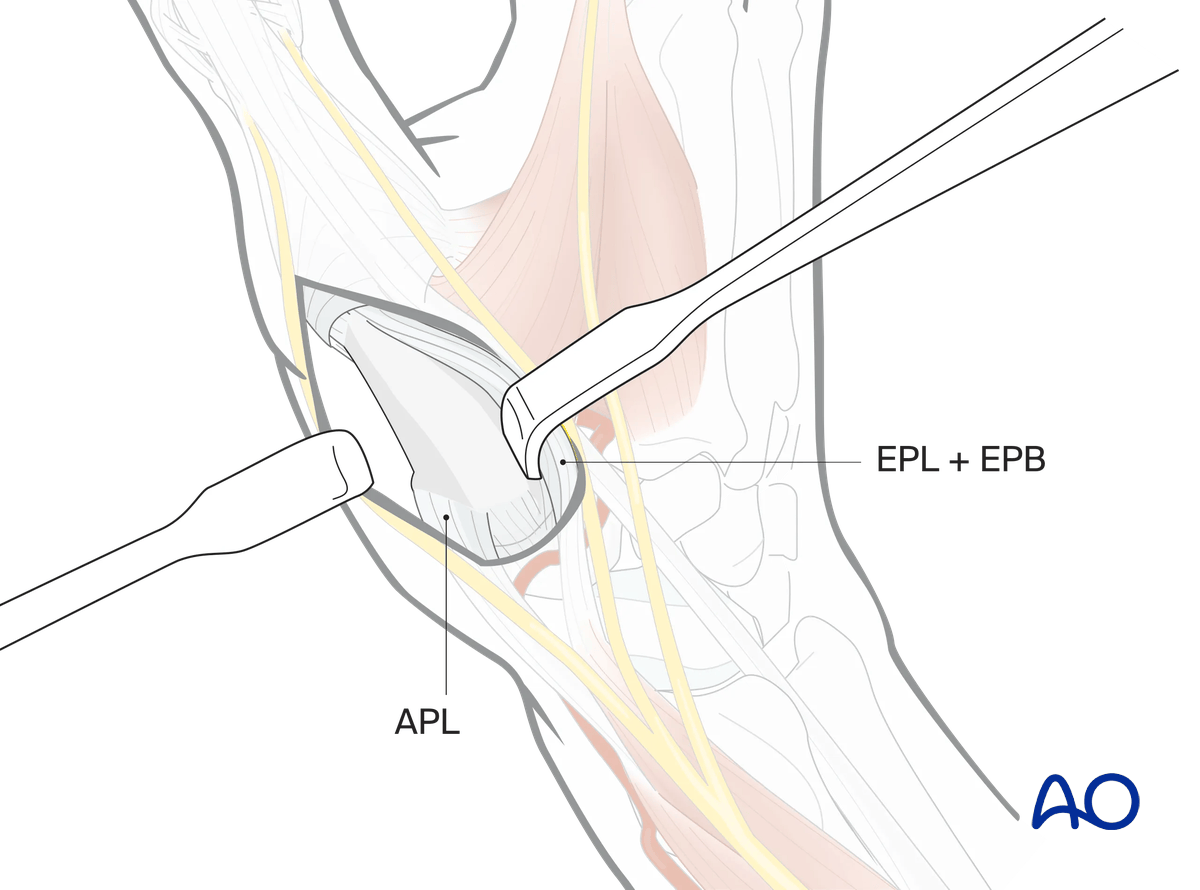
5. Option: capsulotomy
In the case of intraarticular fractures, arthrodesis, or arthroplasty, a capsulotomy of the first carpometacarpal joint is made.
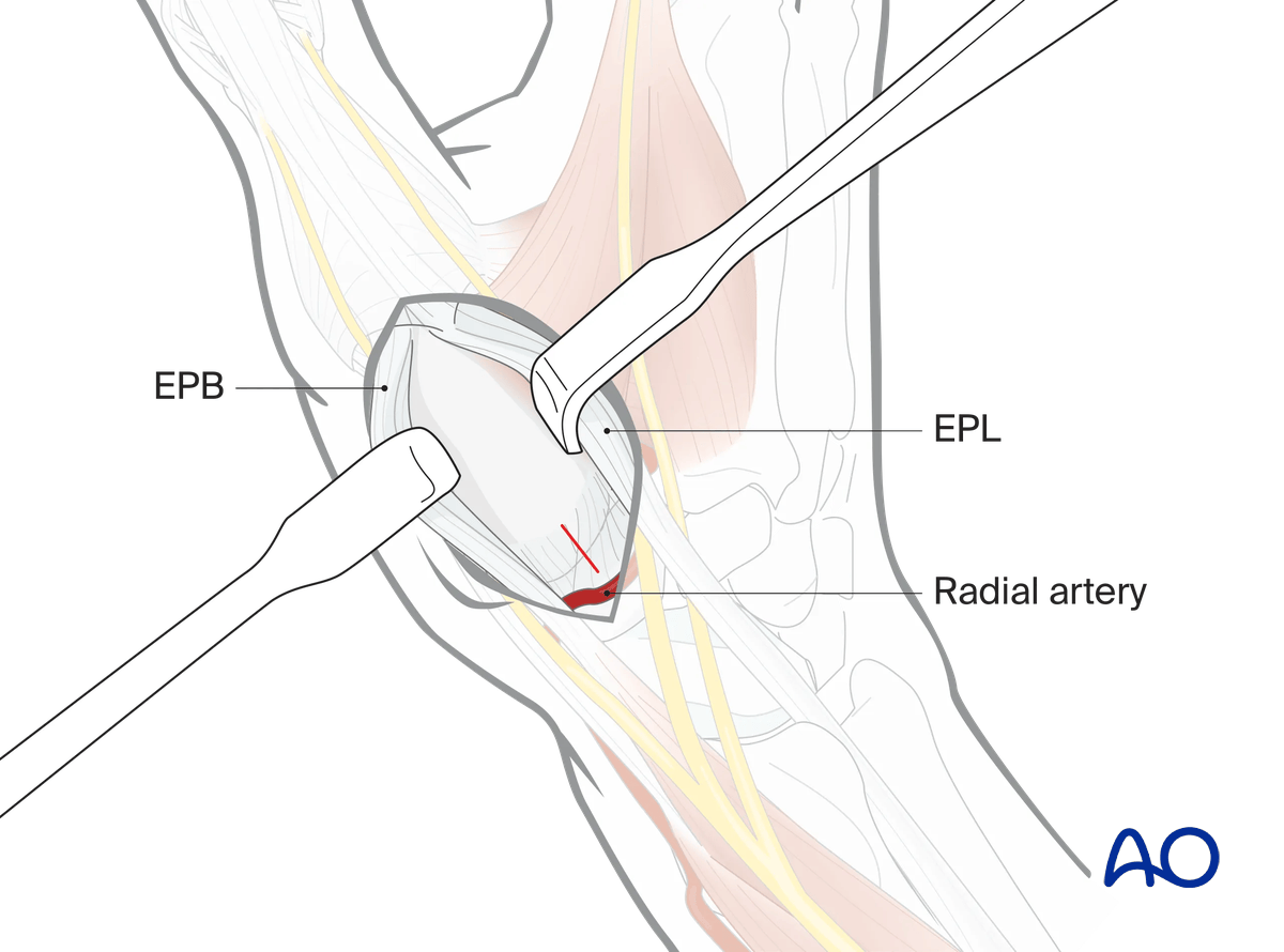
6. Lifting periosteum
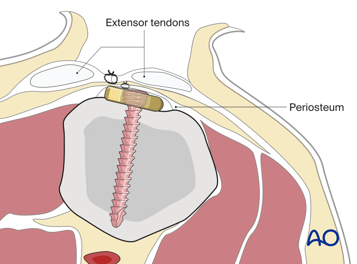
7. Wound closure
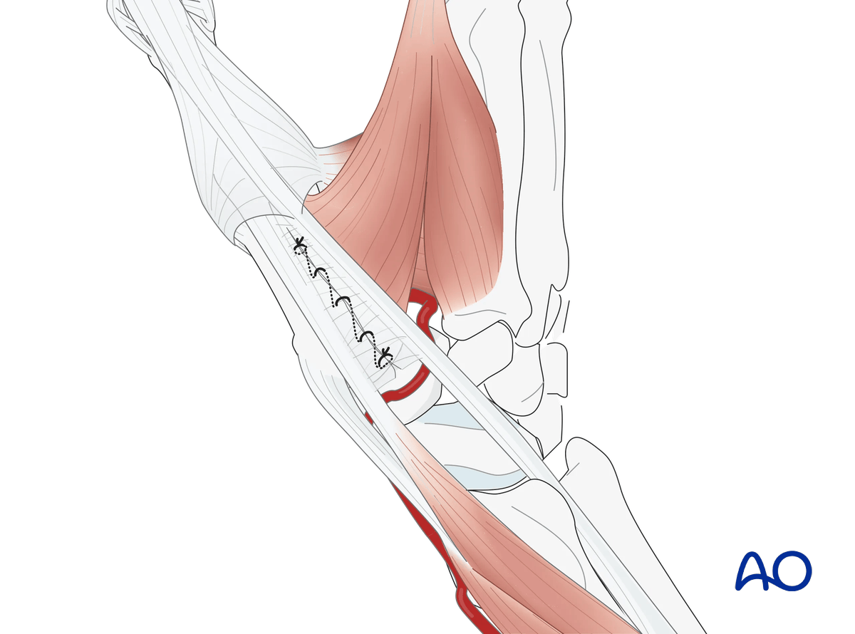
The fascia between the EPB and EPL tendons can be repaired with a resorbable running suture.