Suture and screw fixation
1. Principles
General considerations
Fibular head avulsion fractures are uncommon and may be associated with ligamentous instability of the knee and/or medial tibial plateau fractures. These injuries are often secondary to high-energy forces and represent a fracture-dislocation variant. Varus or varus hyper extension mechanisms are typically implicated. Fibular head avulsion injuries often render the posterolateral corner of the knee incompetent. Significant soft-tissue injury may be associated with these injury patterns. Compartment syndrome, vascular and nerve injuries, particularly peroneal, should be excluded.
Main steps in treatment include:
- In the setting of gross knee joint instability, temporary reduction and immobilization using an external fixator may be required
- Associated bone and soft tissue injury may require advanced imaging using CT scan and MRI
- Centro-medial impaction of the lateral plateau may be identified
When associated with clinically significant posterolateral corner instability, fibular head fixation may be considered.
Timing of surgery
Surgery should be delayed until full recovery of soft tissues (swelling, blisters, abrasions, etc). This usually takes 3–10 days.
Until surgery, the leg should be maintained, immobilized using a knee immobilizer in most cases, or an external fixator in situations with gross instability.
2. Preparation
Patient positioning
In the majority of cases the patient is placed in a supine position. A lateral position may also be used.
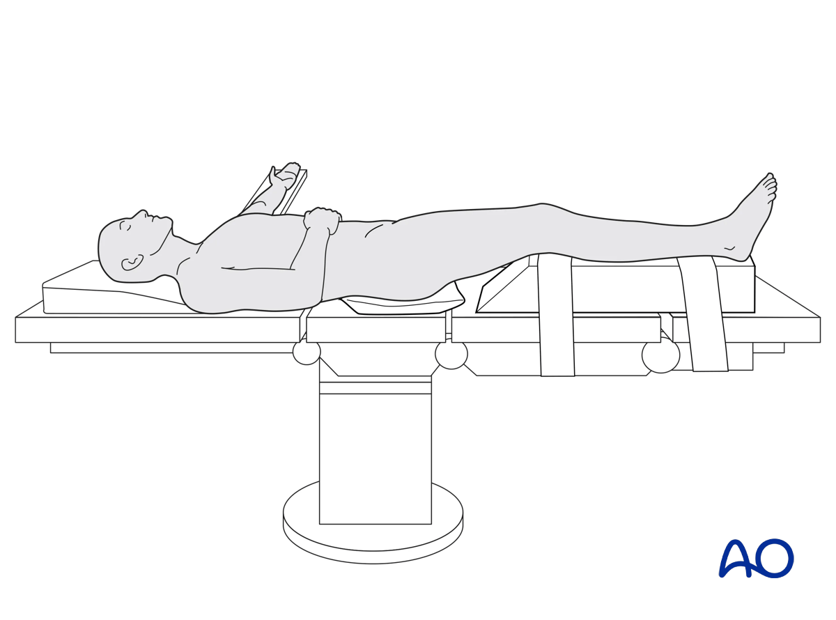
A tourniquet is helpful in most cases. Whether a tourniquet is used depends on the amount of bleeding. Exsanguinate the limb by elevating it.
To allow for intraoperative radiographic control of reduction and fixation, the use of a radiolucent table is mandatory.
3. Approach
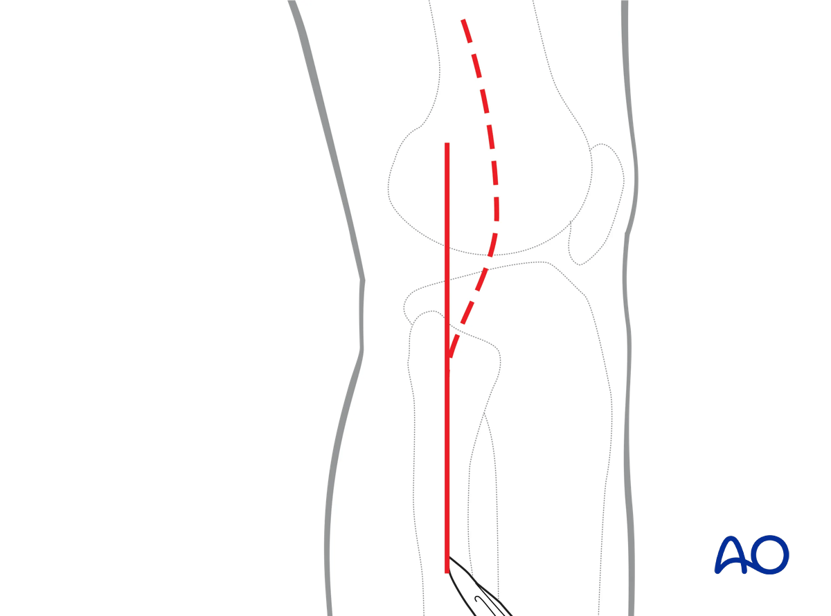
A straight lateral approach is used for the vast majority of cases.
4. Preliminary reduction
General considerations
The injured knee should be examined under anesthesia and a sense of frontal and sagittal plane instability should be identified.
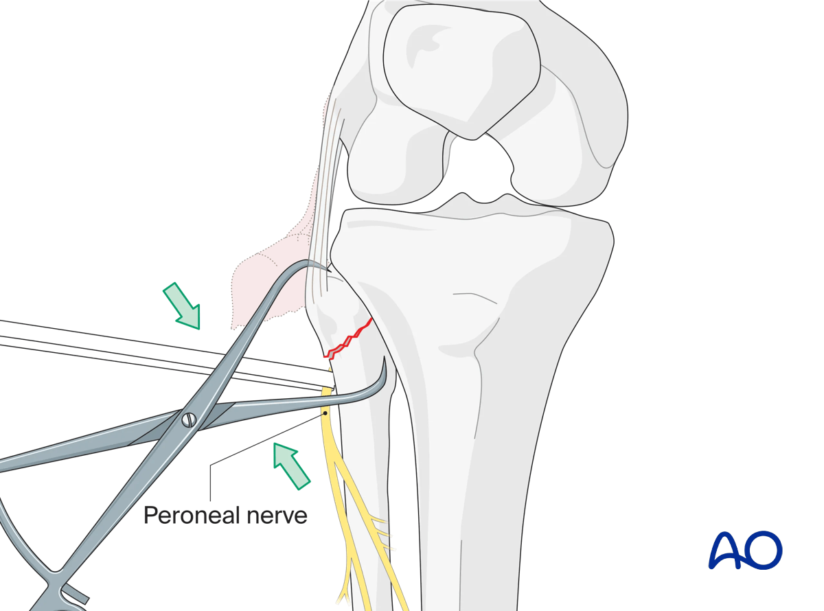
The peroneal nerve must be identified and protected.
The surgeon should be prepared to identify capsular disruption from the lateral tibial plateau.
Reduction of the proximal fibula
Reduction and stabilization of the proximal fibula will restore the insertion of the biceps tendon, lateral collateral ligament +/– popliteal fibular ligament. Significant displacing forces will require neutralization using a variety of techniques including soft-tissue suture repair, tension band repair, and medullary screw fixation.
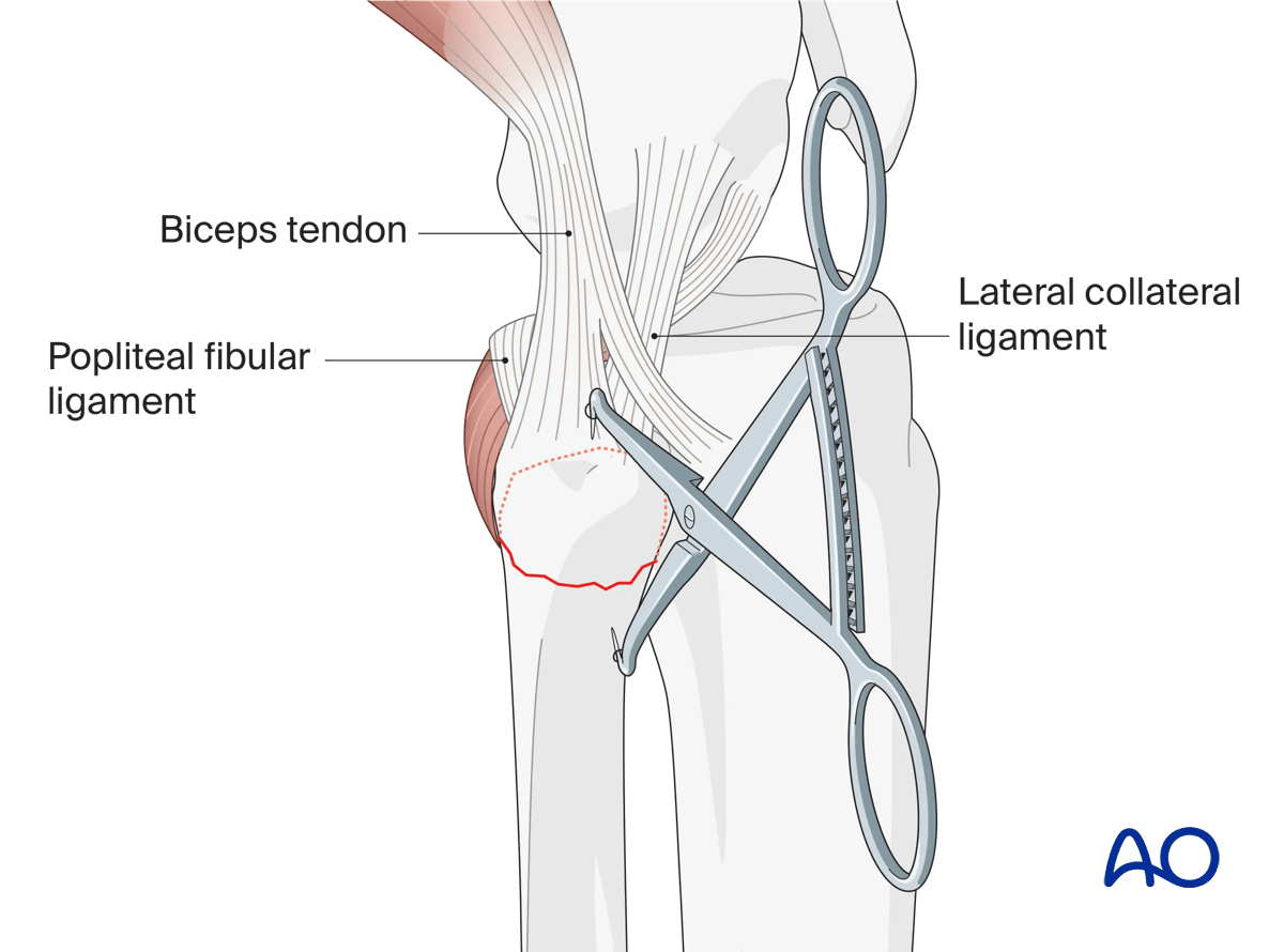
5. Fixation of the proximal fibula
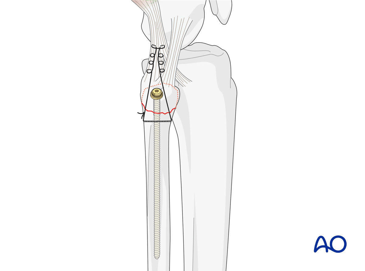
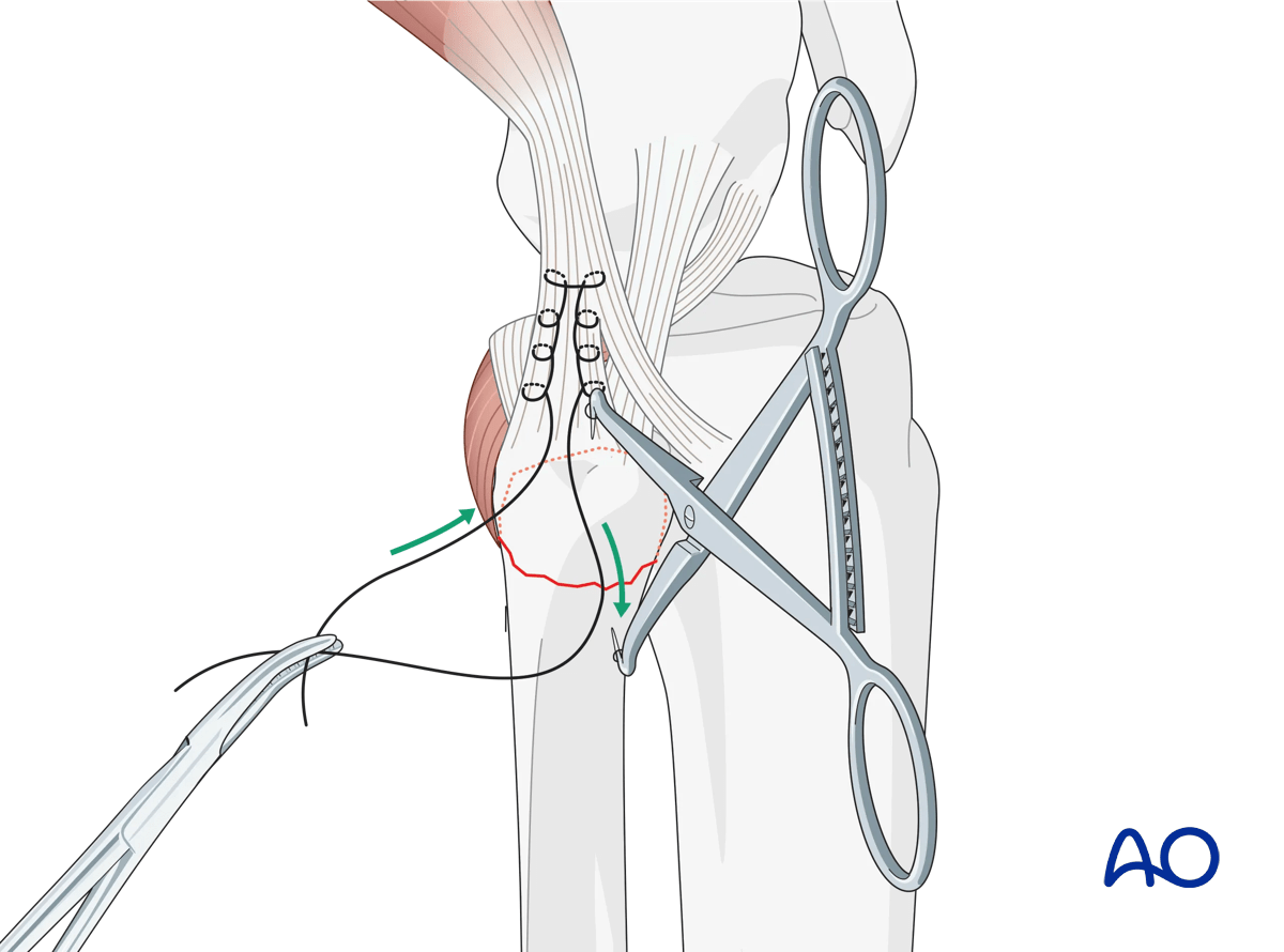
The important components of the injury should be identified including the proximal fibula, and insertion of the biceps tendon and lateral collateral ligament. Capsular avulsion from the lateral tibial plateau is frequently identified, with obvious visualization of the lateral joint. Correct sequencing of the repair is important for successful management. In situations where the capsule is disrupted, suture anchors are placed into the proximal lateral tibial cancellous bone, and the sutures passed through the distal aspect of the capsule disruption. The sutures should be placed on hemostat clamps for later tying. Next, the fibular head is reduced to the distal fibular fragment and held with a modified clamp. The biceps tendon should be identified, and a modified Krackow non-absorbable suture is placed from distal to proximal, starting at the fibula head. After 2–3 cm the suture is brought back from proximal to distal, ending at the fibula head and placed on another hemostat clamp.
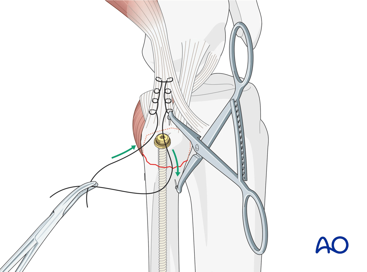
At this point, under C-arm guidance and with the peroneal nerve protected, a long 2.5 mm drill bit is placed on the fibula head inline with and parallel to the medullary canal of the fibula. Pass the drill bit through the fibula head across the fracture and down the proximal fibula medullary canal for several centimeters. Place a long 3.5 mm screw (70–100 mm), with an associated spiked soft-tissue washer down the drill path. Avoid premature grasping of soft tissues by the soft-tissue washer as this may lead to over-tightening of the soft tissues. Secure screw purchase is obtained by endosteal grip. Finally, the Krackow suture is anchored to the proximal fibula distal to the fracture line through a separate drill hole and capsular sutures are tied.
6. Final osteosynthesis
Once osteosynthesis is completed, make a final check with the image intensifier. If all is well, remove the external fixator if present. The knee should then be placed through a full range of motion to ensure fracture stability, and frontal and sagittal plane stability must be reexamined. At this point significant varus instability and/or hyperextension associated intrasubstance cruciate injury may require delayed reconstruction.
Closure
The wound is closed in layers. There should be no fascial closure over the peroneal nerve.
7. Aftercare
Compartment syndrome and nerve injury
Close monitoring of the tibial compartments should be carried out, especially during the first 48 hours after injury and again after surgery to rule out compartment syndrome. More information is provided here:
The neurovascular status of the extremity must be carefully monitored. Impaired blood supply or developing neurological loss must be investigated as an emergency and dealt with expediently.
Consideration for DVT prophylaxis
Oral or subcutaneous administration of DVT prophylaxis for six weeks should be strongly considered.
Functional treatment
Optimal stability should be achieved at the time of surgery, in order to allow early range of motion exercises. Unless there are other injuries or complications, mobilization may be performed on post OP day 1. If available, continuous passive motion (CPM) splints can be very helpful in the early phase of rehabilitation. Static quadriceps exercises with passive range of motion of the knee should be encouraged. Afterwards special emphasis should be given to active knee and ankle movement.
The goal is to achieve as full range of motion as possible within the first 4–6 weeks.
Weight bearing
Weight-of-leg weight bearing is initiated depending on patient comfort. Depending on the severity of the articular displacement, weight bearing can begin as early as 6 weeks postoperatively. In situations where articular displacement was significant weight bearing should be delayed for 10–12 weeks.
Follow up
Wound healing should be assessed within the first two weeks. Subsequently, a 6- and 12-week follow-up with radiographic assessment is usually performed. If a delayed union is recognized, further surgical care may be necessary and should be carried out as soon as possible. Residual knee instability may require delayed reconstruction.
Implant removal
Implant removal is not mandatory and should be discussed with the patient.


