ORIF
1. Principles
General considerations
As the medial fracture is an intra-articular injury, it should be fixed anatomically.
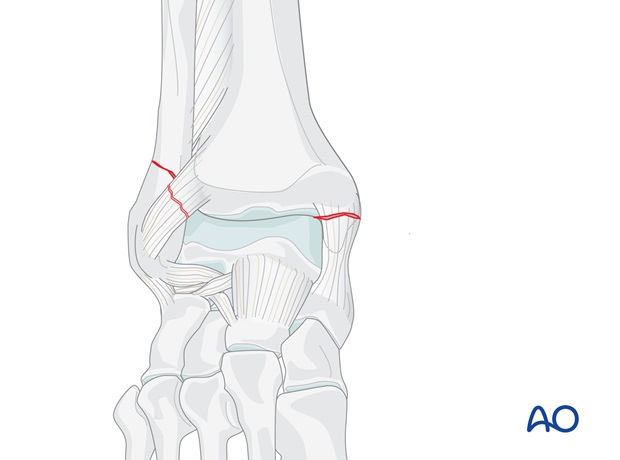
Order of fixation
The choice of fixing the medial or lateral side first may be dictated by the surgeon's preference.
Choice of implant – Lateral fixation
As this is a simple fracture a lag screw and neutralization plate is the most appropriate method of fixation.
Anatomic plates are available, and their lower profile may reduce postoperative discomfort due to prominent hardware. As these plates use locking screws, they may provide more secure fixation in osteoporotic bone.
Choice of implant – Medial fixation
Medial malleolar fractures are usually fixed with lag screws. If the fragment is too small, K-wires and cerclage compression wiring may be better.
Note: “Cerclage compression wiring” was referred to as “Tension band wiring”. We now prefer the term “Cerclage compression wiring” because the tension band mechanism cannot be applied consistently to each component of the fracture fixation. An explanation of the limits of the Tension band mechanism/principle can be found here.
2. Patient preparation and approaches
Patient preparation
Depending on the approach, the patient may be placed in the following positions:
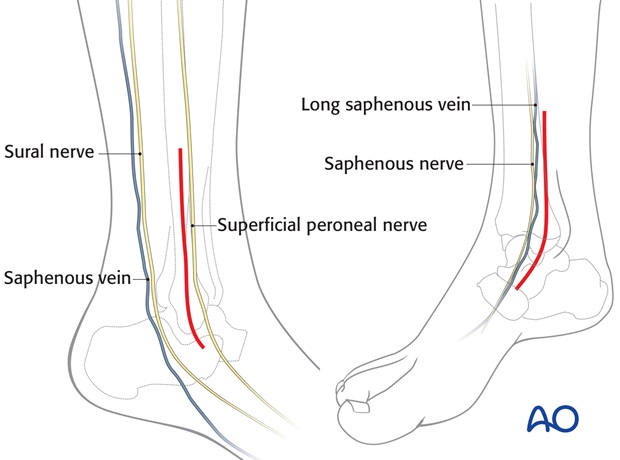
Note on approaches
The two following approaches are used:
3. Fixation
Medial fixation
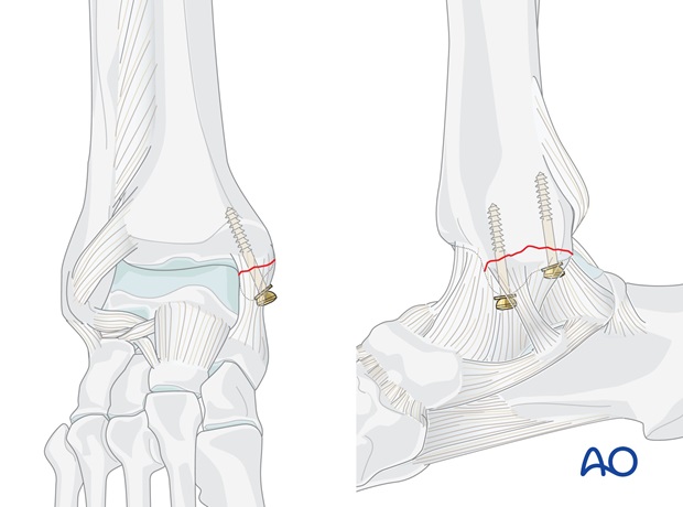
Most medial fractures are fixed with lag screws, which should be inserted perpendicular to the plane of the fracture.
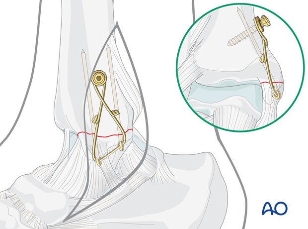
If the fragment is too small or in poor quality bone, K-wires and cerclage compression wiring may be better.
Lateral fixation
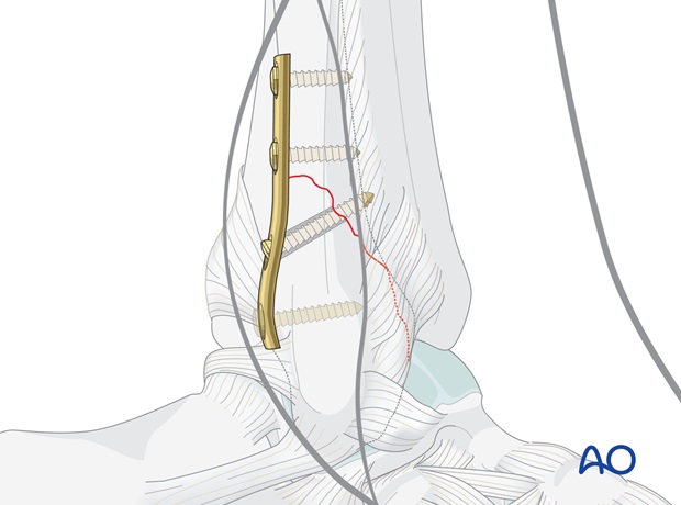
The lateral side is usually a simple oblique fracture, and is fixed with a lag screw and neutralization plate.
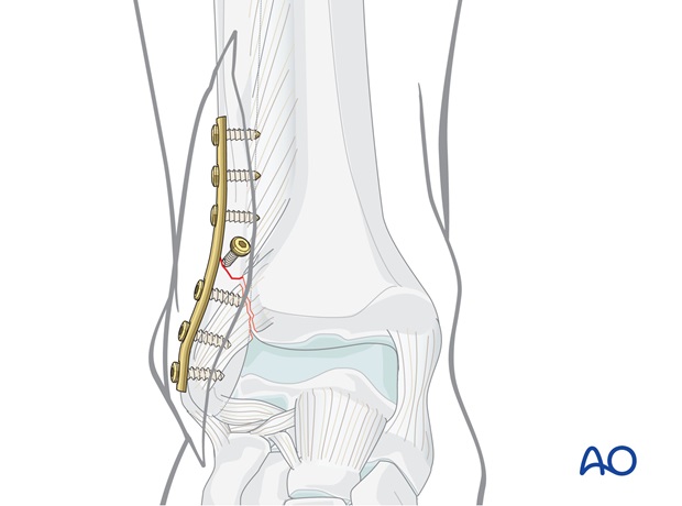
Alternatively, and in particular if there is poor bone or a small fragment, an Antiglide plate may be used.
4. Check of osteosynthesis
Check the completed osteosynthesis by image intensification.
Make sure the intra articular components of the fracture have been anatomically reduced.
Make sure none of the screws are entering the joint. This needs to be confirmed in multiple planes.
5. Postoperative treatment of infra- and trans-syndesmotic malleolar fractures
A bulky compression dressing and a lower leg backslab, or a splint, are applied, and the limb is kept elevated for the first 24 hours or so, in order to avoid swelling and to decrease pain.
In anatomically reconstructed, stable malleolar fractures, early active exercises and light partial weight bearing are encouraged after day one. In osteoporotic bone, weight bearing should be postponed.
X-ray evaluation is made after 1 week and then monthly until full healing has occurred. Progressive weight bearing is recommended as tolerated.


