CAS: Virtual planning and intraoperative imaging (ORIF with orbital reconstruction)
1. Introduction
When using computer assisted surgery, the reduction (and fixation if necessary) is performed according to standard procedures as described in the AO Surgery Reference. It should be considered an adjunct to surgical treatment.
Computer assisted surgery in treating fractures allows virtual preoperative planning of the desired reconstruction using preoperative CT scans and appropriate software.
Intraoperative imaging combined with image fusion of preoperative and intraoperative CT scans and the virtual plan allows verification of proper reduction.
In complex orbital floor fractures radiopaque material for orbital wall reconstruction can easily be visualized by intraoperative imaging allowing intraoperative or postoperative verification of proper reconstruction.
With this technique, insufficient fracture reduction can be can be identified and corrected, eliminating the need for secondary procedures which may be necessary if only postoperative imaging is performed.
Intraoperative imaging requires an additional 10-15 min.
Computer assisted orbital reconstruction planning
Reconstruction of complex orbital wall defects may benefit from preoperative virtual insertion of anatomically preformed orbital implants.
Some complex zygomatico-orbital defects may require patient specific implants for alloplastic reconstruction of orbital walls. This can be performed by transferring virtual preoperative planning into CAD-CAM tools to create patient specific alloplastic implants or by pre-bending standard implants with the use of stereolithographic models either from patients' anatomy or from virtually preplanned reconstruction.
Useful additional reading
2. Virtual planning
Preoperative imaging
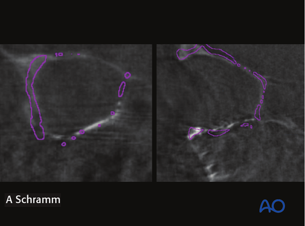
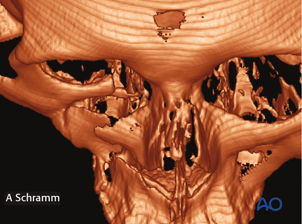
The preoperative CT scan shows a severely displaced fracture of the zygoma. Complex fractures of the orbital floor, medial wall, and nasal bone are also present.
Virtual zygoma reduction
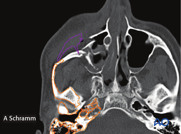
Virtual simulation of the zygoma reduction is performed by mirroring the unaffected contralateral zygoma after auto segmentation. The purple line shows the outer surface of the virtually repositioned zygoma and becomes the surgical goal for reduction.
In this specific case, the virtual zygoma reduction indicates a displaced fracture of the paranasal buttress (arrow). This may be missed in the preoperative examination, leading intraoperatively to malpositioning of the zygoma.
Virtual orbital wall reconstruction
Virtual orbital wall reconstruction is shown in purple. This was achieved by mirroring of the unaffected contralateral side after auto segmentation.
3. Intraoperative assessment of reduction
To verify that the zygoma has been properly reduced, a CT scan is performed intraoperatively.
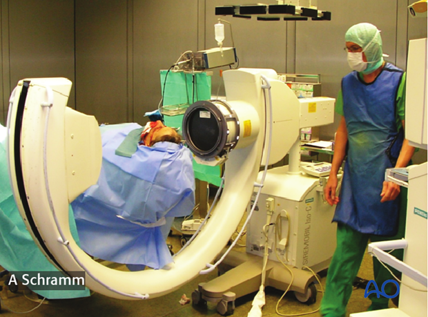
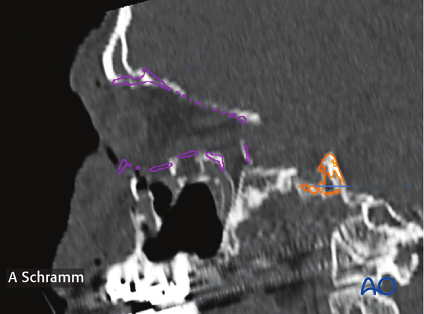
Virtual planning software allows automatic image fusion of preoperative and intraoperative CT scans along with the virtual plan for a better visualization of the achieved reduction.
Note that the reduced zygoma and paranasal buttress fit perfect to the planned position (purple).
If the zygoma is not properly reduced, corrections have to be performed followed by an additional intraoperative CT-scan.
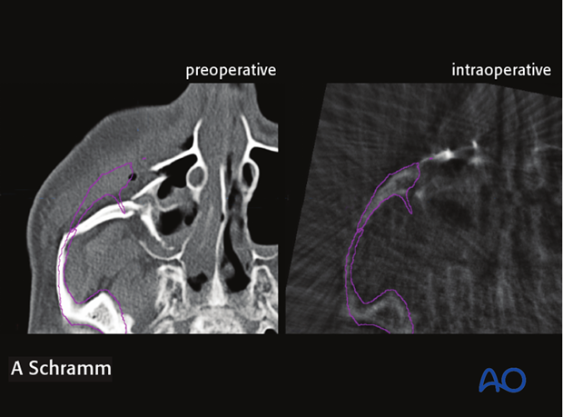
The correct anatomic shape of the inserted titanium mesh can be verified in the intraoperative CT scan when virtual reconstruction in the preoperative CT scan is transferred to the intraoperative scan via image fusion.
Note that the reconstructed orbital floor fits perfectly to the planned position (purple).
In case the orbital walls are not properly reconstructed, correction of the mesh position has to be performed followed by an additional intraoperative CT-scan.
