K-wire fixation
1. General considerations
Introduction
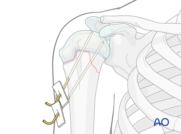
Proximal humeral fractures may require temporary internal fixation that crosses the physis to produce adequate stability.
Stabilization is usually performed with two slightly divergent or parallel K-wires.
The following should be considered to minimize secondary damage to the physis:
- Manipulation of the fracture must be gentle
- Multiple passes across the physis with a K-wire should be avoided
- Select smooth, appropriately sized K-wires
Closed vs open reduction
Due to the remodeling potential, even in an older child, anatomical reduction is not necessary.
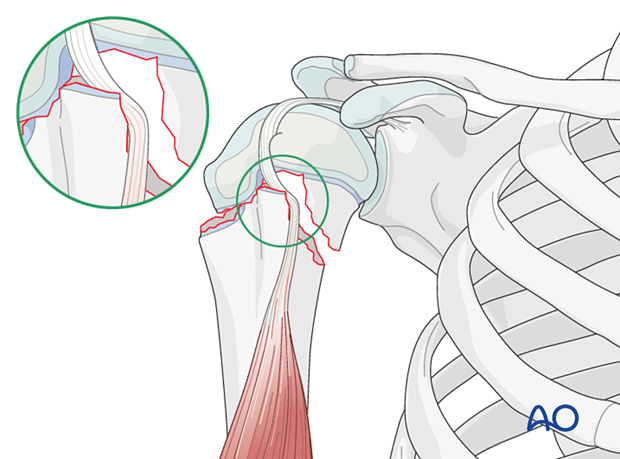
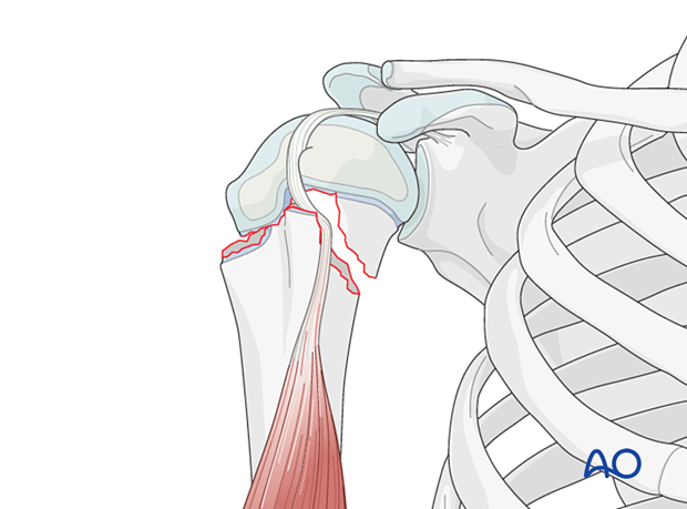
Adequate reduction may not be possible due to soft-tissue interposition. This includes biceps tendon entrapment and buttonholing through the deltoid.
In this case, proceed with an open reduction.
2. Instruments and implants
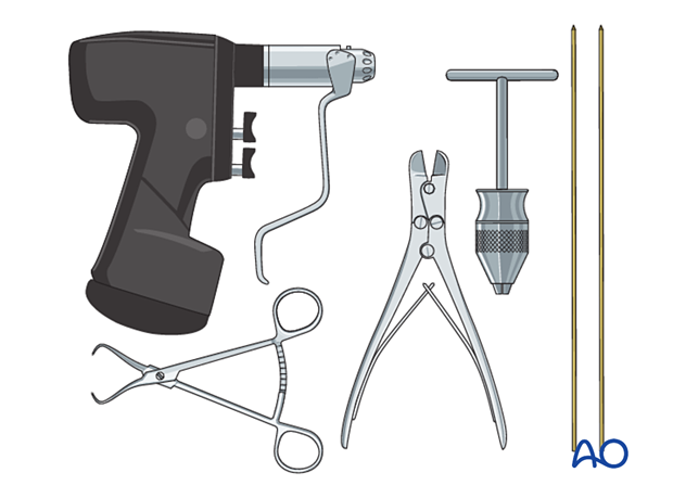
The following equipment is used:
- K-wires of appropriate sizes
- Drill or a T-handle for manual insertion
- Wire cutting instruments
- Standard orthopedic instrument set
3. Patient preparation
This procedure can be performed with the patient in:
4. Approach
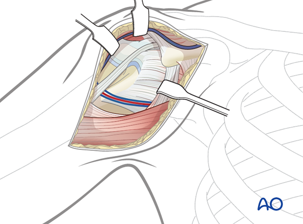
For open reduction, a deltopectoral approach is normally used.
5. Closed reduction
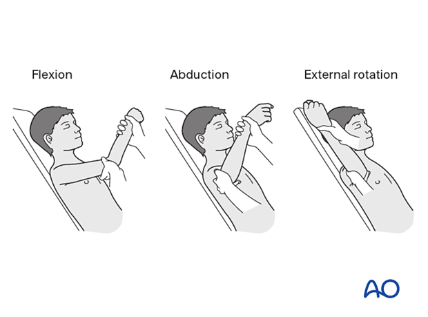
Perform closed reduction with longitudinal traction followed by flexion, abduction, and external rotation.
Forceful reduction maneuvers should be avoided in growth plate injuries to prevent growth arrest.
6. Open reduction
Reduction can usually be achieved by surgical release of interposed soft tissues followed by longitudinal traction and digital pressure on the metaphyseal fragment.
Forceful reduction maneuvers should be avoided in growth plate injuries to prevent growth arrest.
7. Fixation
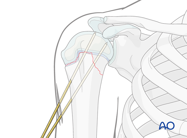
Make a lateral stab incision at the level of deltoid insertion. This should be confined to skin, to avoid injury to the axillary nerve.
Bluntly dissect to the lateral cortex and pass two K-wires distal-lateral to proximal-medial crossing the fracture line up to but not crossing the articular surface.
K-wire cutting and dressing
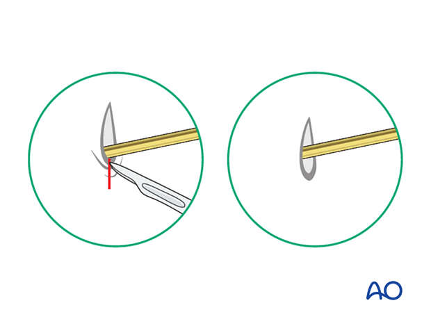
Bend the K-wires approximately 1 cm from the skin to allow for swelling.
Cut the K-wires and apply a dressing to protect the skin.

Release tethered skin around the K-wire by extending the incision.
8. Final assessment
Recheck the fracture alignment clinically. Confirm correct implant position with multiple image intensifier views to exclude perforation of the articular cortex.
Confirm stability of the fixation by moving the arm through a range of motion.
9. Aftercare
Immobilization of the shoulder
The shoulder should be immobilized with a sling until K-wire removal.
Follow up
Follow up after a week for pin site check and an x-ray to confirm that reduction has been maintained.
K-wire removal
K-wires can be removed at 4 weeks after radiological confirmation of fracture healing.
Mobilization
Once the K-wires are removed mobilization can start.


