Open reduction; K-wire fixation
1. General considerations
K-wire fixation provides less stability than ESIN fixation. It needs additional cast or splint immobilization to prevent elbow movement during healing causing elbow stiffness. Other potential disadvantages include infection and cross-union.
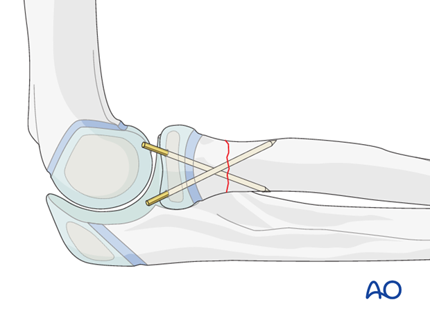
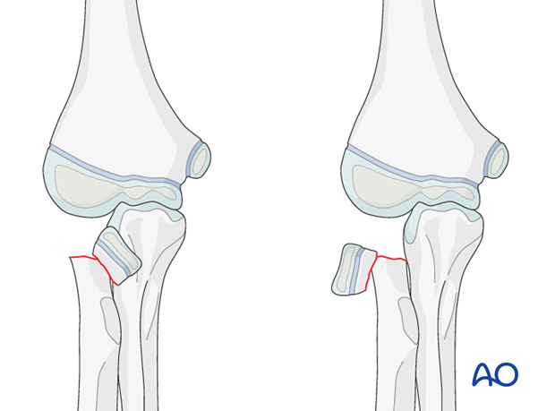
This treatment may be used for reduction and fixation of a radial head that is severely displaced or if an image intensifier is not available.
2. Instruments and implants
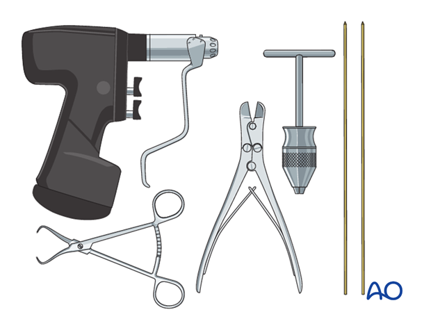
The following equipment is needed:
- K-wires of appropriate sizes
- Drill or a T-handle for manual insertion
- Wire cutting instruments
- Standard orthopedic instrument set
3. Patient preparation
This procedure is normally performed with the patient in a supine position.
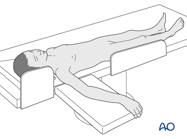
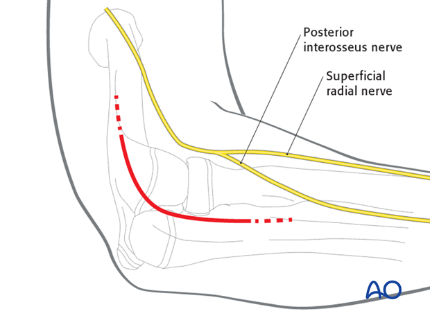
4. Approaches
A direct lateral or a posterolateral approach may be used. This is associated with a high risk of disruption of the remaining blood supply.
5. Reduction
Protect the remaining periosteum throughout the reduction maneuvers.
Pearl: minimizing additional vascular damage to the radial head
To minimize the risk of additional vascular damage to the radial head the following procedure is recommended:
1. Attempt initial reduction of the fracture through the closed capsule.
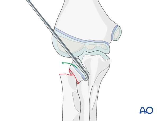
2. If unsuccessful, perform a dorsolateral arthrotomy with irrigation of the joint. The displaced radial head can usually be seen , irrespective of the direction of displacement.
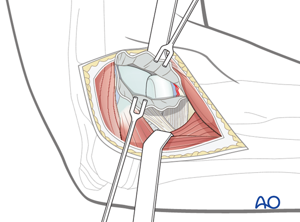
3. Digitally reduce the head fragment .
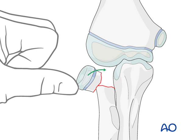
4. If the head is entrapped/displaced in an unusual direction use a dental hook or an identical shaped K-wire push/pull the radial head to an appropriate position .
6. Fixation
Preliminary fixation
After anatomical reduction the head fragment, placed between the metaphysis of the radius and the capitellum, is usually stable. Preliminary fixation is therefore not necessary.
General K-wire principles
Use K-wires with a sharp tip.
Powered insertion of K-wires generates heat in the tissues. Insert wires with a slow-running drill or by hand.
If multiple attempts are made to insert any one K-wire the bone may be weakened or the physis may be damaged. In general, only two attempts of insertion of any K-wire are advisable.
Insertion of K-wires
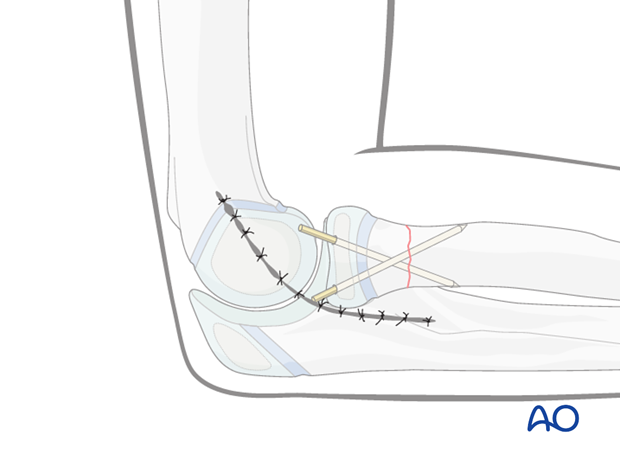
The K-wires must provide adequate spread at the fracture site on any view.
If the K-wire spread is inadequate, the fixation is likely to be rotationally unstable.
Pitfall: The K-wires should engage the far cortex but not protrude into the soft tissues.
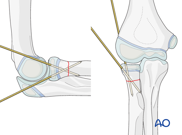
Confirm the position of the K-wires on both AP and lateral views.
If the position is inadequate, adjust the K-wires.
Trimming of K-wires
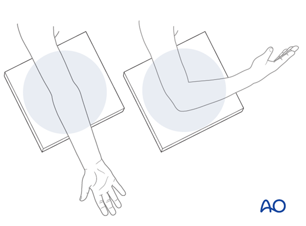
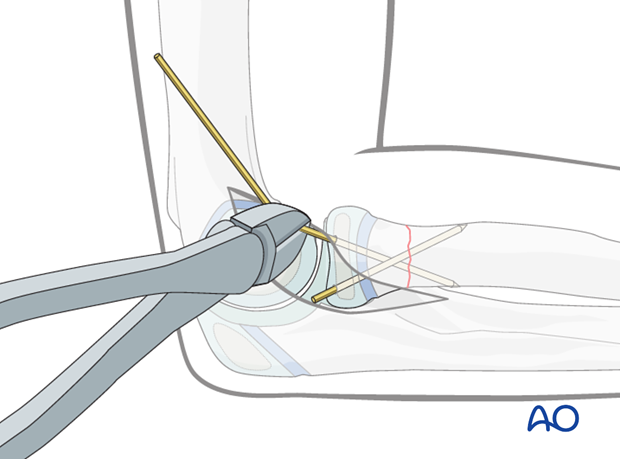
Whether the K-wires are left outside the skin or cut and buried beneath the skin depends on the surgeon’s preference.
K-wires may be left protruding, but there is a risk of pin-track infection. The advantage is that the K-wires can be removed without anesthesia.
Wound closure
Close the wound in layers with resorbable sutures.
Final radiological documentation
Take standard x-rays in lateral and AP view.
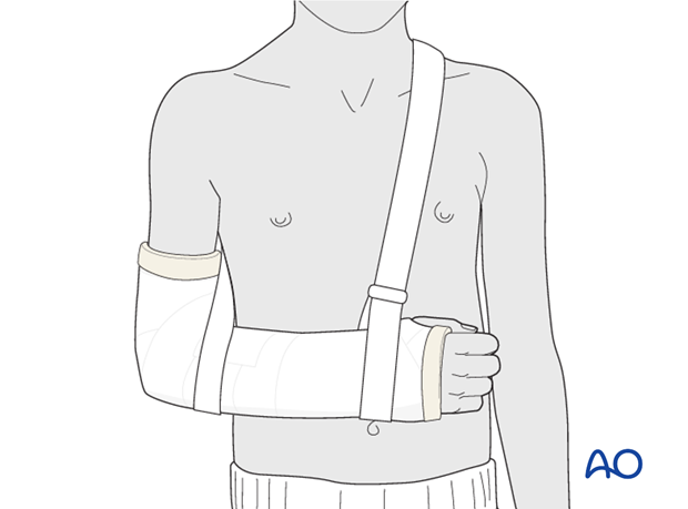
7. Additional immobilization
8. Aftercare following K-wire fixation and immobilization
Duration of immobilization
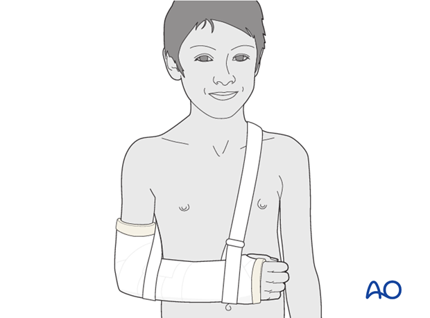
Radial head and neck fractures usually require 3-4 weeks of cast immobilization for adequate callus formation.
Analgesia
Ibuprofen and paracetamol should be administered regularly during the first 24-48 hours after surgery, with opiate analgesia for breakthrough pain.
Opiates should not be necessary after 48 hours and regular ibuprofen and paracetamol should be sufficient until 4-5 after injury or surgery.
The child should be examined if the level of pain is increasing or prolonged analgesia is needed.
Neurovascular examination
The child should be examined after casting/splinting, to ensure finger range of motion is comfortable and adequate.
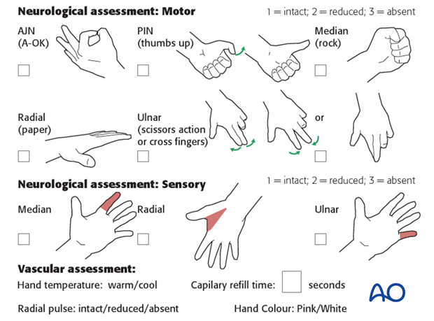
Neurological and vascular examination should also be performed.
Compartment syndrome should be considered in the presence of increasing pain, especially pain on passive stretching of muscles, decreasing range of active finger motion or deteriorating neurovascular signs, which is a late phenomenon.
See also the additional material on complications and postoperative infections.
Compartment syndrome
Compartment syndrome is a possible early postoperative complication that may be difficult to diagnose in younger children.
The presence of full passive or active finger extension, without discomfort, excludes muscle compartment ischemia.
If there are signs of a compartment syndrome in a child in a cast or splint:
- Remove or split constrictive dressings or casts.
- Elevate the limb.
- Encourage active finger movement.
- Reexamine the child after 30 min.
If a definitive diagnosis of compartment syndrome is made, then a fasciotomy should be performed without delay.

Discharge care
Discharge from hospital follows local practice and is usually possible after 1-3 days.
The parent/carer should be taught how to assess the limb.
They should also be advised to return if there is increased pain or decreased range of finger movement.
It is important to provide parents with the following additional information:
- The warning signs of compartment syndrome, circulatory problems and neurological deterioration
- Hospital telephone number
- Information brochure
For the first few days, the elbow and forearm can be elevated on a pillow, until swelling decreases and comfort returns.
Removal of cast or splint
Remove the cast after 3-4 weeks after the injury before taking the control x-ray.
Clinical assessment and x-rays without cast are used to judge adequate healing.
Follow-up
AP and lateral x-rays may be taken at 3-4 weeks following injury to assess position and healing.
See also the additional material on complications.
K-wire removal
Protruding K-wires can be removed immediately after follow-up x-ray.
Buried K-wires can be removed as a day case under anesthesia.
A small portion of the old incision is opened directly over the palpable tips of the bent K-wires. Wires are extracted with pliers or a heavy needle holder.
Recovery of motion
As symptoms recover, the child should be encouraged to remove the sling and begin active movements of the elbow and forearm rotation. See also the additional material on elbow stiffness.
The majority of motion is recovered rapidly and within two months of cast or splint removal.
Physiotherapy is normally not indicated.
The older child may take a little longer.
Once the child is comfortable, with a nearly complete range of motion, he/she may incrementally resume noncontact sports.
Resumption of unrestricted physical activity is a matter for judgment by the treating surgeon.
