Posterolateral (Boyd) approach to the pediatric proximal radius and ulna
1. Introduction
Proximal ulnar injuries associated with radial head dislocation or radial neck fractures, can be addressed through a lateral extension of a posterior skin incision.
This combined approach is also suitable for annular ligament reconstruction, a procedure which is rarely required in the acute setting.
This is the ideal approach for the rare fracture where the radial head is fully displaced posteriorly or medially.
2. Skin incision
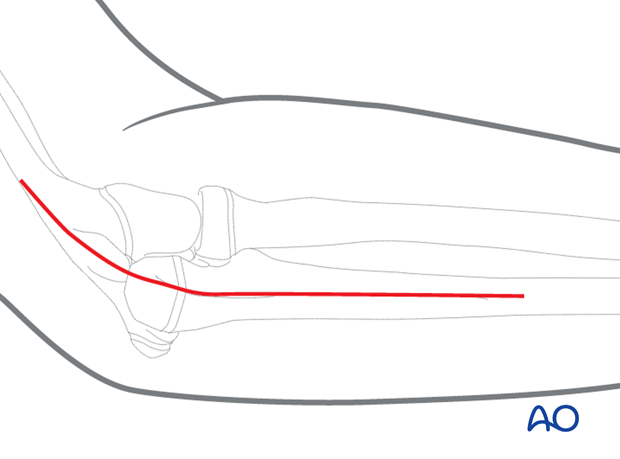
This approach can be performed through a posterolateral incision.
Start the incision posterolaterally behind the supracondylar ridge and continue to the lateral border of the ulna.
Extend the incision as far distally as desired as the direct (subcutaneous) approach to the ulnar shaft.
3. Superficial dissection
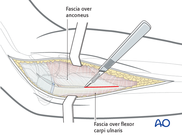
Incise the deep fascia in line with the incision to approach the lateral margin of the ulna.
4. Exposure of posterior surface of the ulna
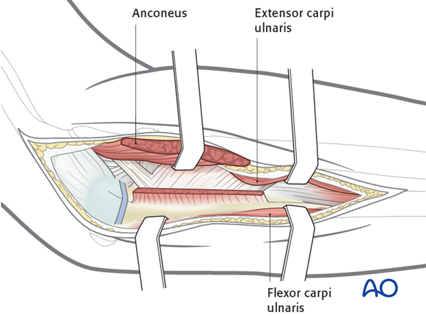
Expose the ulna behind the anconeus proximally and extensor carpi ulnaris more distally.
5. Exposure of radial head/neck
Reflect the anconeus anteriorly/laterally after incising its ulnar insertion.
Detach the supinator near its ulnar origin.
Hold the forearm in a pronated position to keep the posterior interosseous nerve away from the surgical field and limit distal dissection.
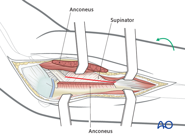
Elevate the anconeus and supinator muscles, carefully protecting the posterior interosseous nerve, which is within the substance of the supinator. It may be necessary to separate supinator from the oblique cord of the interosseous membrane.
Retracting these muscles exposes the posterior joint capsule over the radial head.
Pitfall: Do not place retractors around the radius as this may injure the PIN.
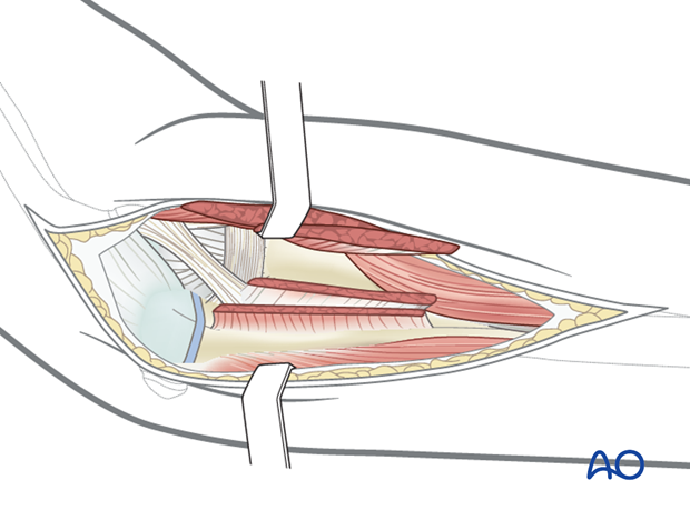
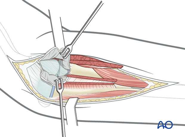
Now that the capsule is exposed, the radial head can easily be visualised.
Note: Preserve the periosteal vessels to the radial head while reducing the fracture.
6. Wound closure
Close the capsule with resorbable sutures (3/0).
Reattach the muscles and fascia with resorbable sutures (2/0 or 3/0).
Close skin and subcutaneous tissue with fine resorbable sutures (this avoids distress to the child when removing nonabsorbable sutures).


