Open reduction, K-wire fixation
1. Preliminary remarks
Introduction
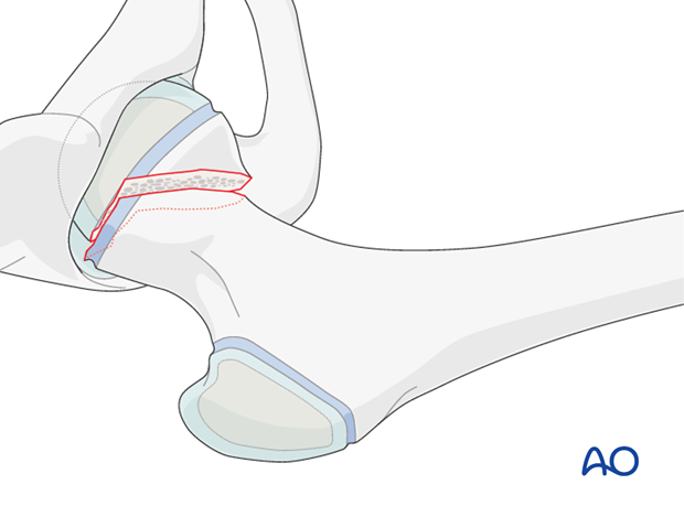
Children's hip fractures have historically had a bad prognosis. Accurate stable fixation has been one of the factors identified to improve the outcome.
Stable fractures have a better prognosis than displaced fractures.
The main problems have been avascular necrosis (AVN) due to disruption of the blood supply, nonunion and malunion due to unstable fixation.
The objective is timely fixation without disruption of the blood supply.
Note: Care must be taken to rule out slipped capital femoral epiphysis (SCFE). Fractures can be distinguished from SCFE by a detailed history supplemented by appropriate x-ray examination and assessment; in unclear situations, an MRI is indicated.
Hardware selection
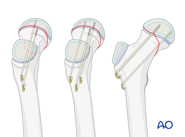
2.0, 2.5, or 3.0 mm K-wires can be selected, depending upon the size of the child.
Larger K-wire sizes are preferred for stability.
Threaded K-wires also increase stability.

Implant insertion
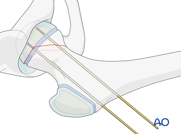
Typically, two K-wires separated vertically are inserted. This configuration may be more suitable in smaller bones.
An alternative is three K-wires used in an apex-distal, triangular configuration, with the lower central K-wire abutting the calcar.
Inserting a single central K-wire does not provide rotational stability.
2. Patient preparation and approaches
Approaches
For this procedure the following approaches may be used:
3. Reduction
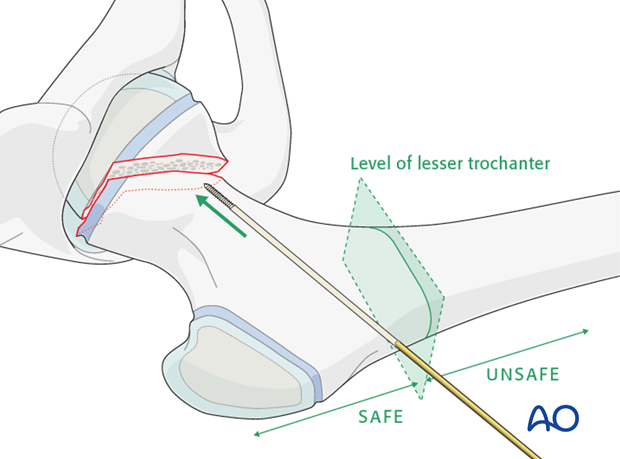
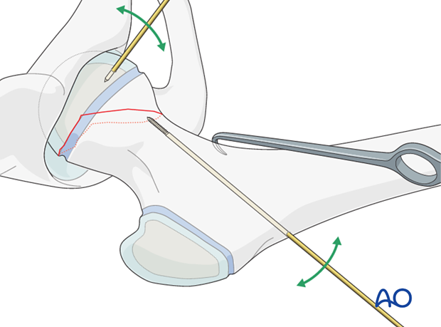
A K-wire is inserted through the lateral cortex of the femur just above the level of the lesser trochanter and advanced to the fracture line.
Pitfall: Secondary subtrochanteric fractures may occur if K-wires are inserted with an entry point distal to the level of the lesser trochanter.
The femoral neck is manipulated gently into position, usually by traction in the line of the femoral neck using a bone hook.
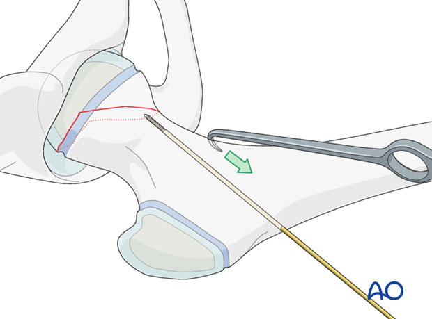
If necessary, manipulation of the femoral head is achieved using a small K-wire, inserted into the femoral head, as a joystick.
Gentle reduction maneuvers are essential, in order to prevent secondary damage to the femoral head blood supply.
4. Fixation
K-wire insertion
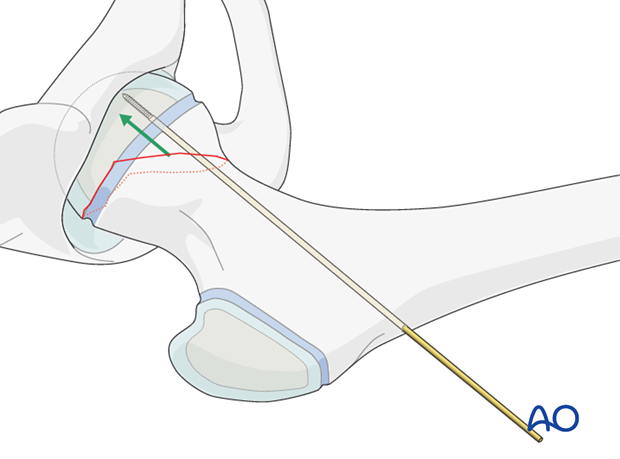
When acceptable reduction has been obtained, the first K-wire is advanced across the fracture and into the femoral head. Overpenetration into the hip joint must be avoided.
Additional wires are inserted under x-ray control, using image intensification.
The K-wire positions are confirmed on AP and lateral x-ray images, using image intensification.
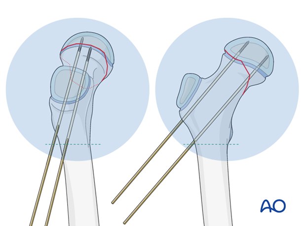
If using a standard radiolucent table, the K-wire fixation is generally stable enough to allow a “frog” lateral view.
The K-wires are repositioned if necessary.
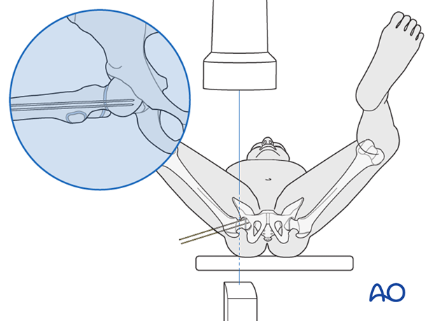
Images at multiple angles are used to confirm that the K-wires do not penetrate into the hip joint.
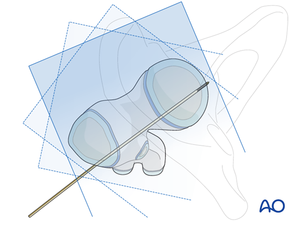
Pitfall: K-wire penetration can occur, especially with eccentrically placed wires, even if not apparent on standard AP or lateral x-rays.
Typically, 5 mm of epiphyseal bone/cartilage should remain between the wire tip and joint surface.
Dynamic, real-time image intensification, with a full range of internal and external rotation at different degrees of flexion, is useful to see how close the K-wire-tip is to the joint surface of the head.
Observing an approach/withdrawal of the K-wire tip helps in judging its position. Some surgeons supplement this examination with 3-D image intensification for confirmation.
Arthrography is useful to confirm correct wire placement in the younger patient.
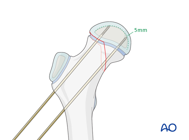
Cutting K-wires
The K-wires can be bent, cut short, and left beneath the fascia lata.
The K-wires are usually removed with a short secondary procedure, once the fracture is healed.

5. Hip spica
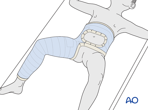
Because K-wire fixation is less stable, and only indicated for younger children, a hip spica cast is generally used to supplement the fixation.
Click here for details of hip spica application.
6. Aftercare
Introduction
Hip spica is only likely to be used in the treatment of small children with proximal femoral fractures.
For larger children, internal fixation should be used even for nondisplaced fractures.

Immediate care
After application, the spica should be trimmed to allow adequate space for bodily functions. The edges of the spica should be padded and waterproofed.
Diaper care
Generally, a hip spica should allow space for a small diaper inside the plaster and a large one outside the plaster. The diapers should be positioned to prevent soiling of the spica.
Washing
The spica is not waterproof. Bathing and showering should not be attempted. Hair washing should be done very carefully.
Skin care
No skin products should be put inside the spica. Only skin that can be seen should be moisturized.
Transport
Both the child and the spica must be lifted. A special car seat will be required. The child may be placed in a stroller or buggy.
In this circumstance, the parents/carers are advised to return to the healthcare provider.
Length of time in spica
The length of time in the spica depends on the age of the child and the healing of the fracture. A proximal femoral fracture in a child aged below 4 years should always be healed in 6 weeks.
Growth
The child will continue to grow and the tightness of the spica should be monitored.
Follow-up x-rays
Nondisplaced fractures being treated nonoperatively should have early radiological follow-up. If the spica is being used for protection of fixation, x-rays are required only when planning removal.


