Supine position
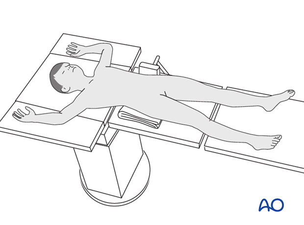
1. Positioning of patient
The patient is placed in a supine position.
A radiolucent support is placed under the ipsilateral buttock. A simple prop against the opposite iliac crest may aid stability.
For the anterolateral approach, some extension of the hip may be required.

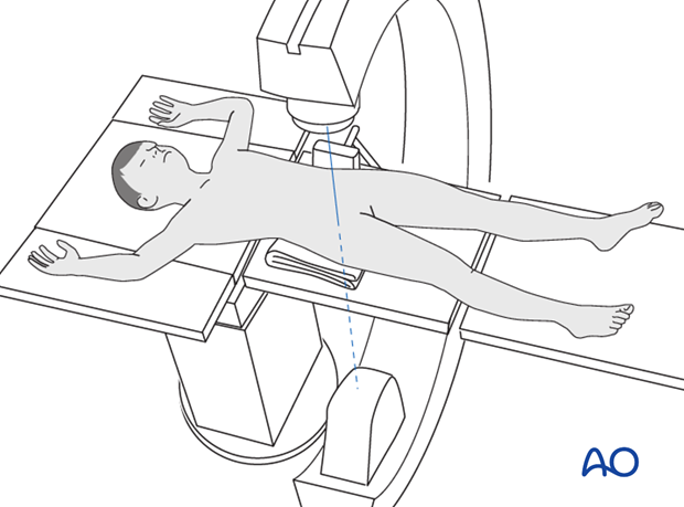
2. Positioning of the image intensifier
The image intensifier is placed on the opposite side of the table.
Lateral views of the proximal femur are obtained by flexing and externally rotating the hip.

3. Draping
The whole leg is prepared and draped with a hip drape to allow free movement of the leg.


