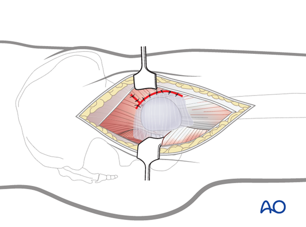
Transgluteal approach to the pediatric proximal femur
1. Preliminary remarks
The transgluteal approach to the proximal femur, between the anterior and medial part of the gluteus medius, provides access to the hip joint at the lateral proximal femur.
This approach is adequate for fracture reduction with K-wire, screw or plate fixation and is indicated in the following situations:
- Open reduction and stabilization of extracapsular neck fractures
- Stabilization of nondisplaced but complete intracapsular fractures
2. Vascular anatomy
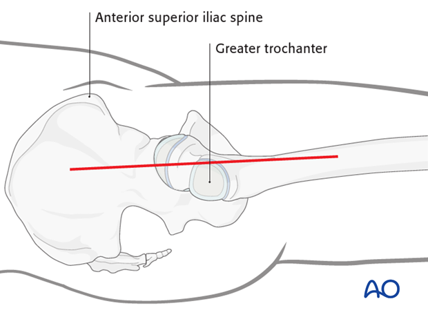
The deep branch of the medial femoral circumflex artery provides the main relevant blood supply to the femoral head.
The medial femoral circumflex artery originates from the deep femoral artery (profunda femoris), courses between the iliopsoas and pectineus muscles, and runs posteriorly between the femur and the pelvis.
During its course, a small branch supplies the inferior retinaculum (ligament of Weitbrecht).
The main branch of the medial femoral circumflex artery is related to the inferior border of the obturator externus muscle and passes posterior to the femur, towards the intertrochanteric crest.
It then crosses posterior to the obturator externus and anterior to the triceps coxae (obturator internus and the superior and inferior gemelli).
Before crossing the triceps coxae, a small branch passes to the greater trochanter.
The vessel enters the joint capsule between the gemellus superior and the piriformis muscles.
Note: The approach must always be cranial to the piriformis muscle. This anatomical detail is crucial when starting to prepare the capsule.
After perforating the capsule, the vessel passes along the superior retinaculum and splits into 3-4 branches.
Provided the obturator externus muscle remains intact, it will protect the medial femoral circumflex artery.
3. Skin incision
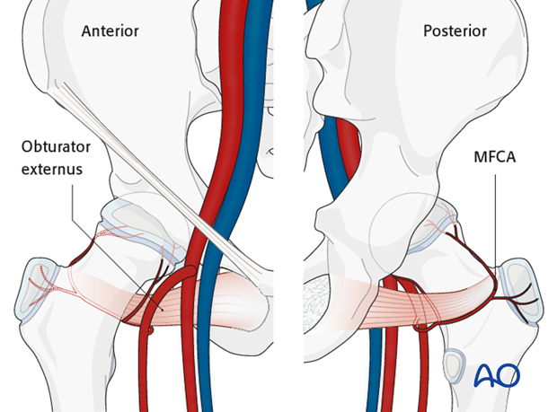
The greater trochanter and anterior superior iliac spine (ASIS) are palpated and outlined with a sterile marking pen.
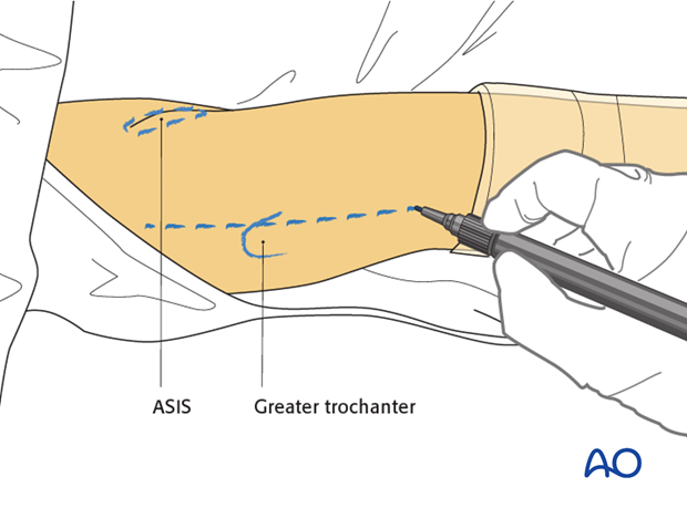
The incision starts a few centimeters proximal to the anterior superior iliac spine and continues along the axis of the femur immediately anterior to the palpable greater trochanter.
The length of the incision will depend on the body mass of the patient and should extend equally inferior and superior to the greater trochanter as shown.
Dissection continues to the fascia lata distally and to the fascia over the gluteus maximus proximally.

4. Dissection of the fascia
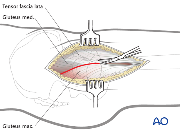
The fascia lata is incised longitudinally and proximally from the most distal extent of the wound up to the greater trochanter.
The incision is continued along the anterior border of the gluteus maximus (Gibson approach).
The gluteus medius is visible proximal to the greater trochanter.
Note: The gluteus medius has three portions arising from:
- The most posterior aspect of the fossa (ala) of the ilium
- The middle part of the fossa
- The anterior aspect of the fossa, almost parallel to the tensor fascia lata
It is important to note that the fascia lata merges directly with the gluteal and the tensor fascia.
Dividing the fibers of the gluteus medius should be avoided.
The transgluteal approach develops a plane between the anterior border of the medial part of the gluteus medius and the posterior border of the anterior part of the gluteus medius.
Pearl: The assistant positioned next to the surgeon, flexes the knee and extends the hip relaxing the gluteus maximus and its fascia. This improves the exposure of the posterior border of the gluteus medius.
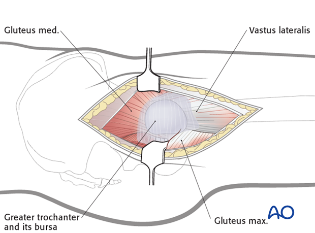
Once the fascia is completely opened, the following landmarks are readily identifiable:
- Greater trochanter and its bursa
- Vastus lateralis
- Gluteus medius
- Anterior border of the gluteus maximus
Note: At this stage, the trochanteric bursa and the loose areolar tissue overlying the short external rotators are still intact.
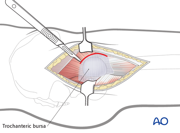
It is recommended that a flap of trochanteric bursa and the attached tissue is prepared with an anterior incision and retracted posteriorly to expose the short external rotators.
This flap is repositioned and repaired when the wound is closed, preventing adhesions between the greater trochanter and the fascia lata.
5. Vastus lateralis mobilization and gluteus medius split
Once the lateral aspect of the greater trochanter and the anterior part of the gluteus medius are clearly visible, the vastus lateralis is mobilized, from the antero-lateral aspect of the femur.
This begins at the most distal part of the incision and extends to the insertion below the greater trochanter.
Note: Care should be taken to ensure that the dissection always remains subperiosteal .
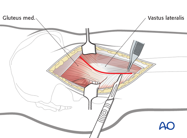
The vastus lateralis and anterior portion of the gluteus medius insertion are dissected from the greater trochanter using a knife.
In younger children, it is recommended that this includes a 1-2 mm thick cartilage portion from the greater trochanter.
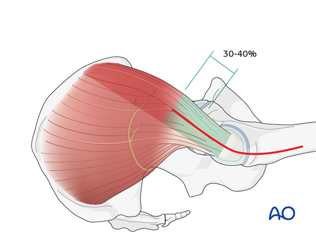
Once this dissection has been completed, the anterior portion of gluteus medius can be split along its posterior border.
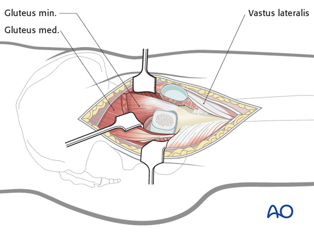
Note: The muscle should not be split more than 30- 40 % of its length to avoid damage to the motor nerve to gluteus medius.
In this illustration, the anterior retractor holds the anterior portion of the gluteus medius and the posterior retractor holds the medial portion of the gluteus medius together with the gluteus minimus.
A third retractor can be inserted into the upper corner of the wound to completely expose the femoral neck and calcar and improve the view of the acetabulum.
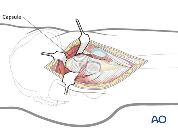
6. Z-shaped capsular incision
Once the anterior and inferior capsule is prepared, the capsule is opened with a Z-shaped incision.
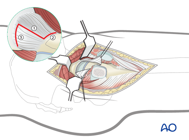
Starting from the distal third of the trochanter, a longitudinal incision is made towards the acetabulum (1).
Care must be taken not to cut into the labrum.
The second cut runs along the distal anterior insertion of the capsule around the calcar (2).
The third cut runs parallel to the edge of the acetabulum in a posterior direction (3).
Note: Depending on the level of the fracture and the degree of displacement, the insertion of the capsule or the capsule itself can be ruptured.
7. Closure
The capsule should be closed loosely to prevent the formation of a hemarthrosis under pressure.
The fascia lata can be closed with a continuous suture.
Pearl: Before the fascia is closed, it is recommended that the layer of the trochanteric bursa is repositioned to prevent the formation of adhesions between the trochanter and the fascia.
Subcutaneous suture and closure of the skin is performed according to the surgeon’s preference.
