Open reduction - Bridge plating
1. General considerations
Introduction
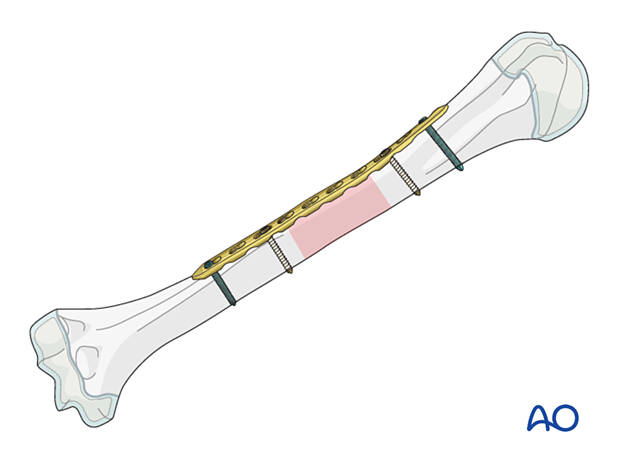
Comminuted humeral shaft fractures may be treated with bridge plating under the following conditions:
- Reduction and stability not achieved with other techniques, particularly in older children
- Open fractures
- Neurovascular injuries
- Polytrauma
The principles of fixation are identical to adult fracture management.
For more details on bridge plating, see the following basic technique:
Throughout this section, generic fracture patterns are illustrated as:
- Unreduced (A)
- Reduced (B)
- Reduced and provisionally stabilized (C)
- Definitively stabilized (D)
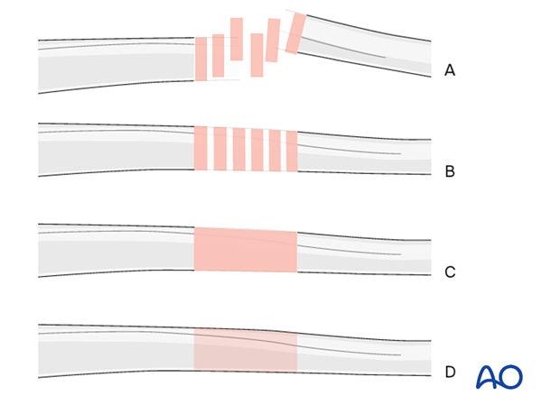
Plate selection
The plate should allow for insertion of a sufficient number of screws on either side of the fracture.
Plates that accommodate the smaller pediatric humerus are available. A 3.5 or 4.5 mm LCP is recommended depending on the bone size. A DCP, without compression, may be used as an alternative.
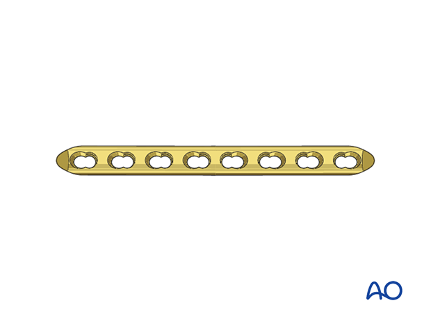
Plate position
Open fractures may dictate the surgical approach and the plate position.
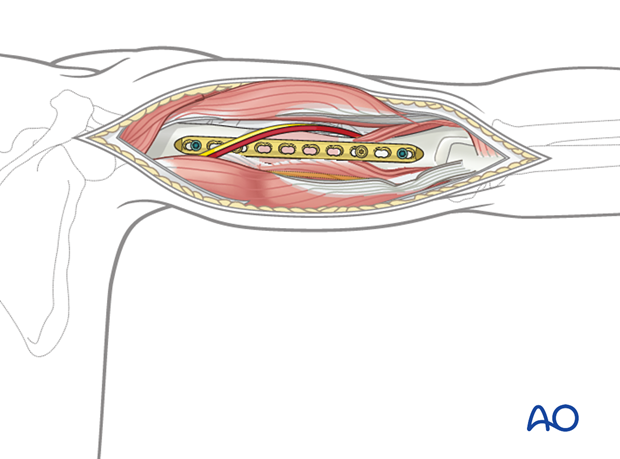
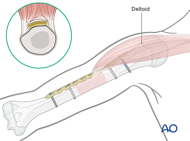
The position of the plate is dictated by the location of the fracture in relation to the soft-tissue attachment and its proximity to the axillary and radial nerves.
The course of the radial nerve in relation to the plate holes should be documented to reduce the risk of nerve injury associated with plate removal.
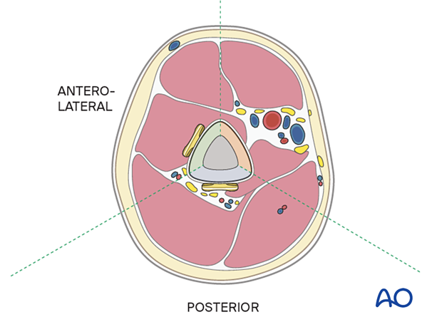
A posterior plate is preferred for middle and distal third fractures.
An anterolateral plate may be selected for proximal and middle third fractures.
2. Patient preparation
Place the patient in a supine position or, alternatively, a beach chair position for an anterolateral plate.
Place the patient prone or in a lateral decubitus position for a posterior plate.
3. Approaches
The approach depends on the plate position.
For an anterolateral plate, the anterolateral approach is used.
The posterior surface is accessed with a posterior approach.
4. Reduction
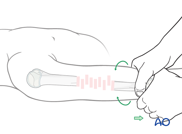
Bring the humerus out to length by traction and hold it until plate application. External fixation or a distractor may be used in older children.
Direct reduction of fracture fragments should be avoided.
The plate can be used as a reduction aid.
5. Plate fixation
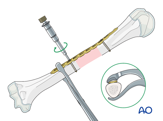
Plate application
Apply the plate avoiding periosteal stripping.
Hold the plate against the proximal fragment, and provisionally attach it with a single bicortical nonlocking screw.
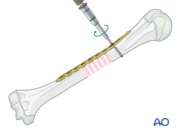
Check the alignment along the shaft and insert a second cortical screw in the distal segment, near the fracture zone.
Finalizing the fixation
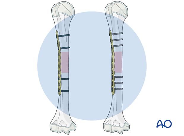
If the alignment is satisfactory, add the remaining screws to both main fragments and reconfirm alignment.
The number depends on the type of screws, the fracture morphology.
Three bicortical screws are usually necessary on either side of the fracture. However, 4 locking screws in a near-far configuration may also be sufficient.
Confirm the reduction, plate position, and screw length under image intensification.
Confirm rotational alignment by physical examination.
6. Aftercare
Initial postoperative treatment
A sling may be used initially, but early mobilization is recommended.
Follow-up
The first clinical and radiological follow-up is usually undertaken within 2 weeks.
X-rays are repeated after 6 weeks.
Implant removal
Implant removal is not mandatory and is associated with a high risk of radial nerve injury.


