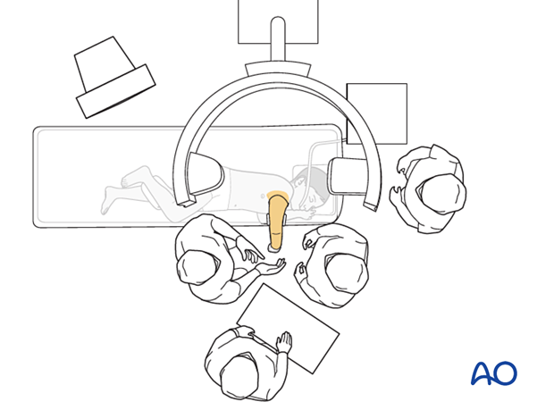
Lateral decubitus position
1. Introduction
The lateral patient position with a hanging arm may be used for posterior approaches.
This position can be more difficult for smaller patients because of the short humeral segment.
2. Preoperative preparation
Consider the additional material on preoperative preparation.
3. Anesthesia
The addition of regional and local anesthesia may reduce postoperative pain.
4. Prophylactic antibiotics
Antibiotics are administered according to local policy and specific patient requirements.
5. Patient positioning
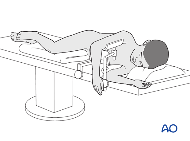
The patient is positioned in a lateral decubitus position on the uninjured side, as close as possible to the edge of the table. Place a pillow between the legs and support the front and back of the trunk. The injured arm should be maintained at 90° abduction with a suitable support.
The contralateral shoulder must be positioned to prevent brachial plexus injury with padding under the axilla.
The injured arm is placed in a gutter support beneath the upper arm, or over a padded arm roll under the elbow.

6. C-arm positioning
The C-arm is introduced from the top of the table.
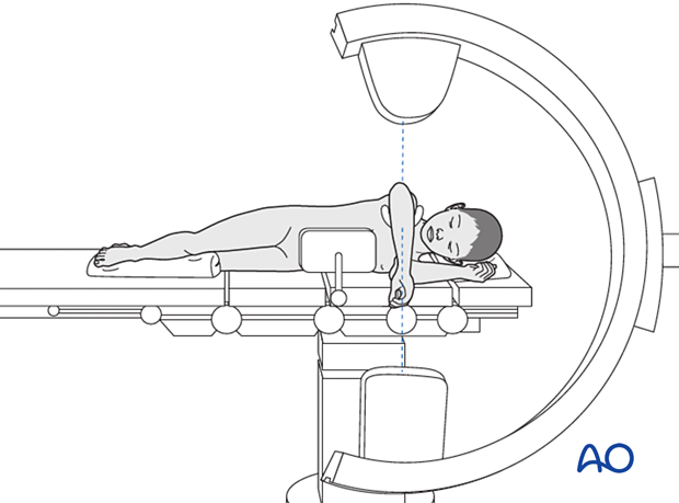
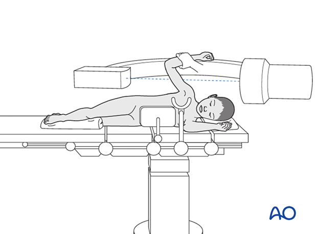
Different imaging positions are usually achieved by rotating the image intensifier if positioned parallel to the table.
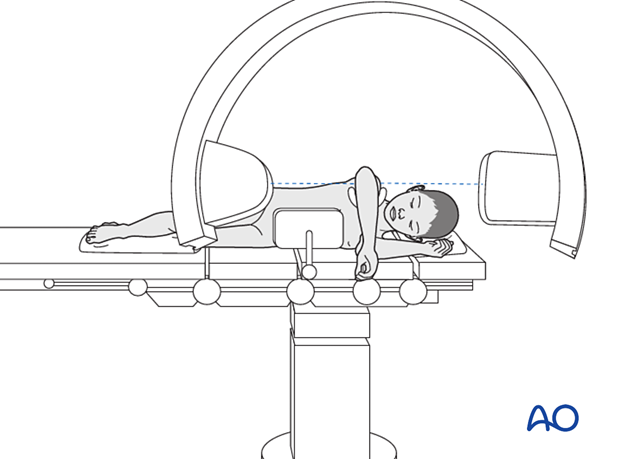
As an alternative, the C-arm can be brought in from the opposite side of the table in a horizontal orientation parallel to the floor. This allows for lateral images to be taken by raising the elbow ...
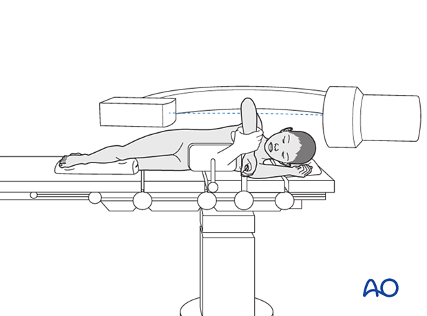
... and AP images by rotating the shoulder. This technique keeps the C-arm away from the surgeons and in a fixed position throughout the operation.
7. Skin disinfecting and draping
It is often useful to lower the table for this part of the preparation to make it easier for the assistant and then adjust the height to suit the surgeon. Holding the arm with gentle traction avoids neurological injury.
Disinfect the exposed area from the shoulder to the hand including the axilla with the appropriate antiseptic.
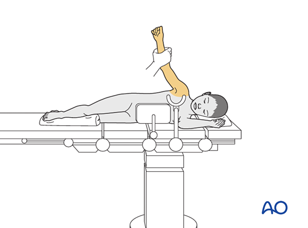
Apply an extremity drape to the affected arm making sure that sufficient coverage is achieved to access the surgical field.
Drape the image intensifier.
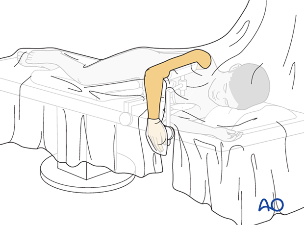
8. Operating room set-up
The surgeon and assistant are positioned on either side of the arm.
The ORP is positioned between the two surgeons.
Place the image intensifier display screen in full view of the surgical team and the radiographer.
