Prone position
1. Introduction
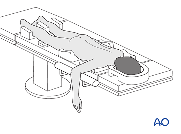
The prone patient position is used for posterior plating the humeral shaft.
2. Preoperative preparation
Consider the additional material on preoperative preparation.
3. Anesthesia
The addition of regional and local anesthesia may reduce postoperative pain.
4. Prophylactic antibiotics
Antibiotics are administered according to local policy and specific patient requirements.
5. Patient positioning
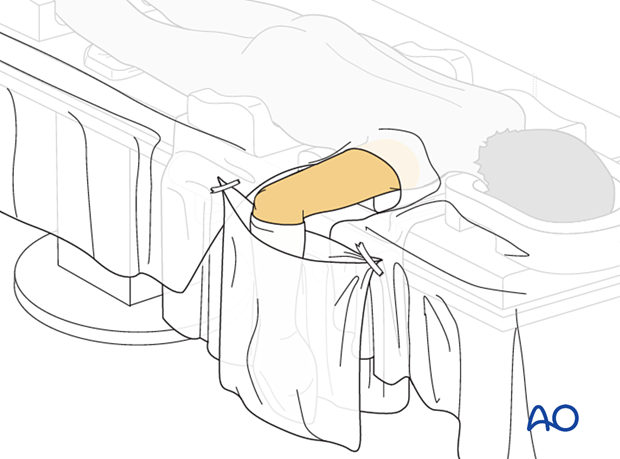
The patient is positioned prone with the elbow hanging over a radiolucent arm roll or short arm/shoulder table.

6. C-arm positioning
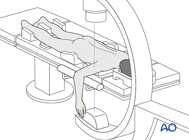
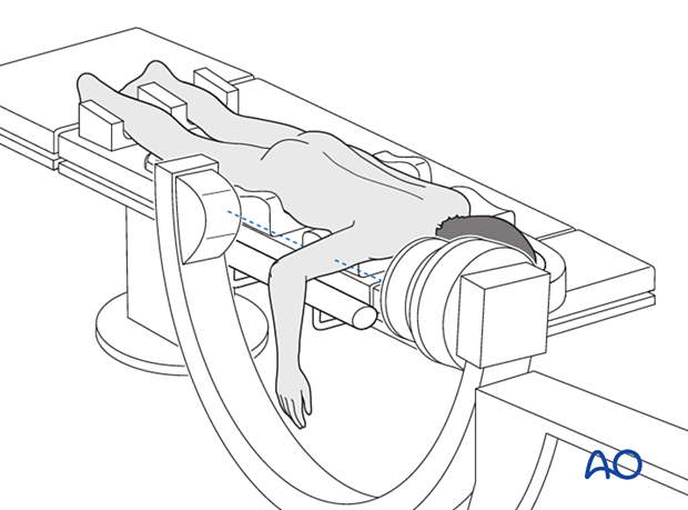
Introduce the image intensifier from the top of the OR table.
The entire humerus including humeral head and elbow must be visible in two planes with the image intensifier. This works best if the C-arm comes from the top of, and parallel to, the table.
C-arm position for AP view
C-arm position for lateral view
7. Skin disinfecting and draping
It is often useful to lower the table for this part of the preparation to make it easier for the assistant and then adjust the height to suit the surgeon.
Disinfect the exposed area from the shoulder to the hand including the axilla with the appropriate antiseptic.
To avoid contamination of the surgical field, the hanging forearm may be protected with a sterile pocket.
8. Operating room set-up
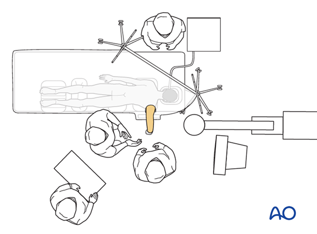
The anesthetist and anesthetic equipment should be situated at the side of the patient.
The surgeon and assistant are positioned on either side of the arm.
The ORP stands between, or behind, the surgeons.
Place the image intensifier display screen in full view of the surgical team and the radiographer.
