

Special considerations in pediatric polytrauma
1. Introduction
Head injuries, injuries to the spinal cord, and burns are severe complications in a pediatric polytrauma patient and need to be considered in primary survey and emergency assessment.
See also the Introduction to pediatric polytrauma.
2. Head injuries
The pediatric population is more susceptible to traumatic brain injury (TBI) due to weaker cervical musculature (in combination with a higher head-to-body mass relationship) and an immature skull as well as ongoing myelination in infants and toddlers.
“Head injury severity is the principle determinant of outcome and mortality in polytraumatized children.”
Indications for CT imaging are described in the chapter on Imaging.
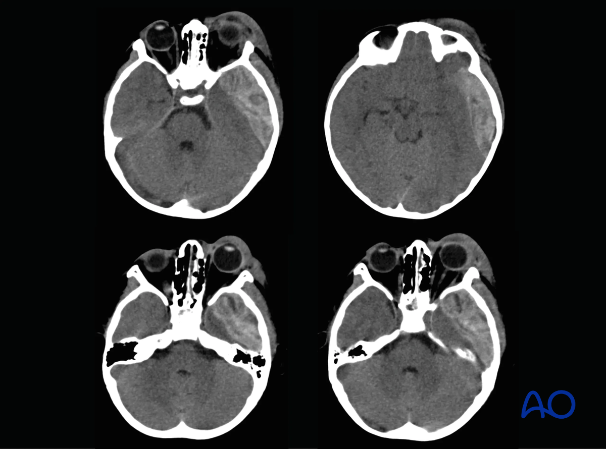
This case shows CT scan images of a 5-year-old child involved in a motor vehicle accident showing extradural hematoma in the temporoparietal region with exophthalmos and periorbital edema.
Acute management
Minor injuries without ongoing bleeding or signs of increasing cerebral edema can be treated conservatively with intensive care, hyperventilation, fluid restriction, barbiturates, and intracranial pressure (ICP) monitoring.
In case of intracranial hematomas with increased ICP, evacuation is indicated.
Cerebral edema or uncontrollable intracranial hypertension require decompressive craniectomy.
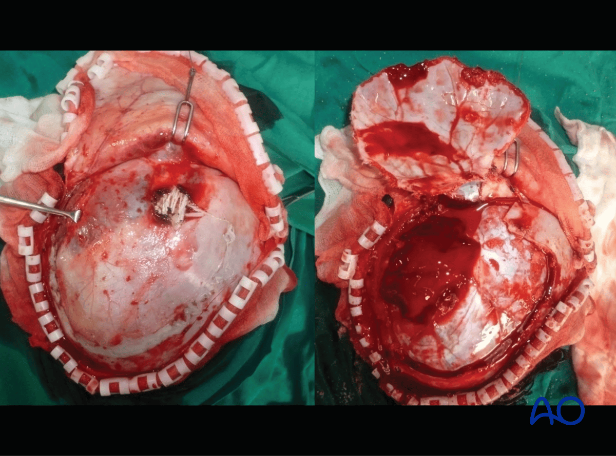
Intraoperative pictures after frontotemporal craniotomy showing the extradural hemorrhage before and after opening the dural layer.
Relationship to fracture care
The child with a severe neurological injury may not be stable enough initially to consider operative fracture management. A sedated child, ventilated in the intensive care unit, is often managed with splints and/or external fixators for temporary stabilization. In this setting, compartment syndrome can be easily missed and enhanced surveillance, including compartment pressure monitoring, should be considered.
Even severe neurological injuries are compatible with excellent recovery and long-term function for children, so the overall indications for fracture care should anticipate the full and functional recovery of the child.
During recovery from traumatic brain injury, there may be a phase with partial consciousness, uncontrolled movements, or spasticity. Ideally, definitive fixation of long bone fractures can be completed prior to this phase of recovery. In that way, the child may be up from bed and safely mobilized rather than having pain and management difficulties related to prolonged splinting or traction.
Children with traumatic brain injury can heal with abundant callus or heterotopic ossification. Stable internal fixation may reduce, but not completely eliminate this risk.
3. Spinal cord injuries
Spinal injuries are uncommon in children but are frequently associated with high-energy trauma and other severe injuries. Due to the unique anatomical characteristics of the pediatric spine, including ligamentous laxity, incomplete ossification, a relatively large head size, and weak cervical musculature, injury patterns differ from those seen in adults. Younger children are particularly susceptible to injuries of the upper cervical spine (C0–C2), whereas older children and adolescents more commonly sustain lower cervical and thoracolumbar injuries.
A spinal injury should be suspected in any pediatric polytrauma patient with:
- Neck or back pain
- Neurological deficits
- Altered consciousness
- Mechanism compatible with high-energy trauma
Until spinal injury has been excluded, spinal motion restriction should be maintained throughout assessment and resuscitation.
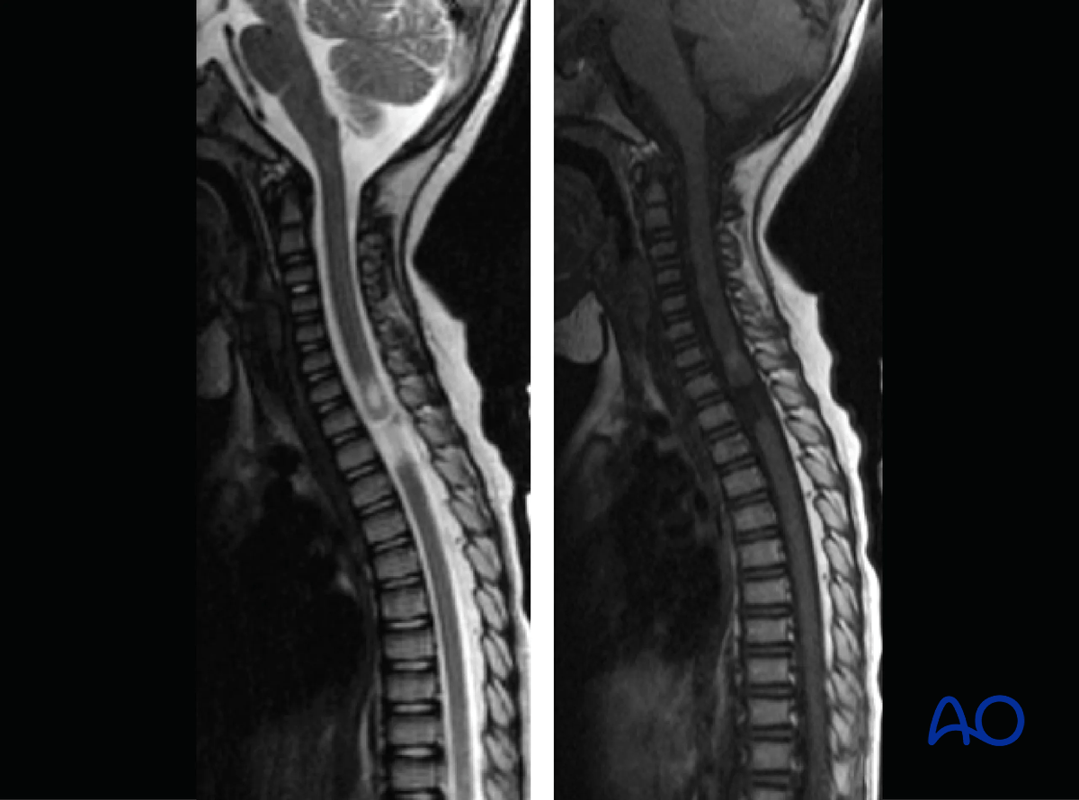
Clinical examination may be challenging in young children or patients with altered mental status. Imaging should therefore be guided by clinical findings, neurological status, and injury mechanism. CT is useful for detecting osseous injuries, while MRI is indicated when neurological deficits are present, when ligamentous injury is suspected, or when spinal cord injury without radiographic abnormality (SCIWORA) is considered. Refer to the chapter on Imaging.
Management
The goals of treatment are:
- Protection of spinal cord
- Restoration of spinal stability and alignment
- Facilitating nursing care and mobilization
- Prevention of secondary neurological injury
As with all aspects of pediatric polytrauma, management should be integrated with the treatment priorities of associated life-threatening injuries.
Treatment depends on fracture stability and neurological status.
- Stable spinal fractures without neurological deficit can usually be managed nonoperatively with immobilization and activity modification.
- Unstable injuries require stabilization to prevent progressive deformity and secondary neurological injury.
- Patients with neurological deficits require urgent specialist assessment and advanced imaging.
- Surgical decompression and internal fixation may be indicated when neurological compromise results from spinal canal narrowing, persistent instability, or progressive deformity.
Relationship to fracture care
Children with complete spinal cord injuries are at high risk of developing scoliosis. This consideration, and the considerations for early and aggressive rehabilitation, help to determine indications for operative management of the spinal column.
Spinal cord injury may make the diagnosis of limb fractures more challenging. The child may be less able to protect the injured limb during recovery and is at risk of later osteopenia. Intramedullary fixation is therefore relatively preferred for long bone fractures where it is anatomically indicated.
4. Burns
Pediatric polytrauma patients with burns need special care. The total body surface area (TBSA) affected by burns can be estimated with the palmar method: The palm including the fingers represents 1% of the TBSA.
Patients with burns >15% TBSA require fluid resuscitation.
Fluid resuscitation in burn and inhalation injuries
Key concepts:
- Over-resuscitation is just as harmful as under-resuscitation (abdominal compartment syndrome, orbital compartment syndrome, the need for escharotomies, impaired gas exchange and prolonged mechanical ventilation).
- Ringer’s lactate (RL) is the preferred resuscitation fluid; children require maintenance (D5 1/2NS) in addition to RL.
- Formulas are just a starting point – titrate to urine output.
- Avoid boluses whenever possible as they may increase the risk of compartment syndrome.
- Avoid vasopressors whenever possible as vasoconstriction may devitalize burned tissue.
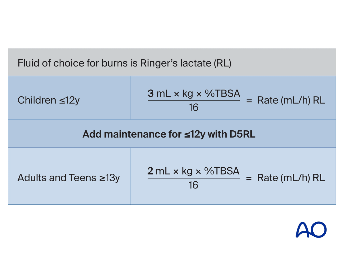
Modified Brooke/Parkland Formula is recommended as a starting point for fluid resuscitation for burns >15% TBSA in children and >20% TBSA in adults.
During the primary survey:
Step 1 – fixed fluids (based on patient age):
- Up to 5 years old: 125 ml/h RL
- 6–12 years old: 250 ml/h RL
- >13 years old: 500 ml/h RL
Step 2 – maintenance intravenous fluids (MIVF) (for <12 years of age):
- D5 + RL using the 4:2:1 rule
During the secondary survey:
Step 3 – Calculate the %TBSA:
- Use the modified formula: 3 mL × patient weight (kg) × TBSA%/16 = rate (mL/h) RL.
- Replace the fixed fluids with this new fluid.
- Titrate and monitor perfusion: Aim for an hourly urine output of 0.5–1cc/kg/h in children.
Relationship to fracture care
Care of a burn complicates the care of underlying fractures in the same limb. Burned limbs are often splinted in a position of function to help prevent contractures. Early mobilization of the limb is also important, depending on the status of the skin. These considerations may favor operative stabilization of fractures to facilitate the care of the burn and the recovery of the limb.
Depending on the specifics of the injury, external fixators or internal fixation, especially with intramedullary devices, can be considered.
Planning for acute care and rehabilitation in collaboration with the plastic surgery service will allow case by case decision making.
5. References
- Cartotto R, Johnson LS, Savetamal A, et al. American Burn Association Clinical Practice Guidelines on Burn Shock Resuscitation. J Burn Care Res. 2024 May 6;45(3):565–589.
- Jakob H, Lustenberger T, Schneidmüller D, et al. Pediatric Polytrauma Management. Eur J Trauma Emerg Surg. 2010 Aug;36(4):325–338.


