

Imaging in pediatric polytrauma
1. Introduction
Imaging is an integral part of the assessment of pediatric polytrauma patients. The imaging strategy aims to rapidly identify life-threatening injuries while minimizing unnecessary radiation exposure.
This chapter reviews the role of imaging during the primary and secondary surveys, indications for CT, and the use of MRI for selected injuries, including spinal cord injury without radiographic abnormality (SCIWORA).
2. Imaging in primary survey
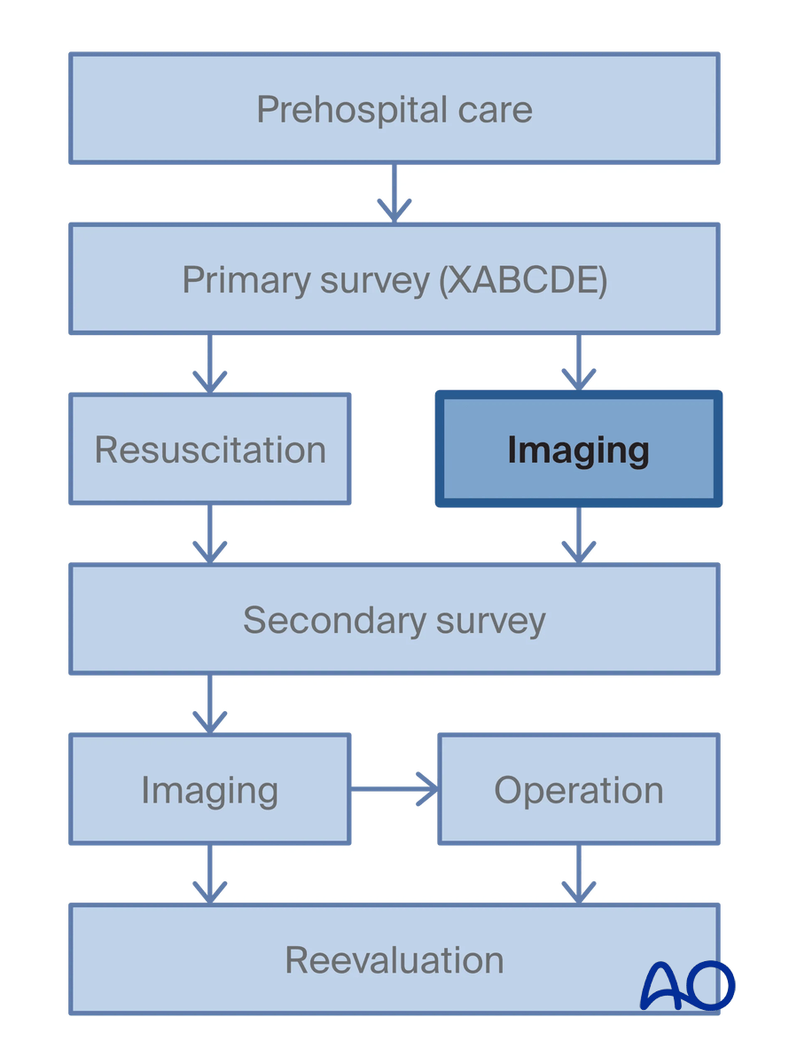
Imaging in the primary survey should allow for the decision on primary operative management or stabilization.
The following imaging should be acquired:
- Chest x-ray
- Pelvis x-ray
- Cervical spine x-ray
- Extended focus assessment with sonography for trauma (E-FAST)
See also the chapter on Emergency assessment of pediatric polytrauma.
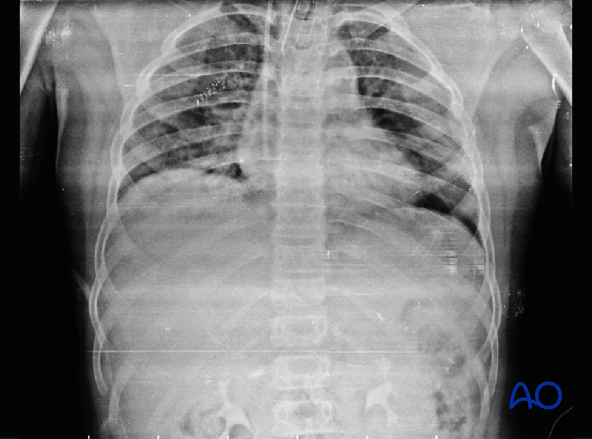
Chest x-rays
An AP view is needed. This will help to identify signs of a pneumothorax and hemothorax which will need emergency intervention.
Signs of pneumothorax:
- Hyperlucency of the lower part of the chest and the upper quadrants of the abdomen
- Lateral deep sulcus sign in the pleural space
- Double-diaphragm appearance
- Sharp appearance of the diaphragmatic edge
- Sharpness of the cardiophrenic fat pad
- Sharpness of the lower surface of the lung
The sign of hemothorax is uniform increase of basal medium density with obliteration of costophrenic angle.
Shift of mediastinum to the contralateral side may indicate hemothorax under pressure.
In this case of a 13-year-old boy with polytrauma following road traffic accident, the chest x-ray shows pneumothorax on the left. The patient had multiple rib fractures on the left along with splenic and liver injury.
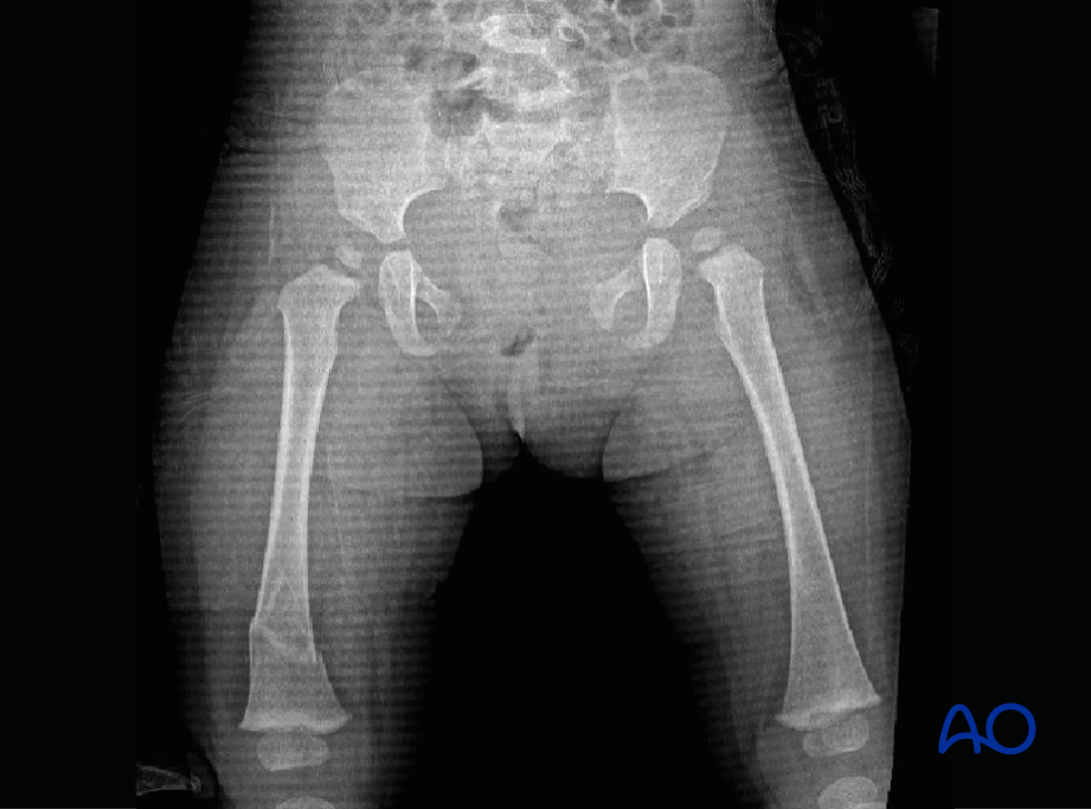
Pelvis x-rays
An AP view is needed to identify unstable pelvic fractures.
This pelvic AP x-ray of a 1-year-old child involved in motor vehicle accident shows pubic symphysis disruption (open-book injury) with distal femoral fracture on the right side.
Cervical spine x-rays
Lateral view can be taken without moving the patient. This view allows identification of any cervical spine fractures and dislocation.
Extended focus assessment with sonography for trauma (E-FAST)
The sonography is essential in the assessment of hemodynamically unstable patients.
It is a fast diagnostic tool which can be placed in the emergency room. It is performed without moving the patient. This helps to identify hemothorax, pneumothorax, and hemopericardium.
The sensitivity and specificity of E-FAST is proven in adult patients, and is likely generalizable to use in older adolescents, with reliability among juveniles and infants less clear.
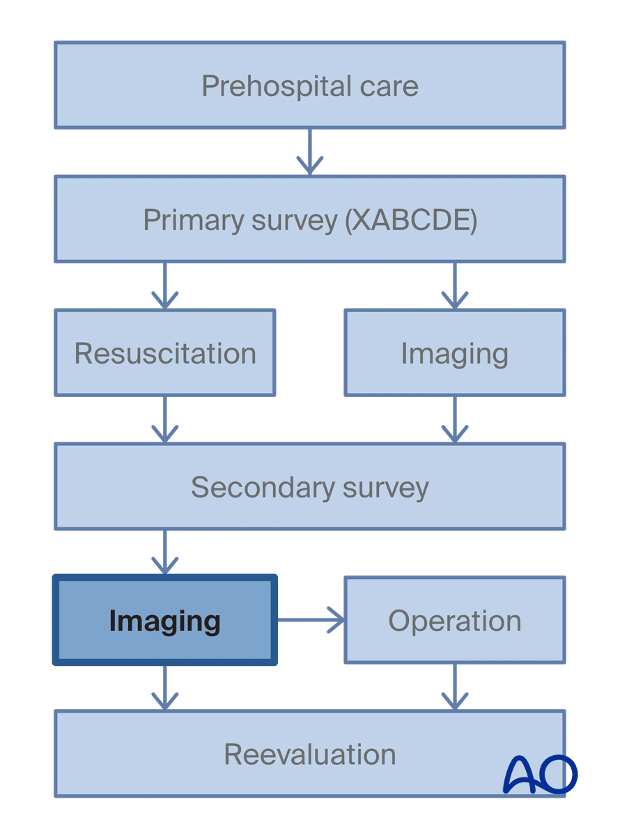
3. Imaging in secondary survey
Musculoskeletal imaging
Appropriate x-rays of the injured limb(s) in two planes have to be taken with the adjacent joints above and below.
See also the section of secondary survey in chapter on Emergency assessment of pediatric polytrauma.
Missed musculoskeletal injuries among trauma patients
It is not uncommon to miss musculoskeletal injuries in the primary survey as the patient is often unconscious, and the priority is to address life-threatening injuries.
Therefore, it is important to do a thorough examination during the secondary survey and have a low threshold for imaging the limbs if there is suspicion for a fracture.
Role of cross-sectional imaging (CT scans)
Computed tomography (CT) should be carried out in accordance with criteria for abdominal trauma and traumatic brain injury, as a team decision and with the use of age-specific low-dose CT protocols.
Pediatric emergency care applied research network (PECARN) criteria for obtaining a CT scan of the abdomen and pelvis for blunt abdominal trauma and definition of an intervention:
- Signs of abdominal wall injury (seat-belt sign)
- Glasgow Coma Scale (GCS) ≤13
- Abdominal symptoms with tenderness, resistance, pain on pressure, and guarding (free fluid in E-FAST)
- Abdominal pain
- Vomiting
- Signs of chest injury/thoracic wall injury
- Diminished breath sounds/change in respiratory rate
According to the PECARN pediatric head injury/trauma algorithm, for children <2 years of age, a head CT scan is required in one of the following cases:
- GCS <15
- Palpable skull fracture
- Altered mental status (ie, agitation, somnolence, repetitive questioning, slow response to verbal communication)
Observation vs CT scan should be evaluated in the following cases:
- Loss of consciousness ≥5 s
- Nonfrontal hematoma
- Not acting normally
- Severe mechanism of injury
For children ≥2 years of age, a head CT scan is required in one of the following cases:
- GCS <15
- Signs of basilar skull fracture
- Altered mental status
Observation vs CT scan should be evaluated in the following cases:
- History of loss of consciousness
- History of vomiting
- Severe headache
- Severe mechanism of injury
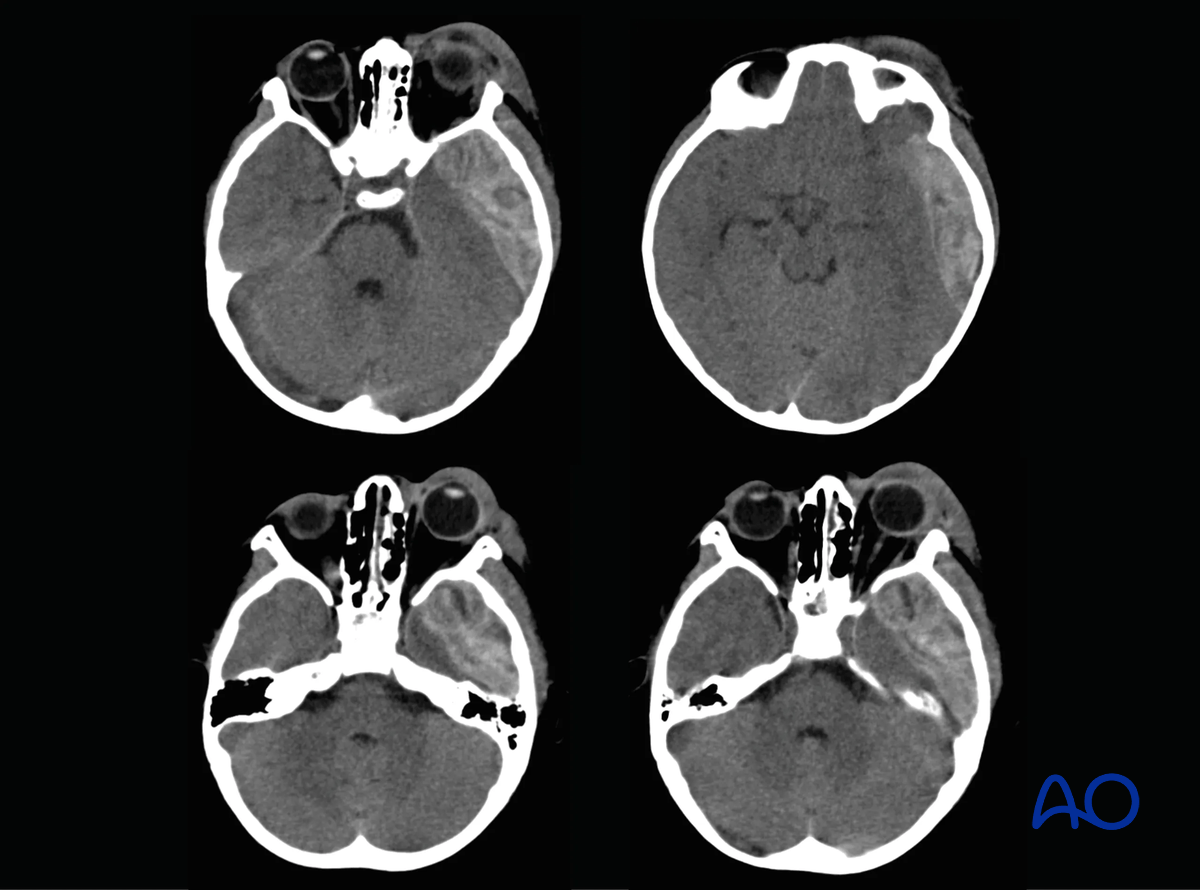
This case shows CT scan images of a 5-year-old child involved in a motor vehicle accident showing extradural hematoma in the temporoparietal region with exophthalmos and periorbital edema.
4. MRI
MRI has a limited role in the imaging of polytraumatized patients. Indications for MRI are:
- Any child with neurological deficit
- Unstable spinal fractures
Spinal cord injury without radiographic abnormality (SCIWORA)
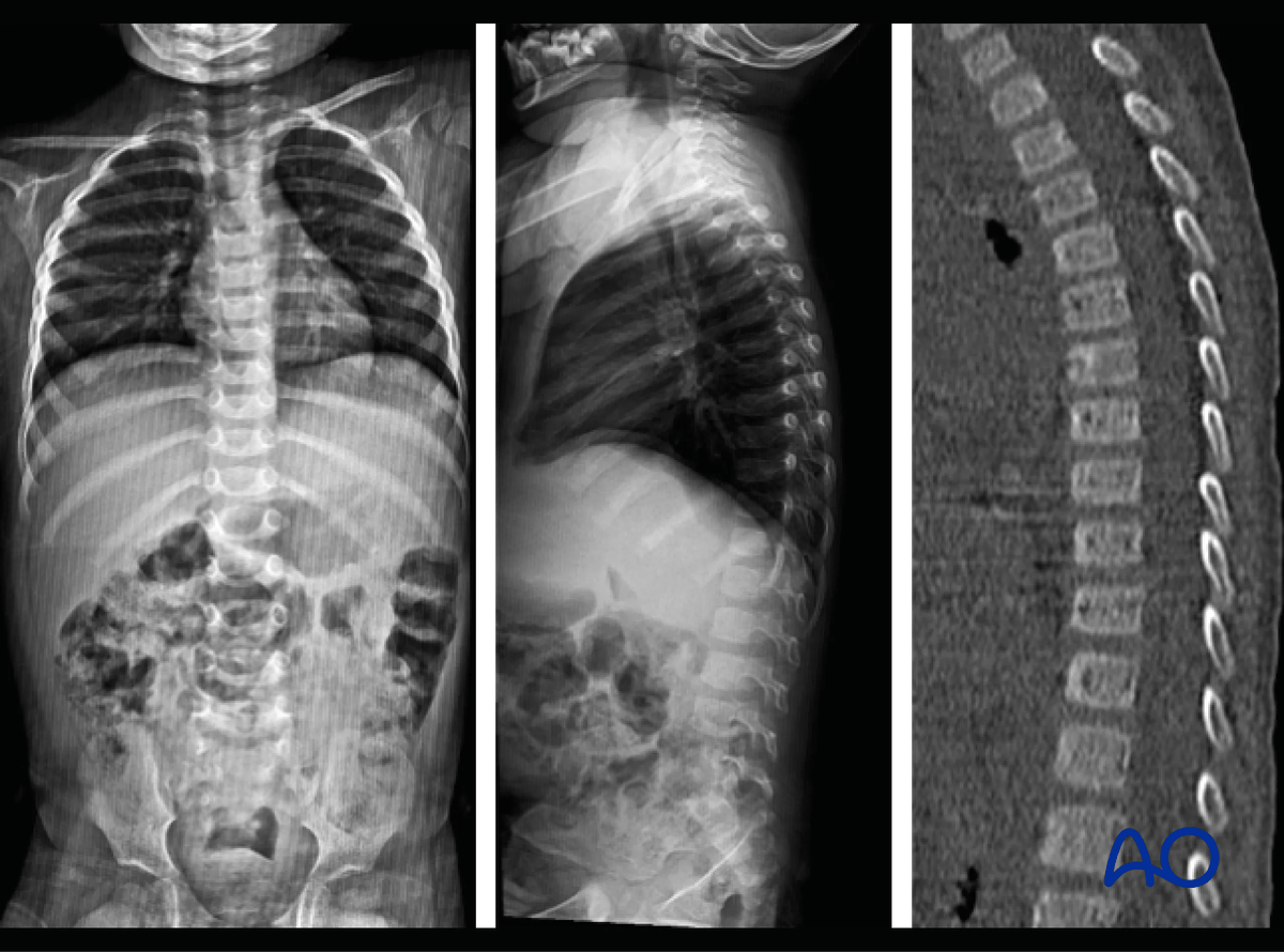
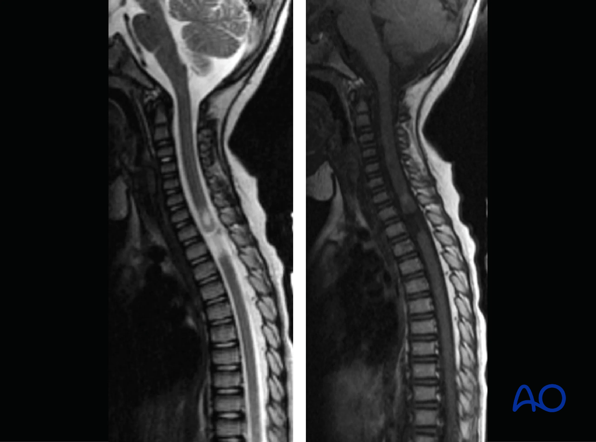
This case shows a 2.5-year-old child with complete paraplegia following motor vehicle accident. The spine X-rays and CT are normal.
MRI spine shows complete discontinuity of cord at T2‒T3 level.
5. References
- Ciorba MC, Maegele M. Polytrauma in children—epidemiology, acute diagnostic evaluation, and treatment. Dtsch Arztebl Int. 2024 May 3;121(9):291–297.
- Dunning J, Daly JP, Lomas JP, et al. Derivation of the children's head injury algorithm for the prediction of important clinical events decision rule for head injury in children. Arch Dis Child. 2006 Nov;91(11):885–891.
- Holmes JF, Lillis K, Monroe D, et al. Identifying children at very low risk of clinically important blunt abdominal injuries. Ann Emerg Med. 2013 Aug;62(2):107–116.e2.
- Kuppermann N, Holmes JF, Dayan PS, et al. Identification of children at very low risk of clinically-important brain injuries after head trauma: a prospective cohort study. Lancet. 2009 Oct 3; 374(9696):1160–1170.
- Sigal AP, Deaner T, Woods S, et al. External validation of a pediatric decision rule for blunt abdominal trauma. J Am Coll Emerg Physicians Open. 2022 Jan 15;3(1):e12623.


