Open reduction; screw fixation
1. Goals
The main goals of treatment of these osteochondral lesions are:
- Fixation of large fragments
- Pain-free and normal elbow function
2. Suitable fracture types
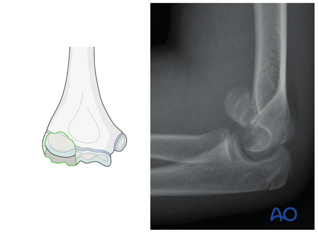
The elbow is a complex joint and has a large area of articular cartilage. Osteochondral fragments can originate from any part of the joint surface. The capitellum is the most commonly affected portion of the joint.
A distinct type of this injury is a coronal shear fracture (see illustration), which is analogous to the adult equivalent separation of the anterior portion of the capitellum (13-B3).
3. Preparation
Instruments and implants
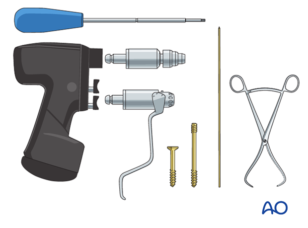
Depending on the fragment size, fragment location, and age, the following equipment is used:
- Small fragment low profile lag screws for anteroposterior fixation
- Standard small fragment lag screws for posteroanterior fixation
- Headless double-pitch compression screws
- Drill
- Standard orthopedic instrument set
Note: All sizes of implants have to correspond with the age of the child and size of the fragment.
Anesthesia and positioning
General anesthesia is recommended for this operation and a sterile tourniquet should be available.
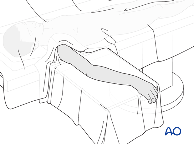
The patient is placed supine with the arm draped up to the shoulder.
4. Approach
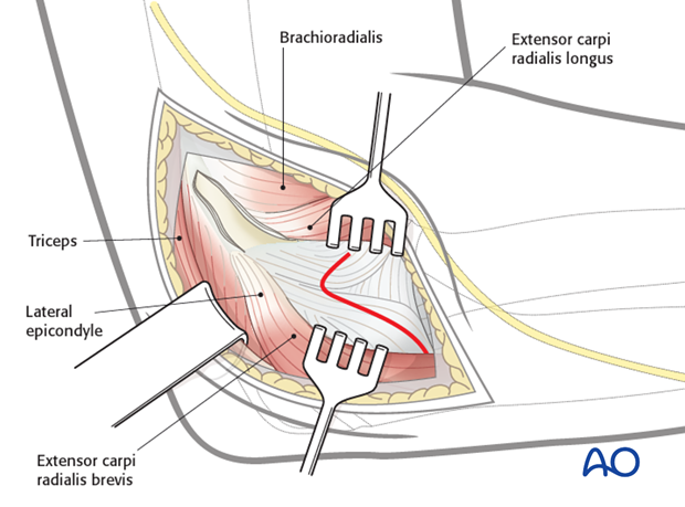
A standard lateral approach is used.
The capsule is opened anterior to the brachioradialis muscle, 1 cm proximal to the radial head. The hematoma is evacuated, and the whole joint irrigated. It is important not to divide the annular ligament.
Care must be taken not to lose any sizeable fragment (through suction or irrigation) during this procedure.
5. Reduction
Options for reduction
There are three possibilities to manipulate and reduce the fragments.
Option 1: Direct digital reduction.
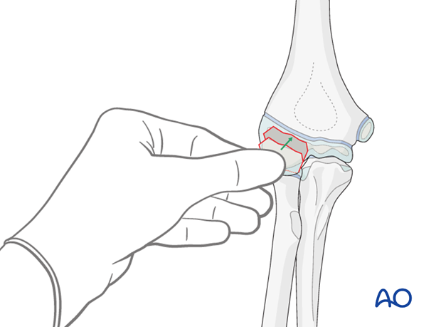
Option 2: Manipulation using a temporary K-wire in the fragment as a joystick.

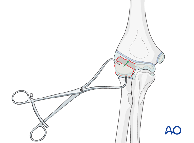
Option 3: Holding and manipulating the fragment with a small towel clamp (only for coronal avulsion fractures), or pointed reduction forceps.
A blunt Hohmann lever retractor is inserted gently into the anterior aspect of the joint, and around the medial articular border.
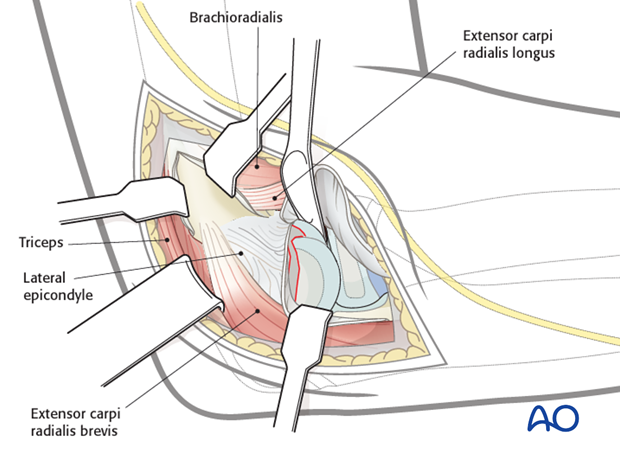
The fragment is manipulated by one of the three options listed above.
Note: In case of delayed surgery, the cartilage fragment can be swollen and bigger than the defect in the main fragment. This can make the reduction difficult and the fragment may need reshaping.
6. Screw fixation
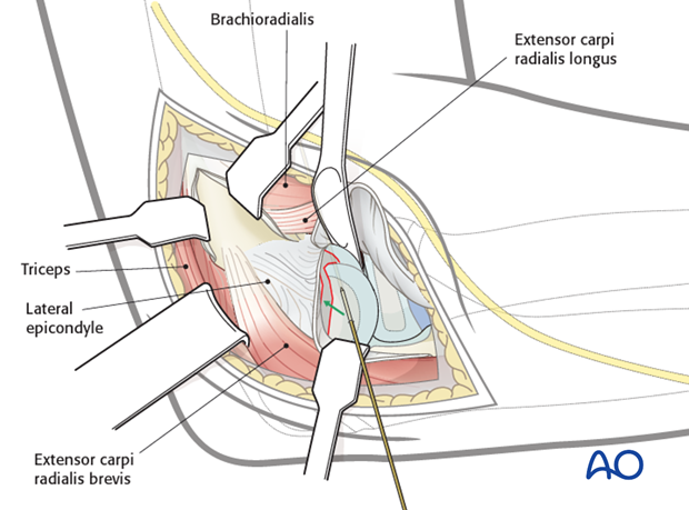
The fragment is held temporarily in place with the help of a pointed reduction forceps, towel clamp, or K-wire and fixed using one of the alternative options below.
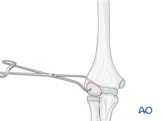
Option 1: Small fragment lag screws from anterior
Depending on the fragment size, 1.5, 2.0, or 2.5 mm screws of 15-20 mm length are used.
The fragment is temporarily fixed with a K-wire. The reduction forceps can be removed once the K-wire is in place and the fracture is reduced.
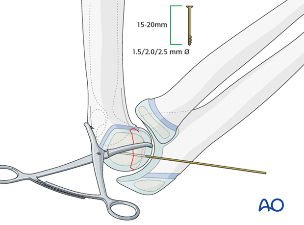
According to the fragment size, one or two pilot holes, corresponding to the size of the screws, are made.
Note: It is recommended to make the holes for the screws with a corresponding K-wire, as a normal drill can damage the cartilage more.
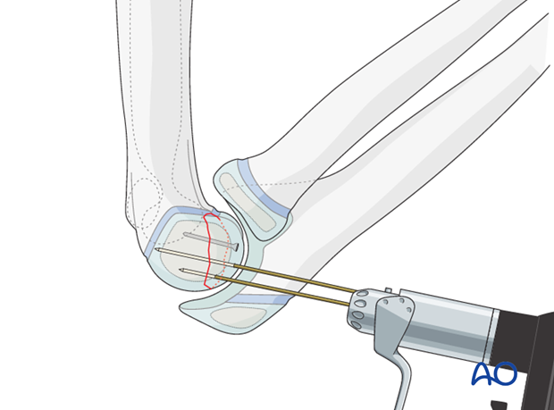
Each screw is inserted so that its head lies deep to the surface of the joint cartilage (prominent screw heads must be avoided).
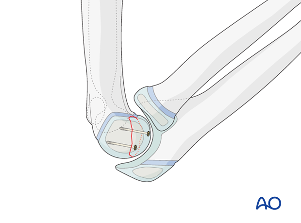
Option 2: Lag screw from posterior
With especially deep fragments, it is possible to achieve fixation by cannulated lag screw fixation from the posterior aspect of the lateral condyle. The guide wire is inserted from the front and protrudes from the posterior aspect. This will then guide the posteroanterior screw of appropriate length.
Depending on the depth of the capitellar fragment, 1.5, 2.0, or 2.5 mm headless, or small fragment cancellous, lag screws are used.
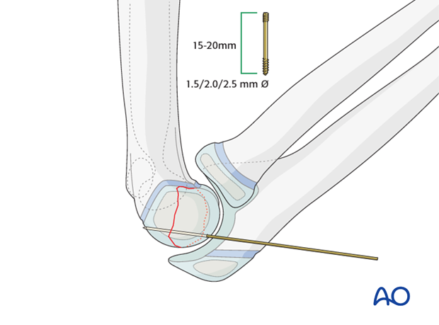
The fragment is temporarily fixed with a K-wire.
According to the fragment size, one or two drill holes, corresponding to the size of the screws, are made.
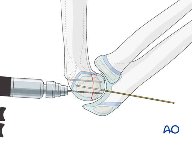
Pitfall: A drill hole made from posterior can perforate the joint surface on the anterior aspect of the capitellum.
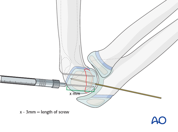
Pearl: The length of the canal can be measured with the help of a depth gauge. The screw length is then calculated by deducting 2-3 mm from the measured length.
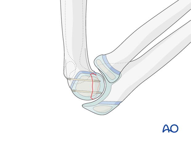
A normal or headless screw is inserted as a lag screw from posterior so that the fragment is compressed.
This procedure allows visual and palpable access to the capitellum, in order to ensure that the screw tip does not penetrate the joint surface.
Option 3: Headless compression screws from anterior
Depending on the fragment size, 1.5, 2.0, or 2.5 mm headless screws of 15-20mm length are used.
The fragment is reduced and temporarily fixed with a K-wire.
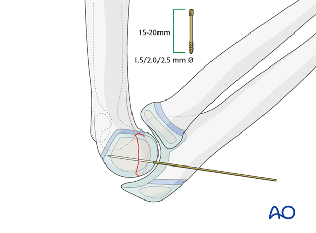
According to the fragment size, one or two pilot holes, corresponding to the size of the screws, are made.
Note: It is recommended to make the holes for the screws with a corresponding K-wire, as a normal drill can damage the cartilage more.
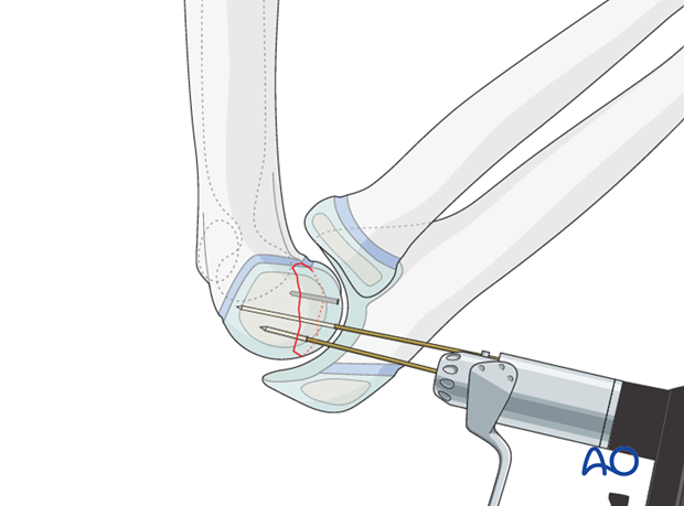
The headless screws are inserted so that they are buried deep to the surface of the joint cartilage.
Note: It is rarely necessary to remove such screws.
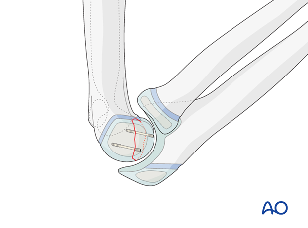
7. Aftercare
It is recommended to apply a posterior splint for pain management. In younger children this also prevents the use of the arm. In older children an arm sling is adequate.
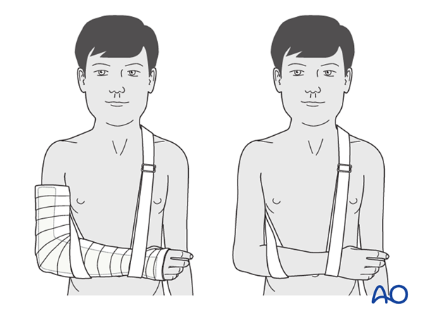
If the child remains for some hours/days in bed, the elbow should be elevated on pillows to reduce swelling and pain.
See also the additional material on postoperative infection and compartment syndrome.
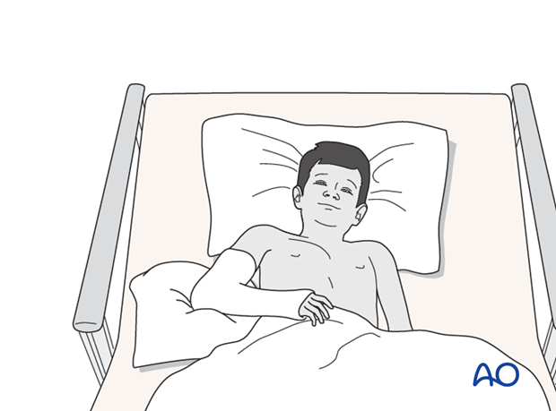
The postoperative protocol is as follows:
- Discharge from hospital according to local practice (1-3 days)
- In osteochondral fractures radiological follow-up at one week is recommended to ensure that the reduction has been maintained
- In all cases, clinical and radiological follow-up, depending on the age of the child, 4-5 weeks postoperatively out of the cast
- In most cases, at this control, the fragment has healed. If the child has had a cast, it is no longer required
- During this clinical follow-up the parents/carers should be informed that implant removal is not necessary in cases of uncomplicated healing
- Physiotherapy is normally not indicated
Screw removal
Screw removal is only indicated if there is a suspicion of prominent screw heads in the joint. Clinical signs of this include repeated swelling of the elbow, joint effusion and/or pain. In this situation, it is recommended to examine the joint arthroscopically, and remove any offending screw if it is visible.


