Supine position
1. Introduction
The most commonly used patient position is supine with the arm either on an arm table, or directly on the draped C-arm. This position can be used for the majority of pediatric fractures of the distal humerus, both supracondylar and articular.
2. Preoperative preparation
Consider the additional material on preoperative preparation.
3. Patient positioning
The patient is positioned with the fractured extremity as close as possible to the edge of the table. To prevent the child from falling down from the operating table during reduction/manipulation. Side support is recommended.
Note: Care should be taken that the upper body is not supported by the arm table especially in younger children with a short arm. Great care must be taken to avoid extremes of neck positioning.
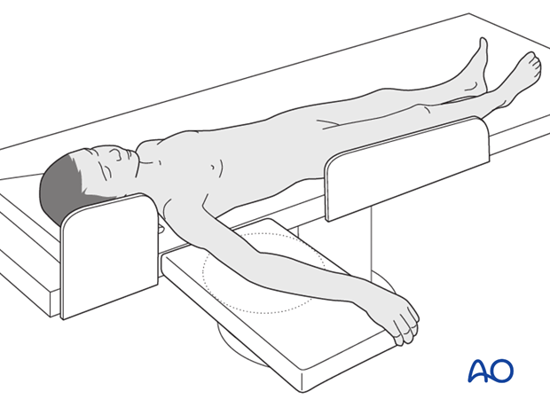
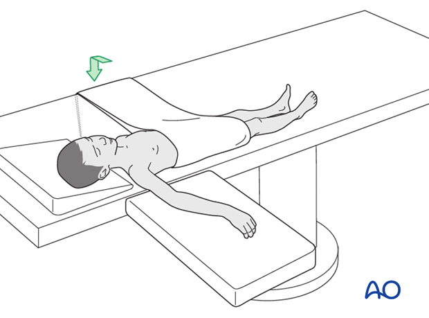
Pearl: Very small/young children (up to 5-6 years) can additionally be fixed using a towel around the thorax that is attached to the contralateral side of the table.
4. Positioning of the C-arm
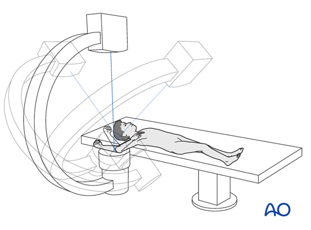
There are two possible positions for the C-arm:
- Parallel to the operating table
- Perpendicular to the operating table
Note: After reduction has been achieved, the arm should be moved as little as possible. Images should be obtained by rotating the C-arm and not moving the arm.
5. C-arm parallel to the OR table
Advantages:
- Unimpeded approach to the arm for the surgeon
- Facilitates reduction
Disadvantage:
- Difficult to rotate the C-arm around the elbow
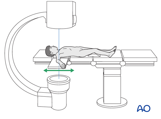
6. C-arm perpendicular to the OR table
Advantages:
- The surgeon has access from both the medial and the lateral side
- Medial/lateral rotation of the C-arm avoids the need to move the arm and thereby risk loss of reduction
Disadvantages:
- No significant disadvantages

7. Positioning of extremity in relation to C-arm
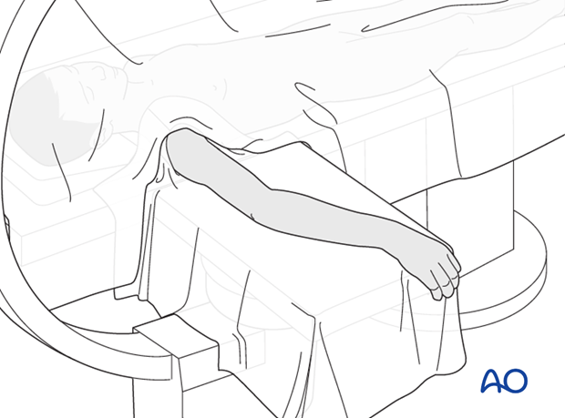
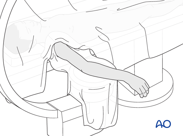
There are two options for positioning of the extremity in relation to the C-arm:
- Arm on the arm table and intensifier below the table
- Arm directly on the draped C-arm
8. Arm on arm table with intensifier below
Advantage:
- Arm more stable
Disadvantages:
- More difficult to positon the arm centrally on the image intensifier
- Higher dose of radiation
- Lower image quality, depending on dose and lucency of table
9. Arm directly on draped C-arm
Advantages:
- Easier to position the arm in relation to the image intensifier
- Better quality x-rays
- Larger bone segment is visible, which facilitates assessment of axis
- Reduced radiation risk
Disadvantages:
- Less stable operating surface
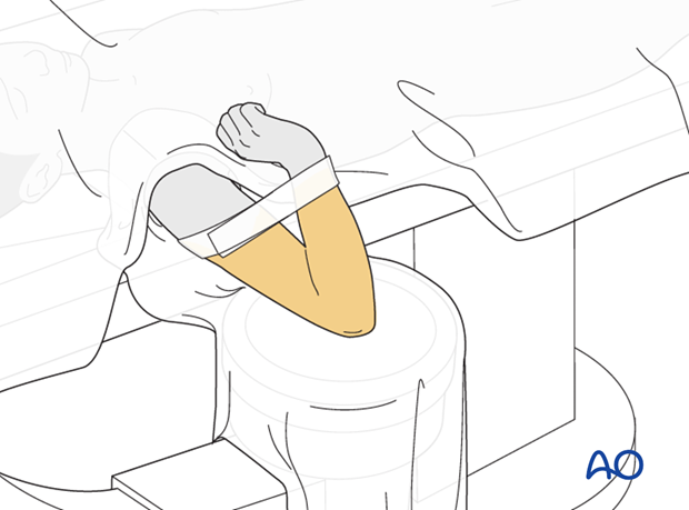
10. Supine with a flexed elbow (supracondylar fractures)
When the preoperative reduction of a supracondylar fracture is undertaken unsterile with minimal assistance, the following steps can be taken:
- Patient is supine with the arm on an arm table, or directly on the C-arm
- Reduction is performed, the elbow is then maximally flexed and the position verified using the image intensifier
- The maximal flexion of the elbow is then held in position with surgical tape (see illustration)
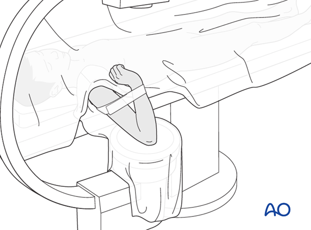
- The skin is sterilized up to the tape
- The arm is draped to exclude the tape from the surgical field
Note: The disadvantage of this position is that the fracture position cannot be altered during the remainder of the procedure.
