Arthrotomy and excision
1. Goals
The main goals of treatment of these osteochondral lesions are:
- Atraumatic removal of small fragments
- Pain-free and normal elbow function
2. Suitable fracture types
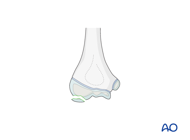
The elbow is a complex joint and has a large area of articular cartilage. Osteochondral fragments can originate from any part of the joint surface. The capitellum is the most commonly affected portion of the joint.

3. Patient preparation
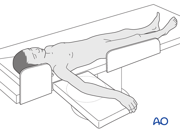
This procedure is normally performed with the patient in a supine position.

4. Fragment excision
If it is appreciated at the time of arthrotomy or arthroscopy that the fragment(s) is too small to reattach it should be removed.
The approach is usually lateral by mini-arthrotomy, but arthroscopy is an alternative.
Multiple fragments might be present and preoperative MRI can help to determine their location. All substantial cartilagenous fragments should be excised.
If the fragment bed is not fresh (osteochondritis dissecans) it may be debrided and then treated with drilling or micro-fractures.
5. Aftercare
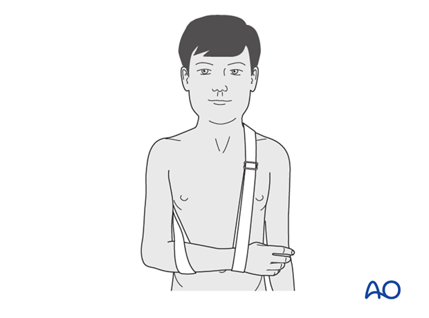
The arm is rested in a sling for comfort, but early motion should be encouraged.

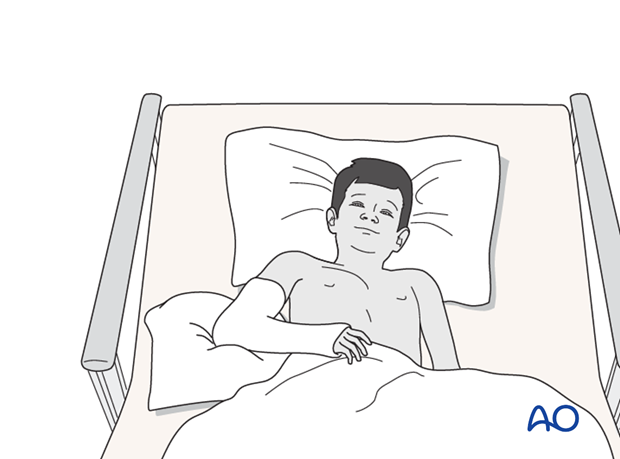
If the child remains for some hours/days in bed, the elbow should be elevated on pillows to reduce swelling and pain.
See also the additional material on postoperative infection and compartment syndrome.

The postoperative protocol is as follows:
- Discharge from hospital according to local practice (1-3 days)
- Clinical follow-up 7-10 days postoperatively
- Return to demanding activities and sports as soon as symptoms allow
- Physiotherapy is normally not indicated


