Open reduction; screw fixation
1. Goals and principles
Goals
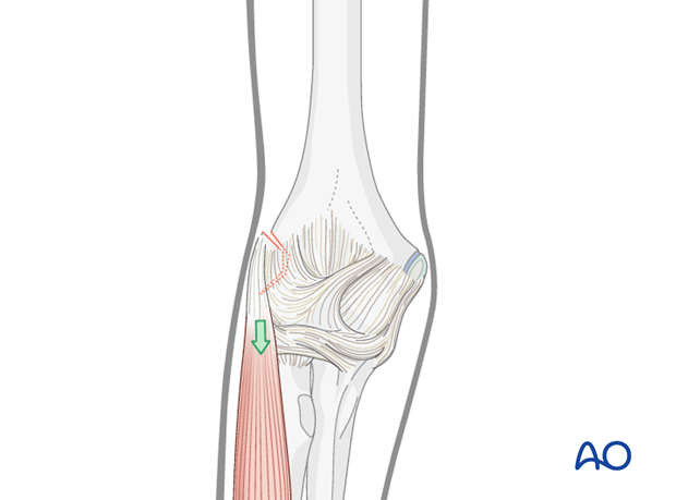
The main goals of treatment (nonoperative or operative) of these ligament avulsions are:
- Restoration of elbow stability
- Prevention of nonunion of the epicondyle
- Prevention of secondary displacement
Principles
The main principles of treatment for displaced injuries are:
- To achieve reduction and stable fixation
- Restoration and maintenance of elbow stability
Note: The lateral humeral epicondyle is intracapsular.
2. Preparation
Instruments and implants
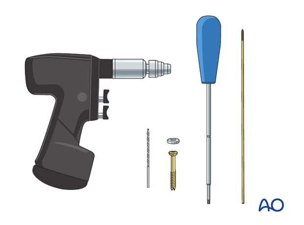
A 2.7 or 3.0 mm lag screw (preferably cannulated) is recommended to stabilize a bony fragment.
Care must be taken to ensure that the implant does not split the bony fragment. If necessary overdrill a larger fragment to prevent this. A washer should be used, particularly in smaller fragments.
Anesthesia and positioning
General anesthesia is recommended and a sterile tourniquet should be available.
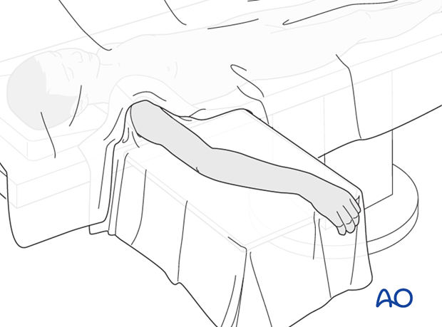
The patient is placed supine with the arm draped up to the shoulder.
3. Approach
A standard lateral approach to the elbow is used.
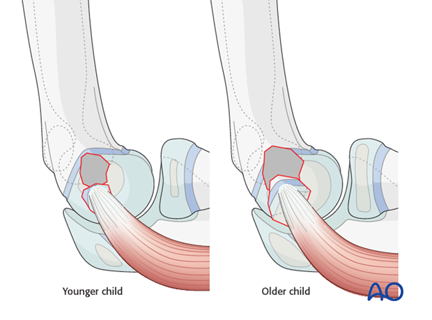
As the lateral epicondyle is visualized the following can be seen:
- In younger children with an isolated cartilage avulsion, the amount of bleeding is minimal
- In older children with a bony avulsion, bleeding is visible from the site of avulsion
Note: In these illustrations, the extensor muscle group is represented by only one muscle.
4. Screw fixation
A hole of appropriate size is made in the center of the epicondylar bed using a 2.0 mm K-wire.
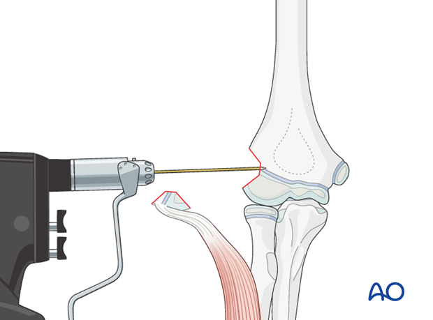
The avulsed ligament and bony fragment is orientated to provide a direct view of the fracture surface.
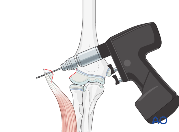
A retrograde drill hole is made in the center (as illustrated).
Pearl: It is useful to make this hole one size larger than the cannulated screw guide wire as this prevents the fragment from splitting during screw insertion.
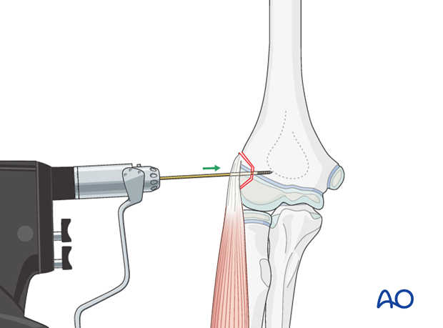
A guide wire for a cannulated screw is inserted by hand through the epicondylar fragment. It is then advanced into the predrilled hole in the metaphysis. The wire is used to guide the fragment into its reduced position.
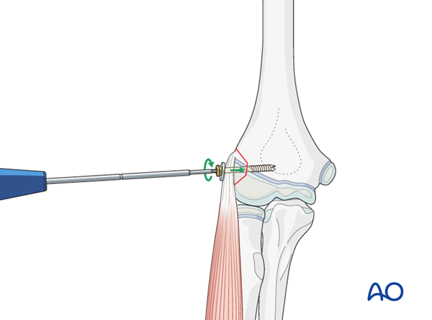
Once the fragment is reduced, a screw is placed over the guide wire and inserted.
Note: Care should be taken not to split the fragment and use of a washer should be considered. A plastic washer with teeth is preferable. The use of a washer increases the likelihood of subsequent screw removal.
5. Aftercare
Note: A cast with the forearm in either neutral or supinated position is required to prevent secondary displacement.
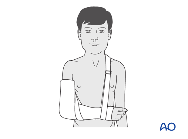
Note: In any case of elbow immobilization by plaster cast, careful observation of the neurovascular situation is essential both in the hospital and at home.
See also the additional material on postoperative infection and compartment syndrome.
It is important to provide parents/carers with the following additional information:
- The warning signs of compartment syndrome, circulatory problems and neurological deterioration
- Hospital telephone number
- Information brochure
If the child remains for some hours/days in bed, the elbow should be elevated on pillows to reduce swelling and pain.
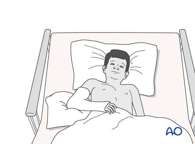
The postoperative protocol is as follows:
- Discharge from hospital according to local practice (1-3 days)
- First clinical and radiological follow-up, depending on the age of the child, 2-3 weeks postoperatively out of the cast
- During the first clinical follow-up parents/carers should be informed about the timing of implant removal
- Physiotherapy is normally not indicated


