Open reduction; screw and K-wire fixation
1. Goals
The main goals of treatment of these fractures are:
- Anatomical reduction and restoration of the joint surface
- Uncomplicated healing Avoidance of malunion or nonunion
2. Preoperative planning
Instruments and implants
- Standard orthopedic instrument set
- Lag screws (if possible cannulated)
- K-wires (preferably threaded at the tip)

Anesthesia and positioning
General anesthesia is recommended and a sterile tourniquet should be available.
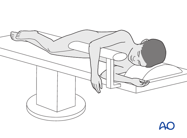
The patient should be placed lateral with the injured arm placed over a padded arm roll or a gutter support. Alternatively a prone positioning may be used.
3. Approach
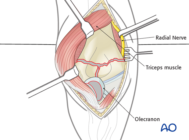
A posterior approach can be used. The triceps may be split or reflected laterally and medially. Olecranon osteotomy is rarely necessary.
Note: Some surgeons prefer a lateral approach as it allows sufficient visualization of the intraarticular component, although access is more limited.
Cleaning of the fracture site
The fracture is cleaned by removing blood clots, loose pieces of bone, and any interposed tissue.
4. Reconstruction of the articular surface
Condylar reassembly
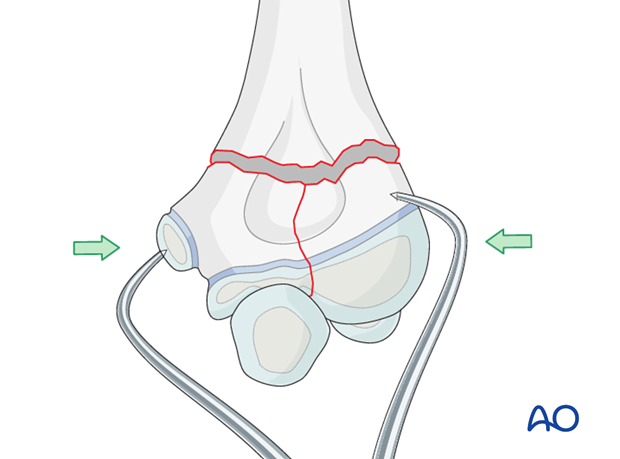
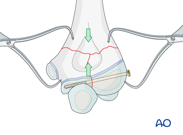
The articular fragments are reduced using pointed reduction forceps, a towel clamp, or with the help of two K-wires.
An anatomical reconstruction of the joint surface (and alignment of the physis) is mandatory.
Definitive interfragmentary fixation
A lag screw is used to obtain interfragmentary compression of the articular fracture plane. A partially or fully threaded screw can be used. If a fully threaded screw is used, it is necessary to overdrill the near fragment.
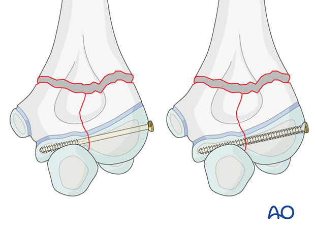
Note: To achieve an optimal mechanical outcome, the physis can be ignored in children approaching skeletal maturity.
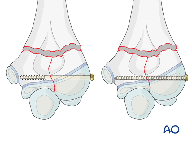
If cannulated lag screws are available, the two reduced fragments are temporarily fixed with a K-wire.
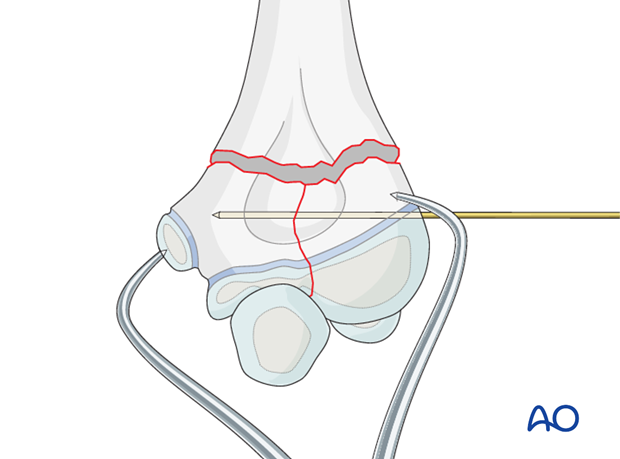
The guide wire for the cannulated screw is inserted through the center of the capitellum, parallel to the joint surface.

An appropriate screw, depending on the fragment size (3.0, 3.5, or 4.0 mm), is inserted and both wires are removed, unless the first K-wire is required for additional stability.
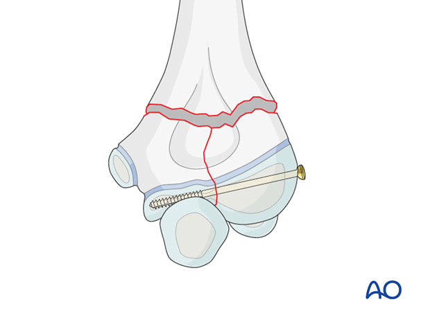
In contrast to adult patients with more unstable lesions, one lag screw, with or without an additional K-wire, is normally sufficient to reassemble the condylar mass in children.
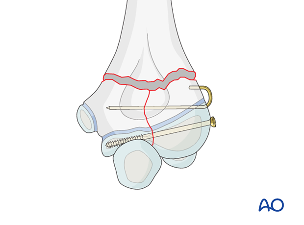
Note: Once the condylar mass has been reconstructed, the injury has been converted to a simple supracondylar fracture type.
Depending on the age of the child, different techniques for the reattachment of the condylar mass can be considered:
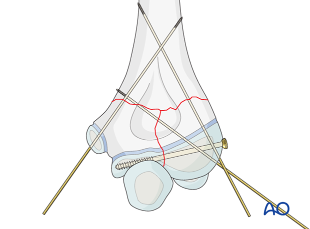
- Divergent lateral 2.0 mm K-wire fixation
- Medial and lateral crossed 1.6 or 2.0 mm K-wire fixation
Note: If additional stability for metaphyseal fracture is thought necessary, the alternatives to K-wire fixation are:
- A small lateral external fixator
- Biplanar plating in older children

5. Condylar reattachment with K-wires
Prerequisite:
- Younger children
- Small bone
The condylar block is reduced anatomically to the metaphysis.
The first K-wire is inserted into the lateral aspect of the condylar mass passing up the lateral supracondylar column.
Depending on the technique (divergent monolateral or cross-wiring), a second K-wire is inserted from the lateral or medial side.

Sometimes, with divergent lateral wires, if the open procedure destroys the posterior periosteum, an additional medial K-wire may be necessary.
Note: Identify and protect the ulnar nerve throughout the medial wiring procedure.
6. Aftercare
K-wire fixation confers only partial stability and therefore, additional cast immobilization is required to reduce the risk of secondary displacement.

If the child remains for some hours/days in bed, the elbow should be elevated on pillows to reduce swelling and pain.
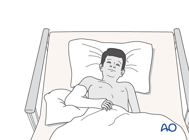
Note: In any case of elbow immobilization by plaster cast, careful observation of the neurovascular situation is essential both in the hospital and at home.
See also the additional material on postoperative infection.
It is important to provide parents/carers with the following additional information:
- The warning signs of compartment syndrome, circulatory problems and neurological deterioration
- Hospital telephone number
- Information brochure
The postoperative protocol is as follows:
- Discharge from hospital according to local practice (1-3 days)
- First clinical and radiological follow-up, depending on the age of the child, 4-5 weeks postoperatively out of the cast
- In most cases, at this first x-ray control, the fracture is consolidated and stable and a cast is no longer required
- Physiotherapy is normally not indicated
K-wire removal
Protruding K-wires can be removed 4-5 weeks postoperatively.
The removal of buried K-wires can be planned with the parents/carers at the time of the first clinical follow-up.


