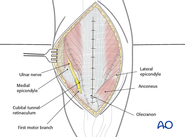
Posterior approach to the pediatric distal humerus without olecranon osteotomy
1. Main indication for posterior approach in trauma
The main indications for a posterior approach in the distal humerus are:
- Y-fractures or T-fractures (13-E/4.2)
Bear in mind that use of posterior approach for extension type supracondylar fractures, compromises the stabilizing effect of the posterior periosteum.
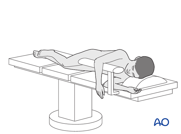
This approach, in combination with the patient positioned in a lateral decubitus position and with the elbow held hyperflexed, provides a free view and approach to the posterior aspect of the olecranon fossa, the distal humerus, as well as the joint.
2. Skin incision
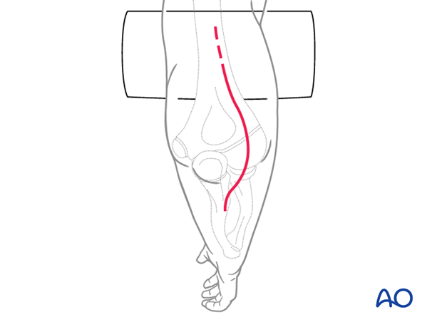
The incision is started approximately the width of the patient's hand proximal to the olecranon (in severe cases slightly longer). It then runs distally curving laterally around the olecranon, continuing some 2-4 cm along the ulnar crest.
An ulnar-based subcutaneous flap is developed.
3. Fascial incision
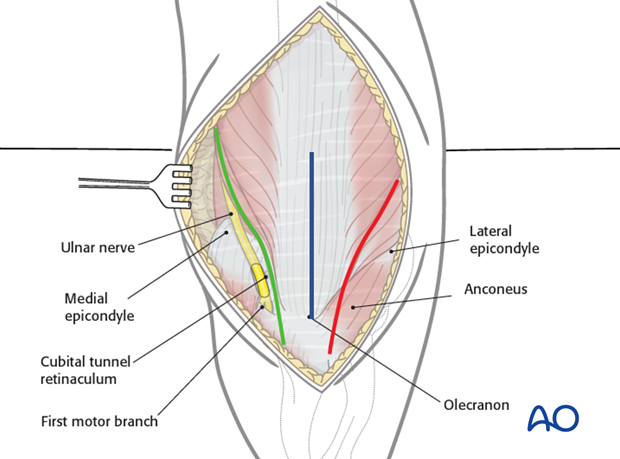
Three different windows can be developed:
- Medial window (green line)
- Lateral window (red line)
- Midline window (blue line)
A detailed description for each window follows below.

4. Medial window – option 1
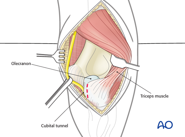
As a first step, the ulnar nerve is isolated and protected with a latex loop.
Proximally, the ulnar nerve is followed along its course on the medial intermuscular septum, and the triceps muscle is retracted laterally.
The insertion of the triceps tendon on the medial side of the olecranon is partially detached tangentially together with a 1-2 mm sliver of cartilage. This does not impair growth of the olecranon, but gives a much better approach to the joint.
5. Medial window – option 2
The posterior fascia is split 3-4 mm from the medial border of the triceps muscle. The muscle is retracted laterally.
The ulnar nerve is protected by the fascia and can be gently moved medially with a smooth round simple retractor.
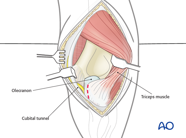
The insertion of the triceps tendon on the medial side of the olecranon is partially detached tangentially together with a 1-2 mm sliver of cartilage. This does not impair growth of the olecranon, but gives a much better approach to the joint.

6. Lateral window
The triceps muscle is freed on its lateral border.
The triceps fascia is split, the muscle is mobilized from the lateral intermuscular septum and humerus, and retracted towards the medial side.
Distally, the anconeus muscle is partially detached from its origin as necessary.
The insertion of the triceps tendon on the lateral side of the olecranon is partially detached tangentially together with a 1-2 mm sliver of cartilage. This does not impair growth of the olecranon, but gives a much better approach to the joint.
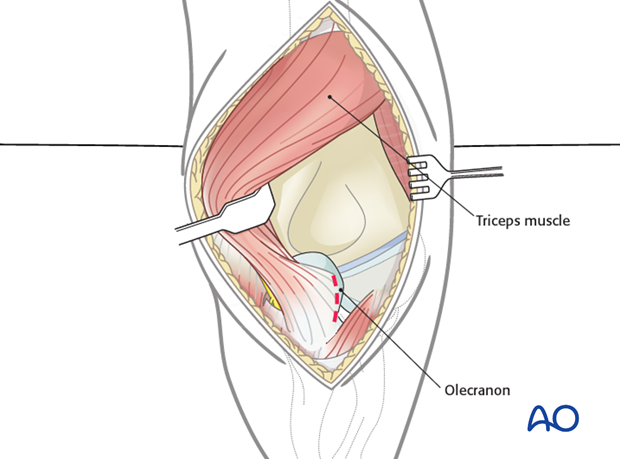
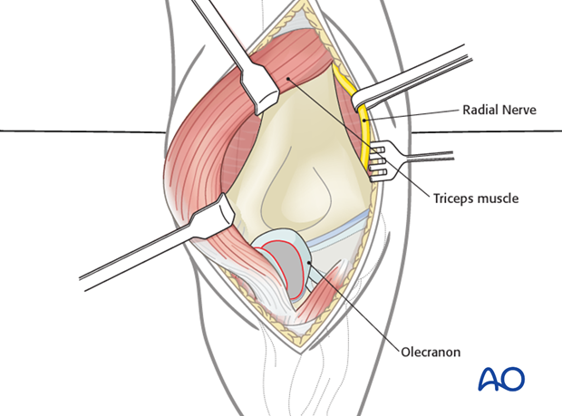
If a more proximal lateral mobilization of the triceps is performed, care must be taken to identify and protect the radial nerve.
Note: The medial and lateral triceps windows can be used together, in order to maximally access the posterior aspect of the distal humerus and elbow joint ("triceps flip"). NB: Be sure to preserve enough triceps tendon, triceps insertion and ulnar periosteum to ensure continuity of the extensor mechanism.
7. Midline window
After the subcutaneous tissue has been dissected from the deep fascia, the triceps tendon is split exactly in the mid line from the tip of the olecranon to the upper limit of the olecranon fossa.
Once the tendon is split, providing a good view of the olecranon fossa, two smooth soft tissue retractors are inserted.
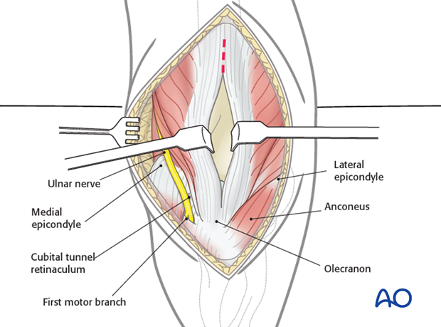
The triceps tendon and aponeurosis are further split proximally (normally 3-4 cm proximal to the fracture line). If a more proximal exploration of the distal humerus is required, the triceps muscle can be split further without causing any damage, but great care must be taken of the radial nerve.
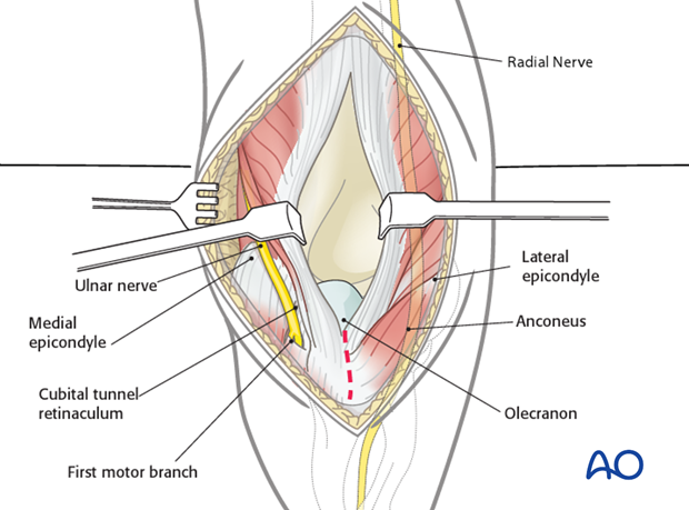
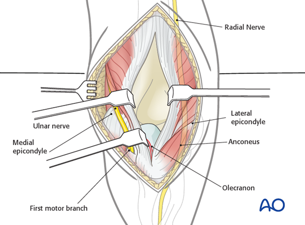
After the triceps tendon has been split distally to the tip of the olecranon, an incision is made along the medial border of the ulna (including cartilage/periosteum). The insertion of the tendon, together with the periosteum or small cartilage flap, is then partially mobilized from the proximal ulna.
Soft tissue retraction will now provide a wide view of the posterior aspect of the distal humerus.
The main advantage of this approach is avoiding damage to the ulnar nerve. In addition, triceps repair is easily performed by continuous suture of the aponeurosis.
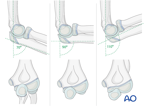
8. Influence of elbow flexion on the view of the joint surface
70° flexion:
- The olecranon overlaps nearly 2/3 of the posterior joint surface
90° flexion:
- The olecranon overlaps only a small part of the medial joint surface
110° flexion:
- Free and complete view to the whole joint surface
9. Closure
The capsule is closed with resorbable sutures (3/0).
Muscles are returned to their anatomical position and the prepared small olecranon flap is reattached to the ulnar periosteum with resorbable sutures (2/0 or 3/0).
The fascia should also be closed with 2/0 or 3/0 resorbable sutures, otherwise muscular hernia might occur.
Skin and subcutaneous tissue are closed with fine resorbable sutures (this avoids any distress to the child in removing nonabsorbable sutures).
