Open reduction; K-wire fixation
1. Goals
The main goals for treatment of these fractures are:
- Uncomplicated healing
- No secondary displacement
- Avoiding overgrowth due to unstable fixation
- Avoiding secondary complications, such as nonunion, leading to additional problems (eg, ulnar nerve irritation)
It is important to be aware that K-wire fixation gives less stability than screw fixation.
2. Suitable fracture types
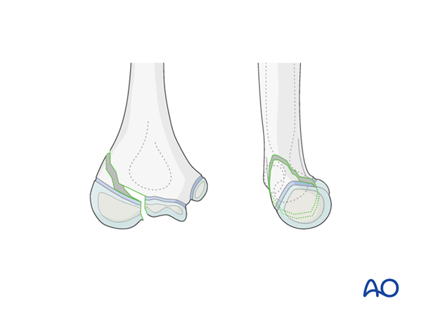
13-E/4.1L with displacement of more than 2 mm in the metaphyseal area.
All lateral condylar fractures carry the risk of avascular necrosis (AVN). It is important to be aware that the blood supply to the capitellar fragment enters posteriorly. If during surgery the posterior dissection is too invasive, this risk is increased.
3. Preparation
Instruments and implants
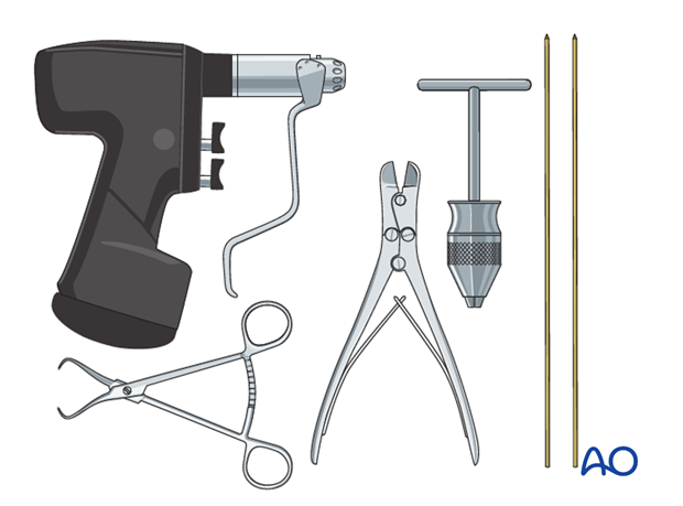
Use 1.6 or 2.0 mm K-wires.
The following equipment is needed:
- K-wires of appropriate sizes
- Drill, preferably oscillating to avoid thermal injury, or a T-handle for manual insertion
- Wire cutting instruments
- Standard orthopedic instrument set
Anesthesia and positioning
General anesthesia is recommended and a sterile tourniquet should be available. Muscle relaxation is not necessary.
The patient is placed supine with the arm draped up to the shoulder.

4. Approach
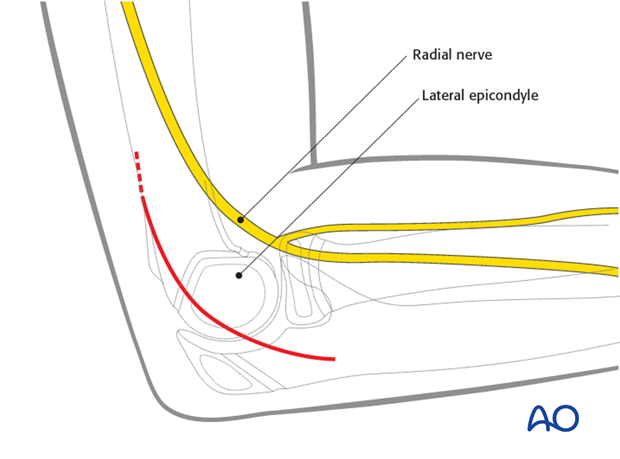
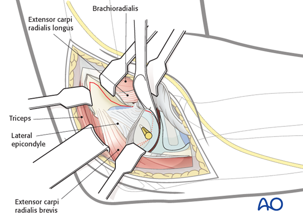
The recommended access is a posterolateral approach to the distal humerus.
5. Reduction and fixation
Options for reduction
There are three possibilities to manipulate and reduce the fragments
Option 1: Direct digital manipulation.
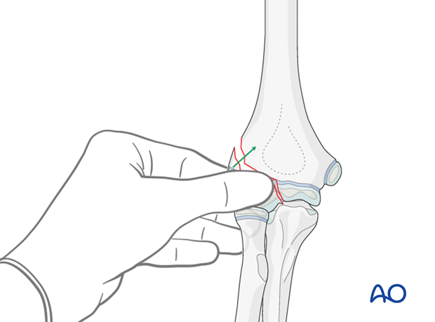
Option 2: Manipulation using a temporary K-wire in the fragment as a joystick.
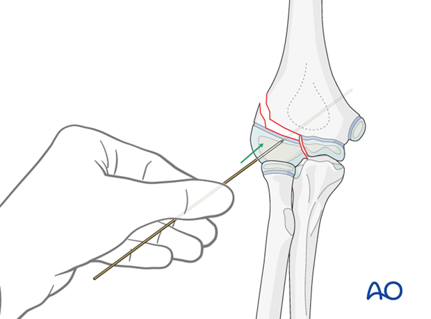
Option 3: Holding and manipulating the fragment with a small towel clamp or pointed reduction forceps.
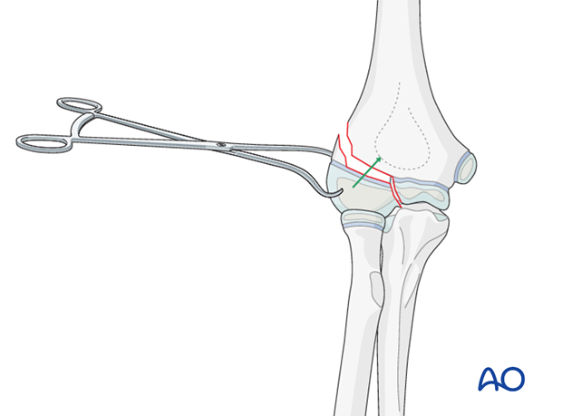
Reduction
A blunt Hohmann lever retractor is inserted gently into the anterior joint, and around the medial articular border.
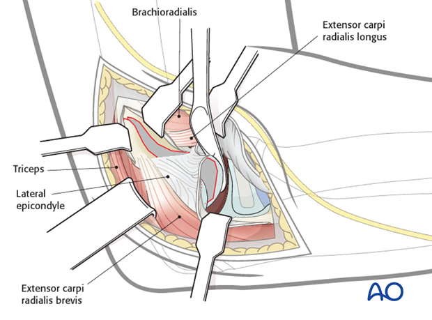
The medial cartilage fracture line is visualized on the joint surface.
The fragment is manipulated by one of the three options listed above.
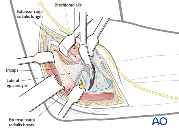
The fragment is then reduced so that both cartilage fracture lines (on the fragment and of the trochlea) are perfectly aligned.
Fixation
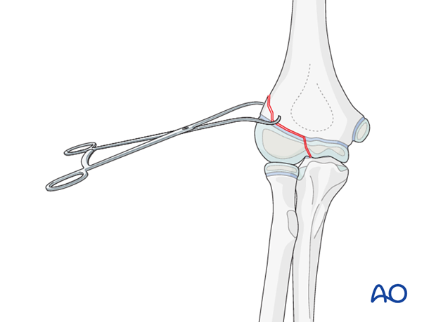
The fragment may be held reduced using a pointed reduction forceps.
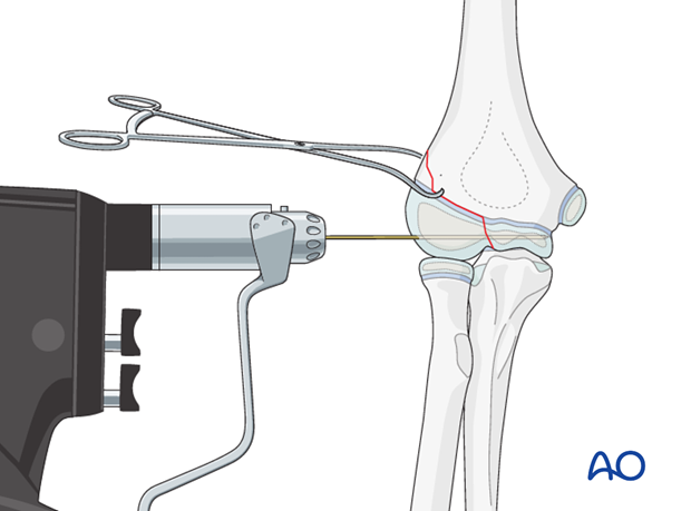
A 1.6 or 2.0 mm K-wire is inserted parallel to the joint line, starting from the center of the capitellum and advanced into the medial part of the trochlea.
Note: Always start with this K-wire to hold the joint surface aligned. The use of an oscillating drill is preferred to avoid the risk of thermal injury to the growth zones.
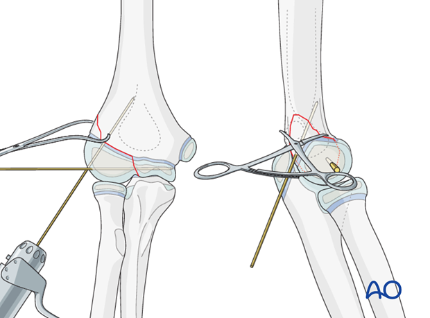
A second K-wire is inserted behind the first K-wire and advanced proximally along the lateral column of the humerus as far as the anterior cortex.
Trimming of K-wires
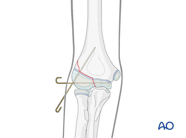
Whether the K-wires are left outside the skin or bent and buried beneath the skin depends on the surgeon's preference.
After cutting the wires obliquely, it is possible to bend them through 180° and impact them further into the bone. This provides slightly more stability and reduces pin-track infection. However, a second anesthetic is required for wire removal.
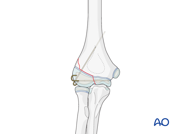
K-wires may be left protruding, but there is a risk of pin-track infection. The advantage is that the K-wires can be removed without anesthesia.
6. Aftercare
Note: K-wire fixation alone confers minimal stability. Therefore, additional cast immobilization is required to reduce the risk of secondary displacement. Recommended immobilization for K-wire fixation is posterior and anterior long arm splints.
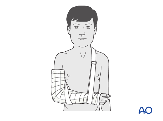
Note: In any case of elbow immobilization by plaster cast, careful observation of the neurovascular situation is essential both in the hospital and at home.
See also the additional material on postoperative infection.
It is important to provide parents with the following additional information:
- The warning signs of compartment syndrome, circulatory problems and neurological deterioration
- Hospital telephone number
- Information brochure
If the child remains for some hours/days in bed, the elbow should be elevated on pillows to reduce swelling and pain.
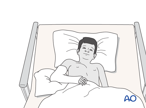
The postoperative protocol is as follows:
- Discharge from hospital according to local practice (1-3 days)
- First clinical and radiological follow up is undertaken, depending on the age of the child, 4-5 weeks postoperatively out of the cast
- In most cases, at this first x-ray control, the fracture is consolidated and stable so that cast is no longer required
- Union is assessed radiologically at about 8 weeks
- Physiotherapy is normally not indicated
K-wire removal
Protruding K-wires can be removed 4-5 weeks postoperatively.
Buried K-wires can be removed after 2-3 months. This can be performed as a day case under light anesthesia.


