Open reduction; K-wire fixation
1. Goals
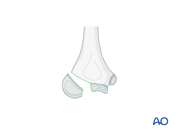
The main goals for open treatment of these fractures are:
- Anatomical reduction
- Uncomplicated healing
- No secondary displacement
- Avoiding joint incongruity
2. Preparation
Instruments and implants
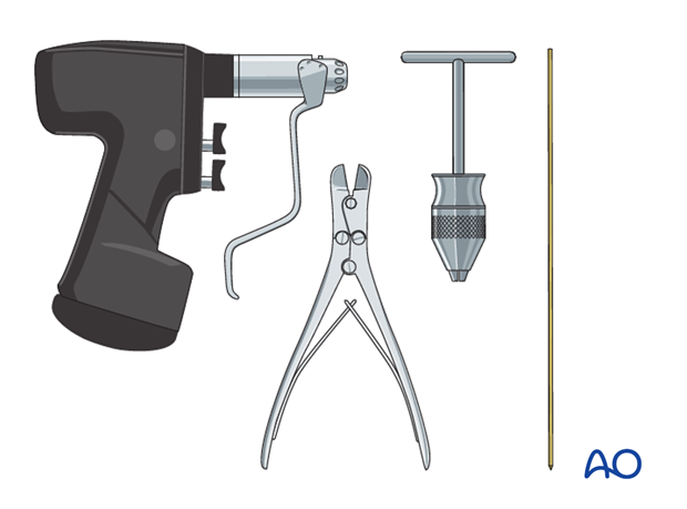
It is recommended to use 1.2 or 1.6 mm K-wires.
The following equipment is needed:
- K-wires of appropriate sizes
- Drill, preferably oscillating to avoid thermal injury, or a T-handle for manual insertion
- Wire cutting instruments
- Image intensifier
- Contrast medium for arthrography
Anesthesia and positioning
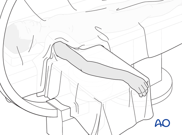
General anesthesia is recommended for this operation and it is helpful to use a tourniquet.
The patient is placed supine with the arm draped up to the shoulder.
3. Approach
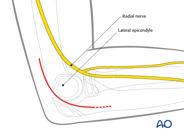
The procedure is performed using a standard posterolateral approach.
4. Reduction
Reduction options
There are three options to manipulate and reduce the fragments.
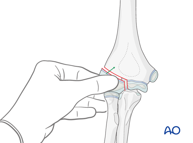
Option 1: Direct digital manipulation.
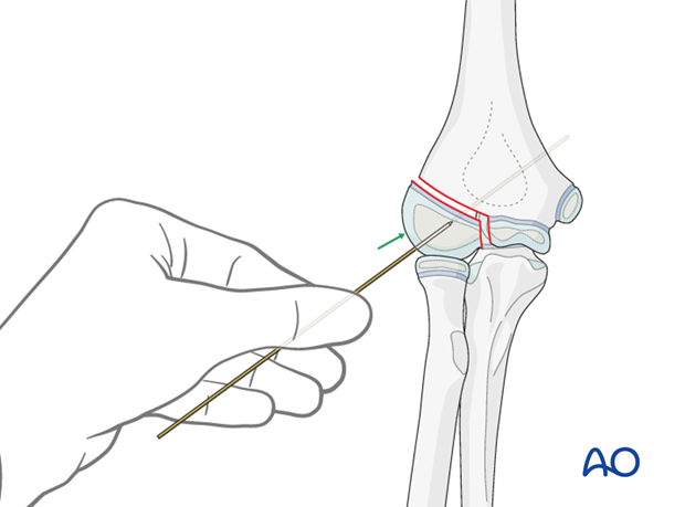
Option 2: Manipulation using a temporary K-wire in the fragment as a joystick.
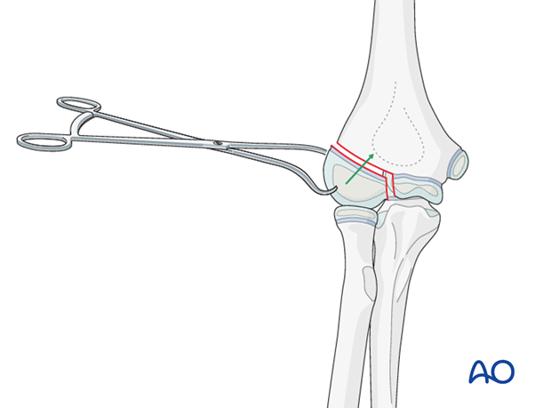
Option 3: Holding and manipulating the fragment with a small towel clamp or pointed reduction forceps.
Reduction
A blunt Hohmann lever retractor is inserted gently into the anterior joint, and around the medial articular border.
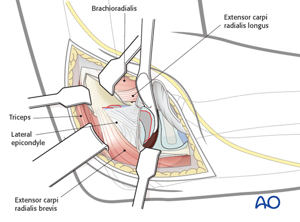
The medial cartilage fracture line is visualized on the joint surface.
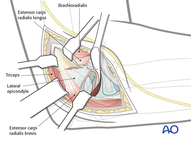
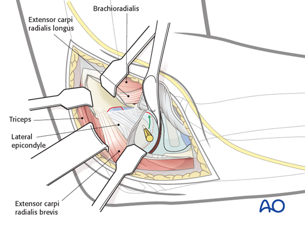
The fragment is reduced by one of the three options listed above, so that both cartilage fracture lines (on the fragment and the trochlea) are perfectly aligned.
5. K-wire fixation
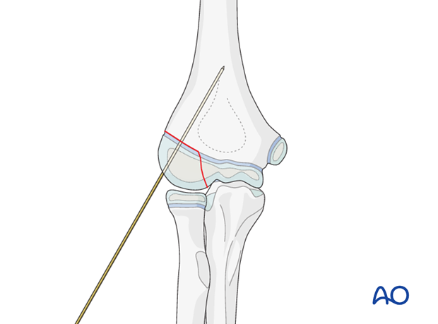
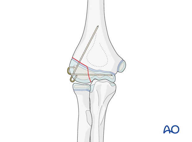
Fix the fragment with two divergent 1.2 or 1.6 mm K-wires.
The first oblique K-wire is used as a joystick to achieve reduction and then advanced into the metaphysis. The oblique K-wire is used first as, once advanced, its position in the metaphysis is less critical, whereas the track of the transverse K-wire must be parallel to the medial physis and totally within the epiphysis.
The fragment is additionally fixed with a second K-wire inserted parallel to the ulnar physis and totally within the epiphysis.
The wires are either buried deep to the skin or cut outside the skin and bent over. The first option requires a second operation for removal, but the second option risks pin-track infection.
Note: In the older child with a visible ossific center in the trochlea, consideration can be given to an intraepiphyseal screw instead of the horizontal K-wire.
6. Aftercare
With K-wire fixation it is recommended to apply a circular cast with posterior plaster splint for pain management and to prevent the use of the arm in younger children. In older children an arm sling is sufficient.
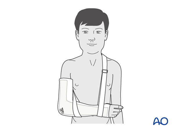
Note: In any case of elbow immobilization by plaster cast, careful observation of the neurovascular situation is essential both in the hospital and at home.
See also the additional material on postoperative infection.
It is important to provide parents/carers with the following additional information:
- The warning signs of compartment syndrome, circulatory problems and neurological deterioration
- Hospital telephone number
- Information brochure
If the child remains for some hours/days in bed, the elbow should be elevated on pillows to reduce swelling and pain.
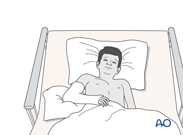
The postoperative protocol is as follows:
- Document results radiologically shortly after operation
- The cast, or splints, should be applied under anesthesia immediately after the operation
- Discharge from hospital according to local practice (1-3 days)
- First clinical and radiological follow-up, depending on the age of the child, 4-5 weeks postoperatively out of the cast
- In most cases, at this first x-ray control, the fracture is healed and stable so that cast is no longer required
- During the first clinical follow-up parents/carers should be informed about the timing of implant removal
- Physiotherapy is normally not indicated
Implant removal
Protruding K-wires should be removed after 4-5 weeks.
Buried K-wires can be removed after 2-3 months as a day case, under light anesthesia.


