Open reduction; K-wire fixation
1. Introduction
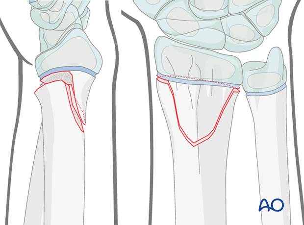
Open reduction is required for these fractures when closed reduction maneuvers fail. The fractures are usually posteriorly angulated (apex anterior). Impediments to reduction are interposed periosteum and pronator quadratus. Anteriorly angulated (apex posterior) fractures are less common. Impediments to reduction are the extensor tendons.
2. Patient preparation
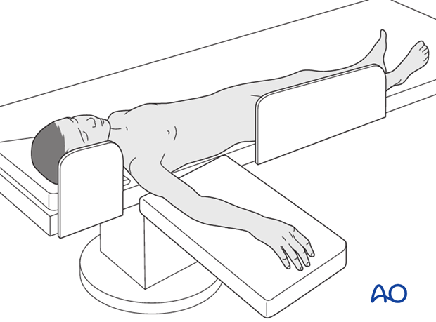
This procedure is normally performed with the patient in a supine position.
3. Approach
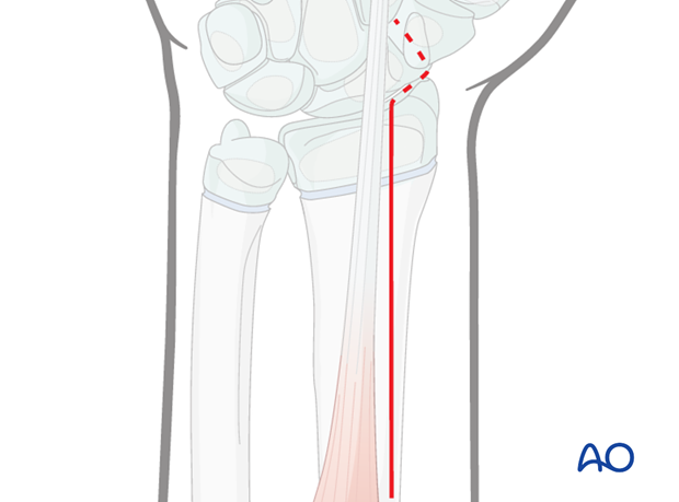
As the majority of these fractures are posteriorly displaced, open reduction is most commonly performed via the anterior approach.
A posterior approach may occasionally be necessary for irreducible anterior displacement.
A thorough knowledge of the anatomy of the wrist is essential. The corresponding additional material gives a short introduction.
4. Open reduction
Removal of impediments
Soft tissue impediments to reduction are removed, eg, pronator quadratus in the illustration.
Once the soft tissue impediments have been removed, the fracture will be reduced under direct vision.
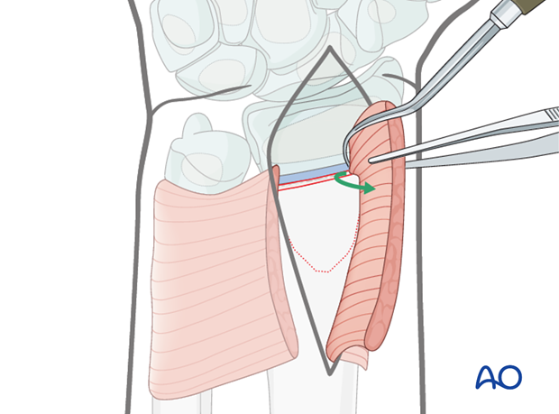
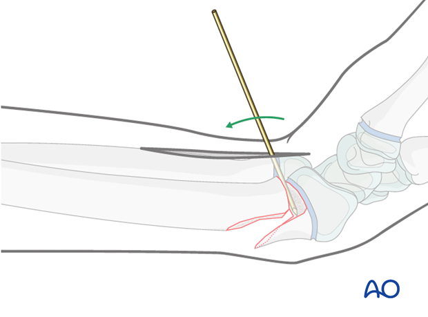
Direct reduction using K-wire
A K-wire inserted into the fracture site and used as a lever can be used to facilitate reduction
5. K-wire fixation
General considerations
For fractures that are unstable after reduction, a single K-wire is usually sufficient to stabilize the fracture.
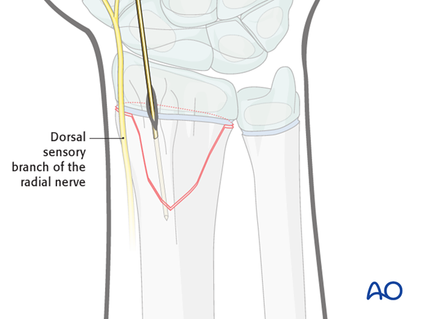
In cases of a more lateral metaphyseal wedge, the K-wire is inserted more in the coronal plane than in the sagittal plane.
Skin incision
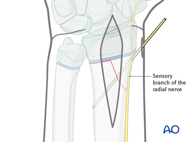
A small, separate skin incision is required for the K-wire insertion.
Care should be taken to avoid the dorsal sensory branch of the radial nerve.
The incision is deepened to the bone using a blunt artery forceps and a protective sleeve inserted.
K-wire insertion
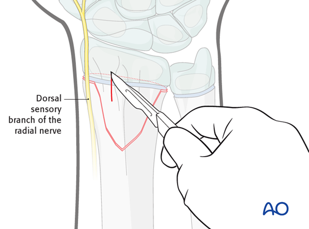
Via the protective sleeve, a single smooth 1.6 mm K-wire is inserted through the dorsal metaphyseal fragment engaging the anterior cortex of the radial diaphysis.
The wire should be inserted with an oscillating drill and cooled with saline to prevent thermal injury. Alternatively, the wire can be inserted manually using a T-handle.
Care should be taken to avoid the dorsal sensory branch of the radial nerve.
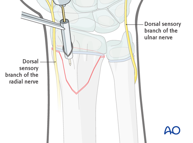
Ideally, wires are inserted using image intensification control, in order to check the trajectory of the wire and to ensure engagement of the far diaphyseal cortex without penetration of soft tissues.
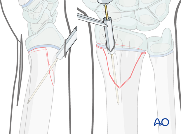
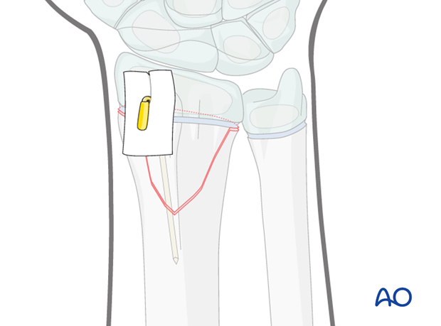
Each K-wire is left protruding through the skin, bent and cut. The skin is protected with sterile padding prior to the application of a cast.
The illustration demonstrates the use of a small section of plastic tubing over the cut end of the protruding wire. This adds further protection for the skin.
Note: Excessive pressure between dressing and skin should be avoided to prevent skin necrosis.
6. Short arm cast
General considerations
The purpose of the cast is protective and for pain relief, as stability is provided by the K-wire(s).
The short arm cast is applied according to standard procedure:
Note: In young, small, or noncompliant patients, it is safer to apply a long arm cast.
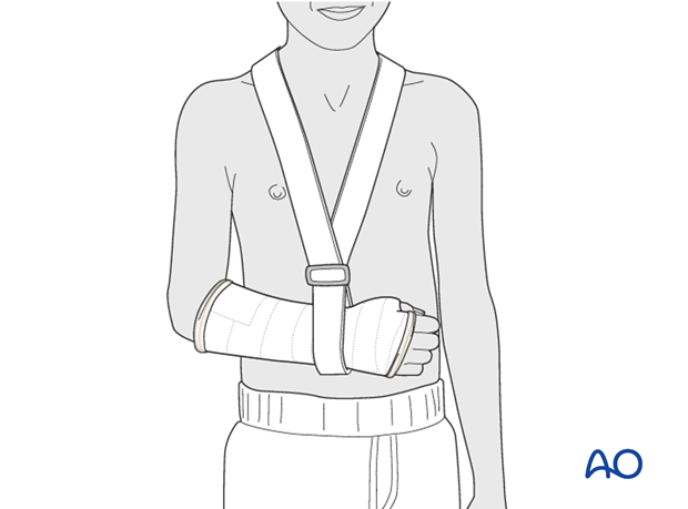
Splitting of the cast
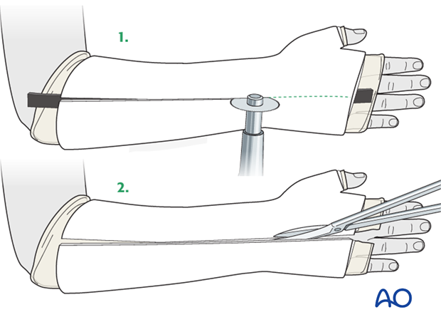
If a complete cast is applied in the acute phase after injury, it should be split over the full length of the cast. The split of the cast must be full thickness and expose the underlying skin.
7. Long arm cast
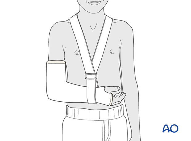
In the event that a long arm cast is necessary (see above) it is applied and split according to standard procedure:
8. Aftercare
Tight cast
Further swelling in a restricting cast can cause pain, venous congestion in the fingers and occasionally a compartment syndrome.
For this reason any complete cast applied in acute phase should be split down to skin.
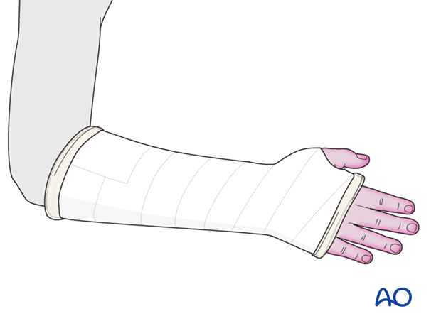
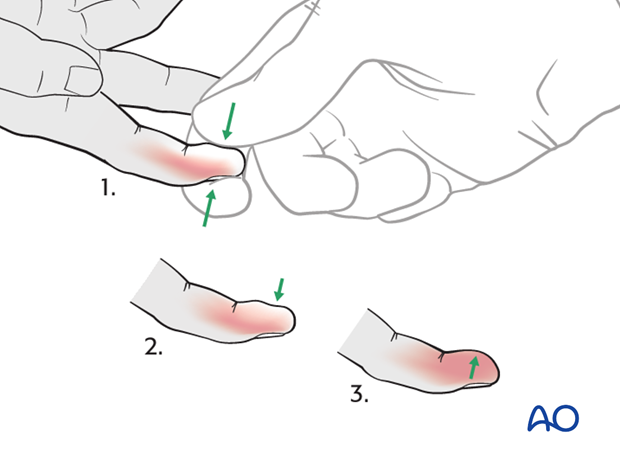
Parents/carers should be instructed how to detect circulatory problems by pressing and releasing the fingertips and watching if the blood flow/color returns to normal (capillary refill), compared to the opposite hand.
Compartment syndrome
Compartment syndrome is an unusual but serious complication after the application of a complete cast and can be difficult to diagnose, especially in younger children. Neurological signs appear late and the main sign is excessive pain on passive extension of the fingers.
It is important to make sure that the parents/carers are aware of the risk of compartment syndrome.
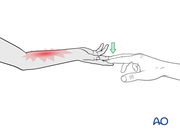
The parents/carers and patient should be informed to take note of increased pain and/or unresponsiveness to normal painkillers.
They should know that this may indicate serious complications. It is important for them to detect these signs as early as possible and report them urgently to the surgeon/nurse by telephone, or to attend the Emergency Room (ER) without delay.
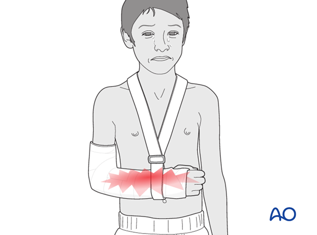
Nerve compression is an occasional complication and the signs include:
- Sensory deficits (numbness)
- Weakness of active finger movement
- Paresthesia
Infections
See also the additional material on postoperative infections.
Cast care
If a normal plaster of Paris cast is used, it is important to keep the cast clean and dry in order to maintain the reduction.
When the swelling has reduced, the cast can become loose. The loss of support can result in loss of reduction.
In this circumstance, the parents/carers are advised to return to the healthcare provider.
Follow-up x-rays
Depending on the fracture pattern, the age of the child and the method of treatment, the patient has to return for follow-up x-rays to monitor the fracture position.
X-rays taken for fracture position can be taken with cast in place. Any x-rays to assess the state of bone healing must be taken without the cast and correlated with clinical examination.
In most cases, it is conventional to obtain follow-up x-rays after reduction to ensure that the position is maintained.
In general, in the child below 5 years of age, the follow-up is usually about 4-5 days after reduction. In the older child with a potentially unstable fracture, an x-ray would normally be taken at 7-10 days.
Further follow-up x-ray is a matter of clinical judgement, the responsibility of the treating surgeon, and tends to be longer in older children (see also Healing times).
For complete fractures of the metaphysis, redisplacement after reduction is not uncommon. It is therefore, important to take early follow-up x-rays in order to detect a possible redisplacement.
See also the additional material on posttraumatic growth disturbances.
K-wire removal
The K-wires can be removed without sedation in the clinic after approximately 3 weeks (depending on the age of the child).


