

Lag-screw fixation with a neutralization plate
1. General considerations
Principles
In short oblique fractures, only one lag screw can be inserted. This is insufficient to produce enough stability, and the lag screw must be protected by a plate. A neutralization plate may also be indicated if screw fixation alone is not providing sufficient stability, eg, in poor quality bone.
As the screw should be inserted perpendicularly to the fracture plane, in most cases, it should be inserted independently of the plate.
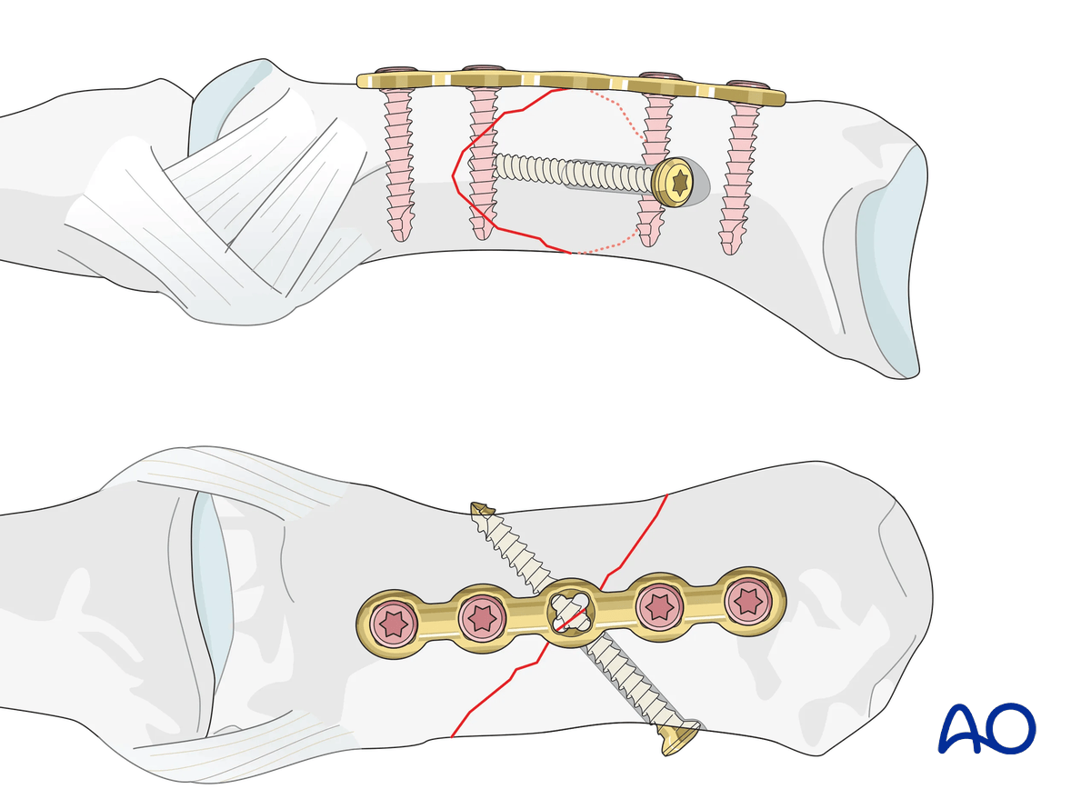
Plate selection
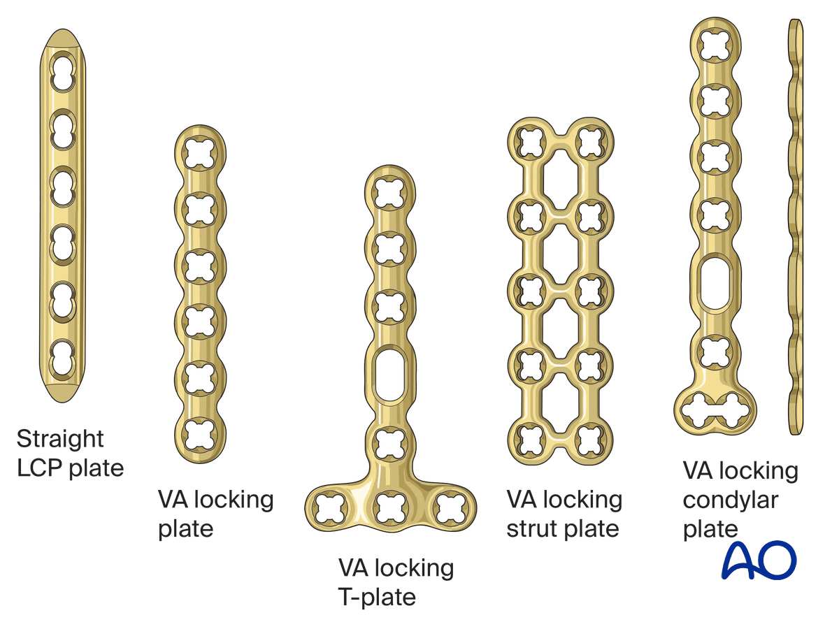
Select a plate (1.5–2.0 mm) according to the size of the bone, fracture geometry, and surgeon’s preference.
The plate length should allow for at least two screws in each main fragment.
The plate is available as a conventional compression plate or with variable-angle (VA) locking-head screws.
2. Patient preparation
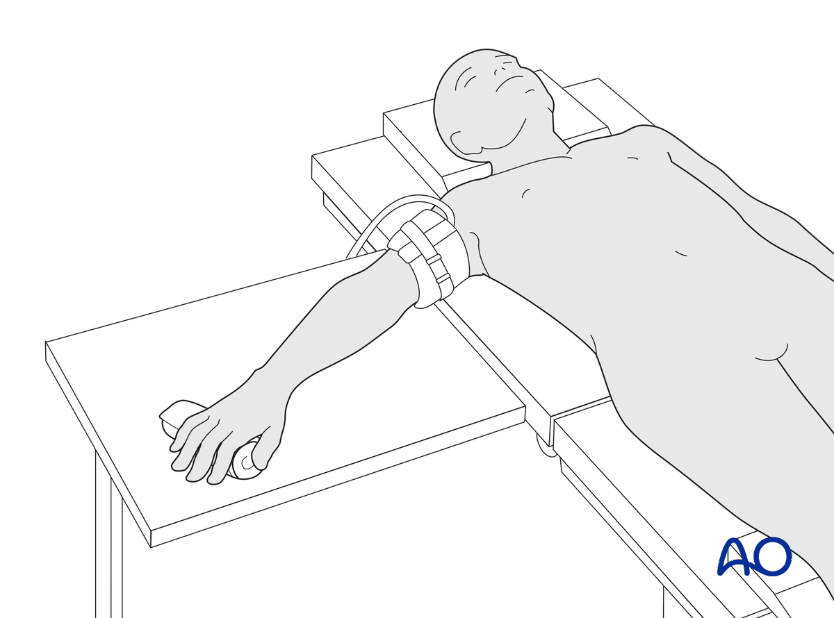
This procedure is usually performed with the patient in a supine position with the arm on a radiolucent side table.
3. Approach
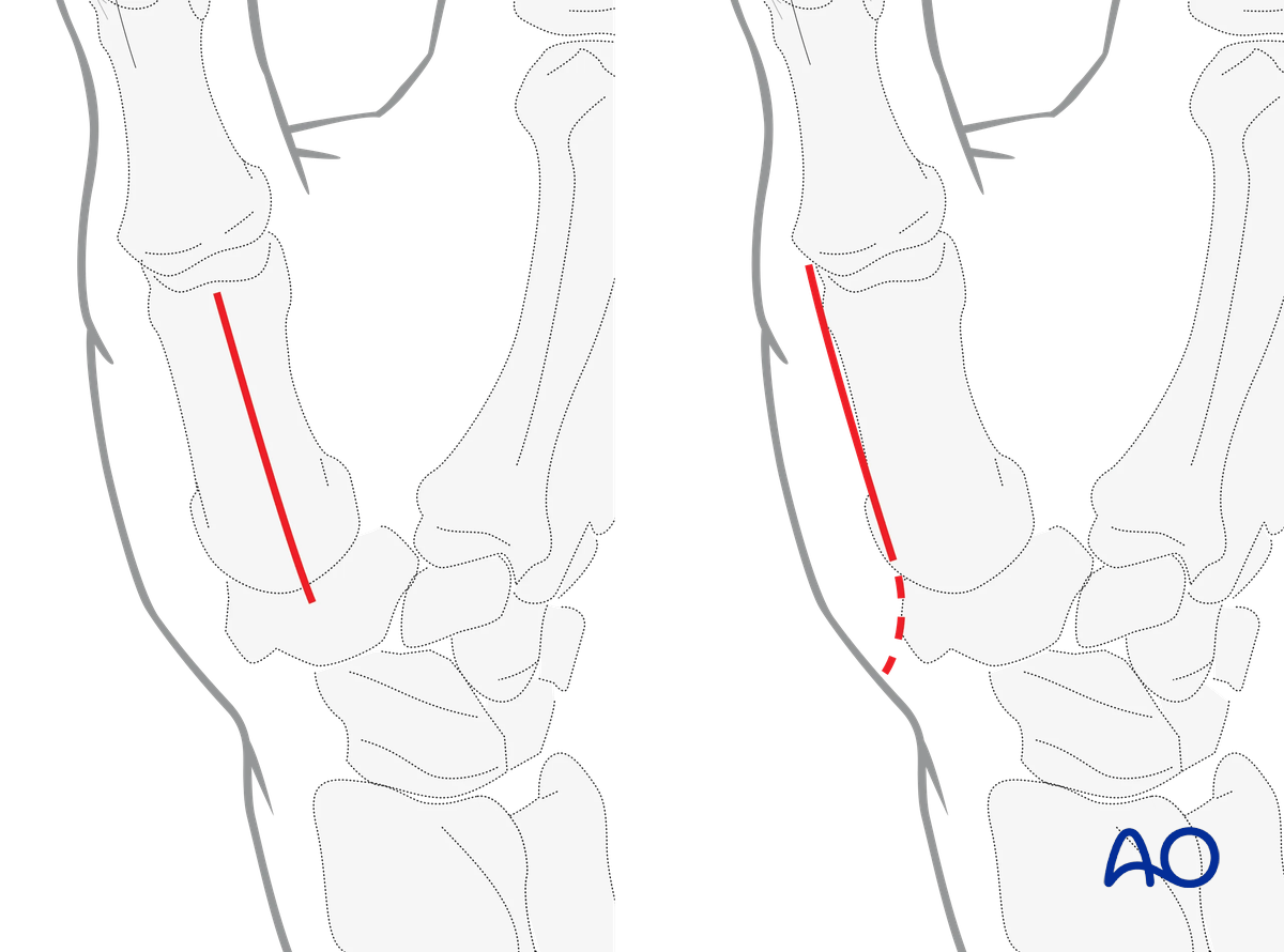
For this procedure, the following approach may be used:
4. Reduction
Any axial displacement should be reduced to gain full length of the metacarpal.
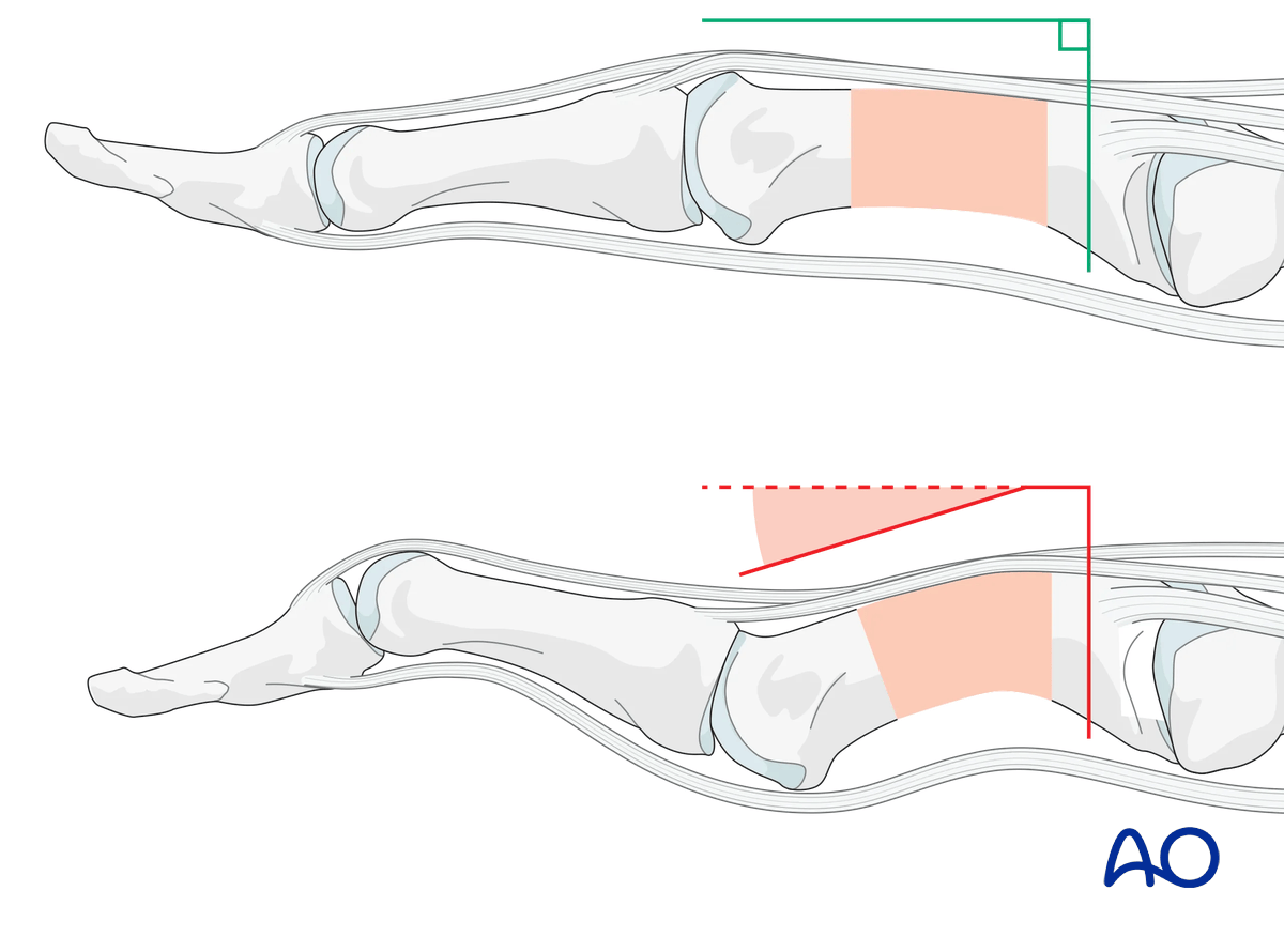
Indirect reduction by traction
Reduce the fracture with longitudinal traction on the thumb and pressure from a periosteal elevator or a dental pick.
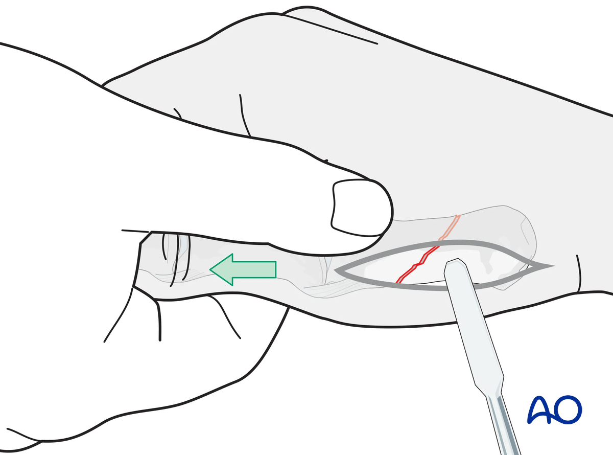
Direct reduction
Secure the reduction with pointed reduction forceps and confirm reduction with an image intensifier. Ensure the reduction forceps do not conflict with the planned screw position.
It is essential to confirm that the apex of each fracture fragment has been properly reduced.
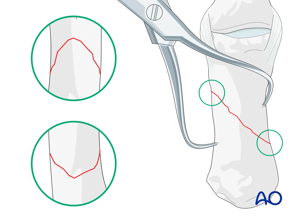
Alternatively, special reduction forceps designed for percutaneous fixation may be used.
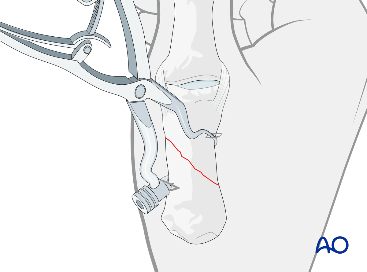
5. Checking alignment
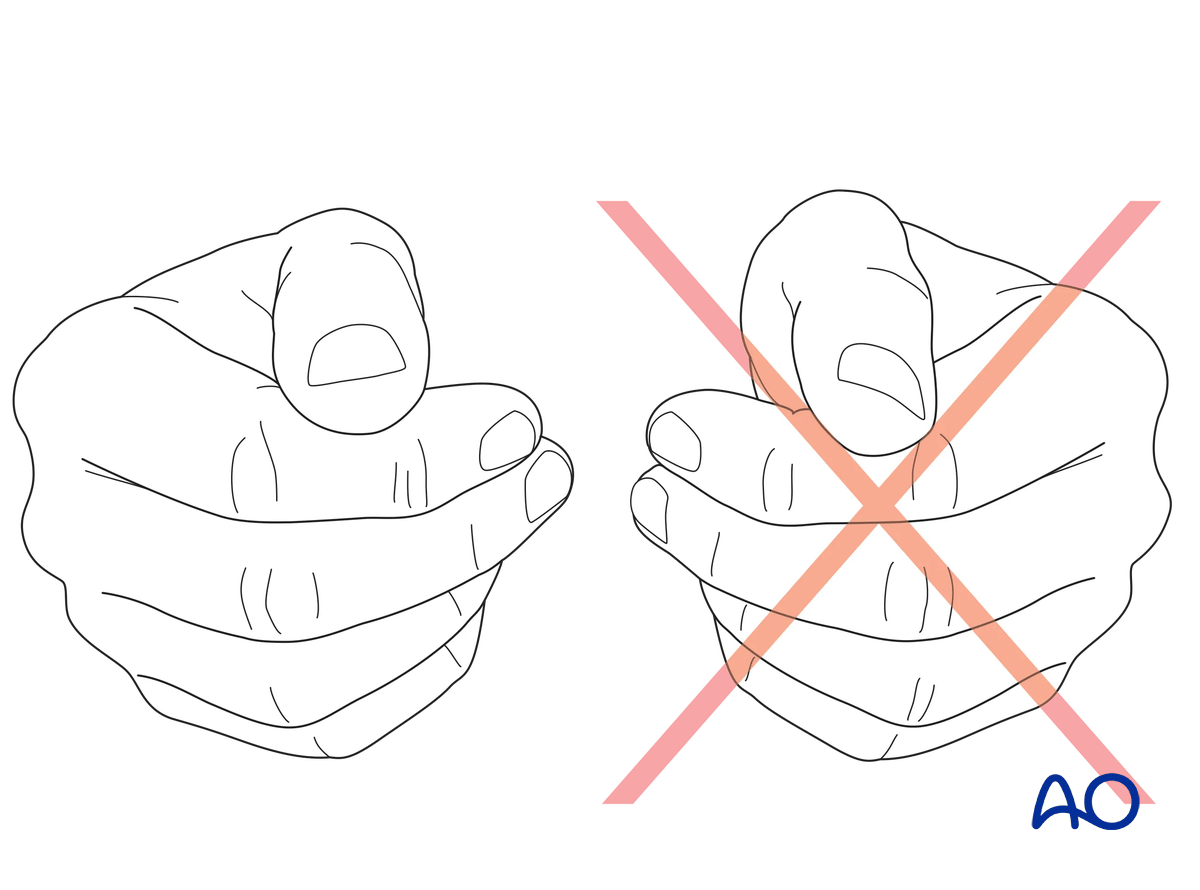
Confirm correct rotational reduction by inspecting the thumb nail plate in relation to the index finger. Compare it to the contralateral side if necessary.
6. Lag-screw fixation
Insertion of the lag screw
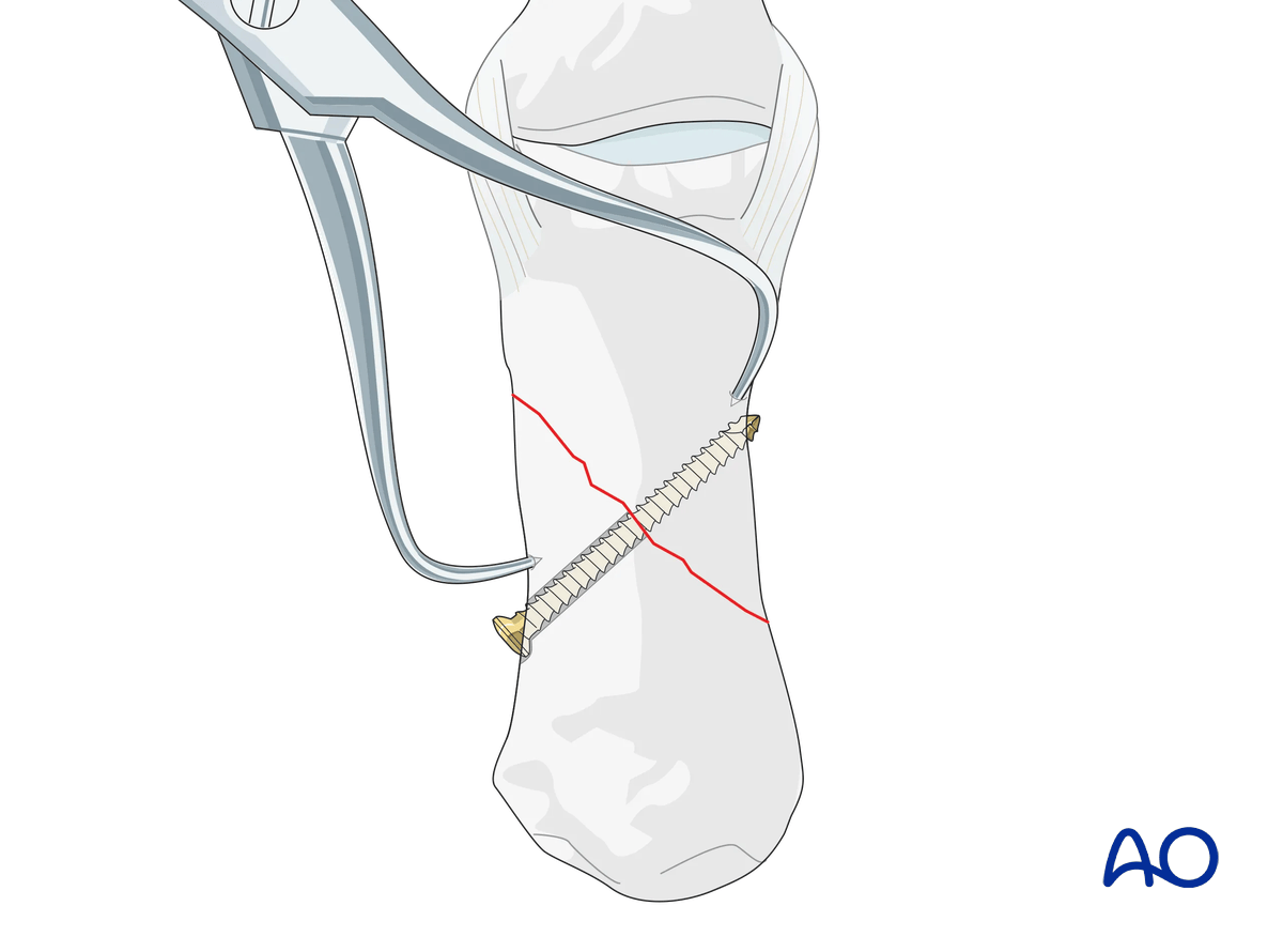
Insert the lag screw in the center of the fracture and perpendicular to the fracture plane. Carefully tighten the screw.
The fracture is now compressed. The reduction forceps may now be removed.
Confirm reduction and correct screw position with an image intensifier.
Screw-size selection
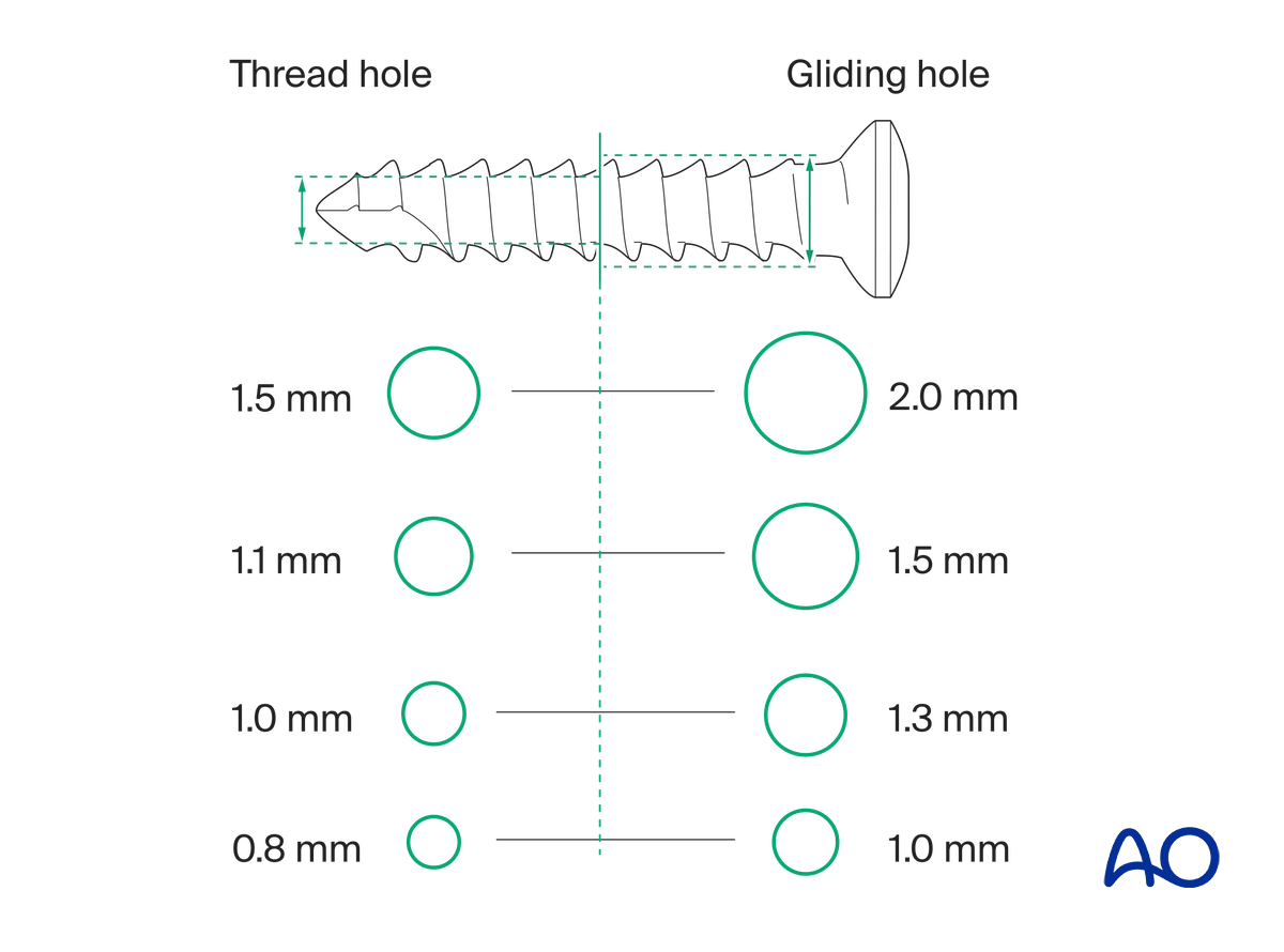
The exact size of the diameter of the screws used will be determined by the fragment size and the fracture configuration.
The various gliding and thread hole drill sizes for different screws are illustrated here.
Screw length pitfalls
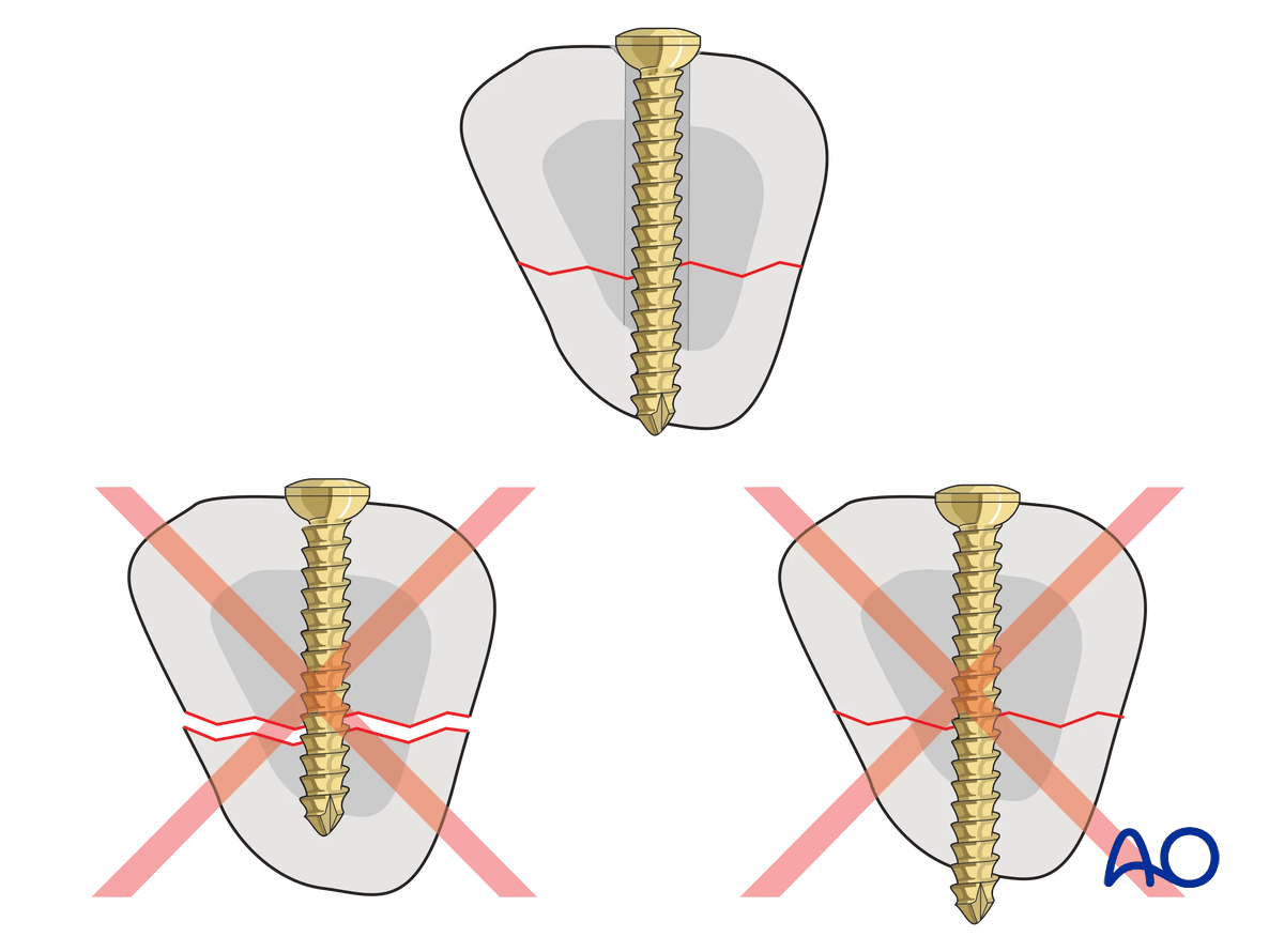
- Too short screws do not have enough threads to engage the cortex properly. This problem increases when self-tapping screws are used due to the geometry of their tip.
- Too long screws endanger soft tissues, especially tendons and neurovascular structures. With self-tapping screws, the cutting flutes are especially dangerous, and great care has to be taken that the flutes do not protrude beyond the cortical surface.
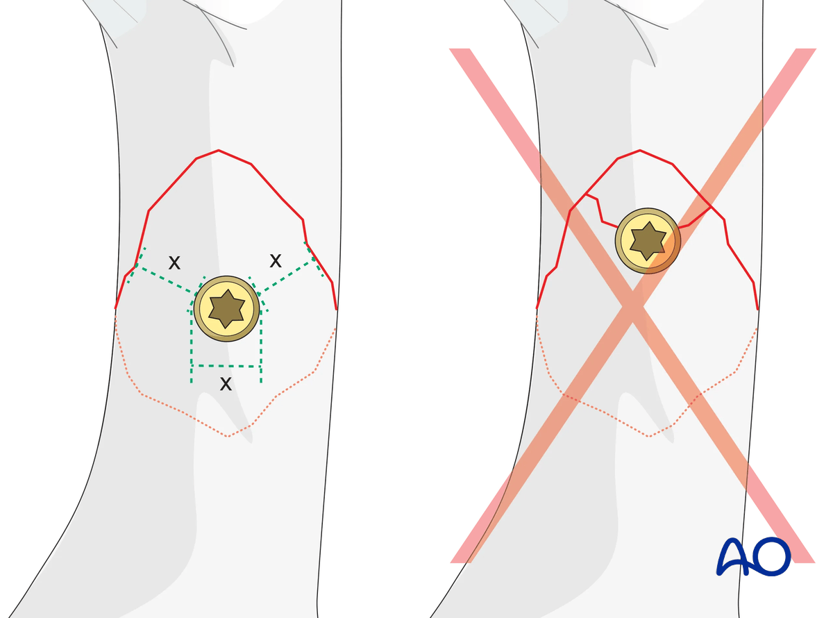
Pitfall: screw too close to the fracture
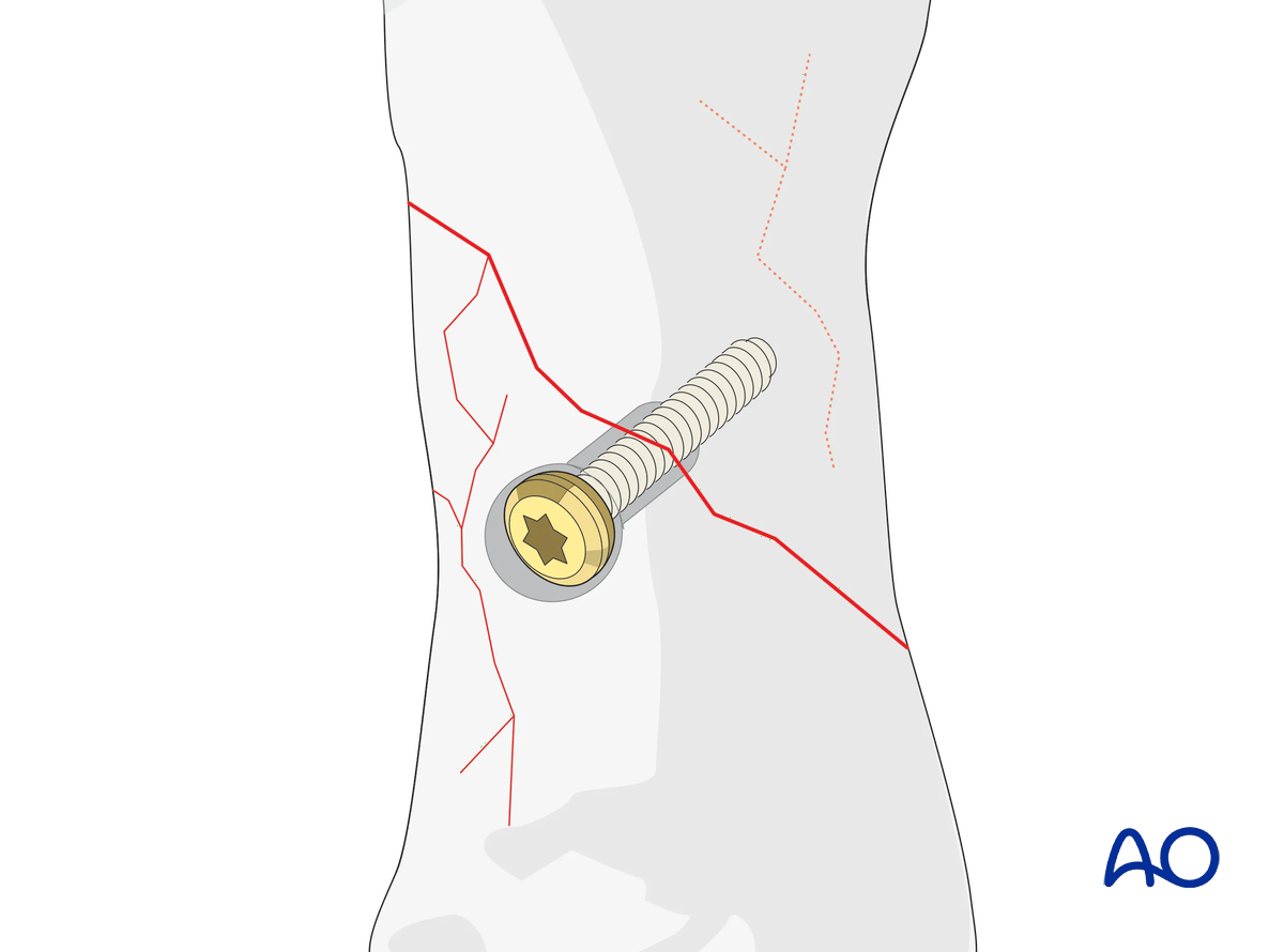
Pitfall: beware of fissure lines
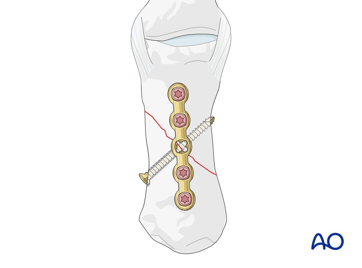
7. Plate fixation
Apply the neutralization plate dorsally.
Insert at least two screws proximally and distally to the fracture in neutral mode.
Cover the plate with periosteum, if possible, to avoid adhesion between the tendon and the implant leading to limited finger movement.
8. Final assessment
Confirm correct rotational alignment by clinical examination.
Confirm anatomical reduction and correct placement of implants in AP, lateral, and oblique views under image intensification.
9. Aftercare
Healing phases
The aftercare can be divided into four phases of healing:
- Inflammatory phase (week 1–3)
- Early repair phase (week 4–6)
- Late repair and early tissue remodeling phase (week 7–12)
- Remodeling and reintegration phase (week 13 onwards)
Full details on each healing phase can be found in the chapter Postoperative treatment of thumb injuries.
Postoperative care
Early range of motion, without load bearing, in the hand is important for optimal outcome.
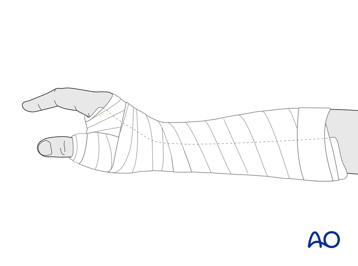
Immediately postoperatively, a temporary plaster splint is applied. This also helps with swelling and pain control.
The plaster splint can be switched to a custom-made thermoplastic splint after a week. The splint should be worn until fracture healing has been confirmed.
Hand therapy should be initiated as soon as possible.
The arm should be actively elevated to help reduce any swelling.
Mobilization
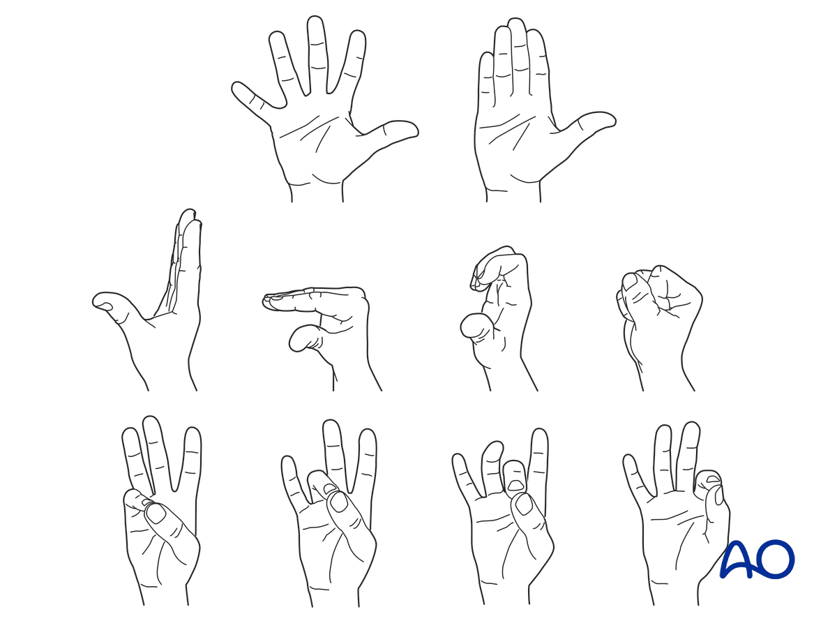
To prevent joint stiffness, the patient should be instructed to begin active motion (flexion and extension) of all nonimmobilized joints of the hand immediately after surgery.
In compliant patients with stable fixation, the splint can be removed under the supervision of a hand therapist, and early active motion exercises started after a few days.
Weight bearing should be avoided until fracture healing is confirmed.
Follow-up
The surgical wound is checked as necessary depending on soft tissue status. The wound is monitored for evidence of infection.
To allow for early joint mobilization and to prevent wound dehiscence, leave the sutures for 2 weeks.
Confirm fracture healing clinically and radiologically after about 5–6 weeks (depending on the fracture configuration).
Implant removal
The implants may need to be removed in cases of soft-tissue irritation.
In case of joint stiffness or tendon adhesion restricting finger movement, arthrolysis or tenolysis may become necessary. In these circumstances, the implants can be removed at the same time.


