ORIF - Plates without angular stability
1. Principles
Anatomical reduction
Depression fractures are intraarticular, therefore they need anatomical reduction.
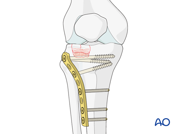
The plate in this procedure acts as a buttress to neutralize the axial forces on the tibial plateau and protects the weakened or fenestrated (windowed) medial cortex from failing.
The operative procedures for lateral condylar fractures and medial condylar fractures are comparable. A lateral condylar fracture treatment is shown here.
2. Patient preparation and approach
Patient preparation
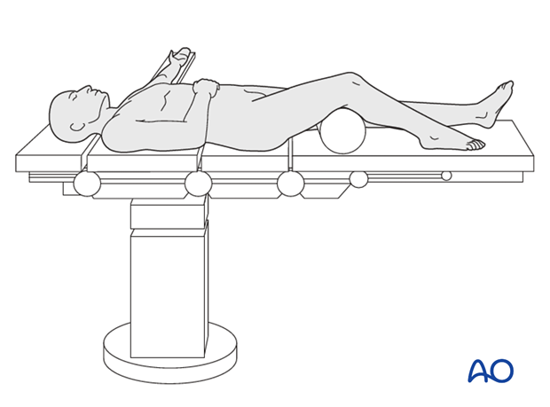
This procedure is normally performed with the patient in a supine position.
Approach
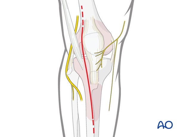
For this procedure an anterolateral approach is used.
3. Reduction
Reduction of the impacted fragment(s)
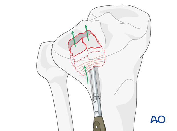
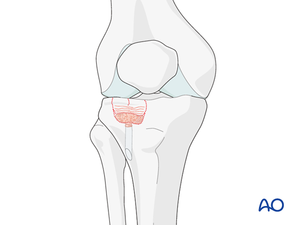
Make a window in the anterolateral cortex of the tibial condyle lateral to the tibial tuberosity and about 5 cm distal to the joint line.
Introduce a curved impactor and elevate the impacted bone until the articular fragments are reduced and the joint is congruent again. Slight overcorrection will compensate for a slight loss in height of the reduced articular fragment(s) which may occur following surgery.
The articular surface may be inspected directly through a standard submeniscal articular exposure or by means of an arthroscope inserted through a medial portal.
Temporary fixation of the elevated articular fragments with K-wires may be helpful.
Filling of defect
The defect which is created once the impacted articular fragments are reduced must be filled with an autologous cancellous autograft or a corticocancellous block graft to support the elevated fragments. Alternatively the use of bone substitutes may be considered.
4. Fixation
K-wire fixation
K-wire fixation is useful in maintaining the articular fragments reduced until the metaphyseal defect is bone grafted.
Positioning of the knee is important for correct reduction and fixation. If the knee has a valgus injury, then the knee should be held with more varus positioning to ensure a good reduction. If the knee has a varus injury (medial condyle) then valgus positioning during reduction is important.
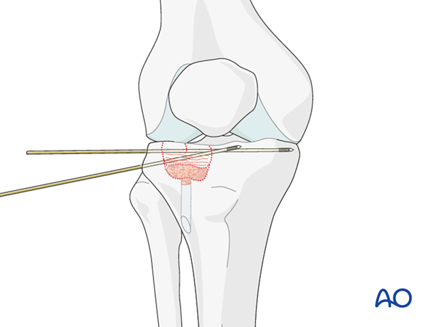
Plate osteosynthesis
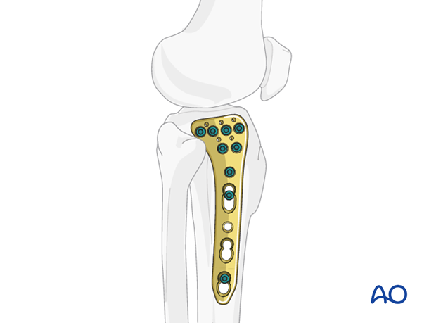
In the purely depressed fracture, the plate acts as protection for the weakened fenestrated lateral cortex.

A raft plate is both stronger and more efficient in supporting the articular surface. Especially in comminuted and osteoporotic cases.
The plate and screws are placed perpendicular to the longitudinal and axial force in the tibial plateau fracture, resisting the displacement forces.
5. Fixation
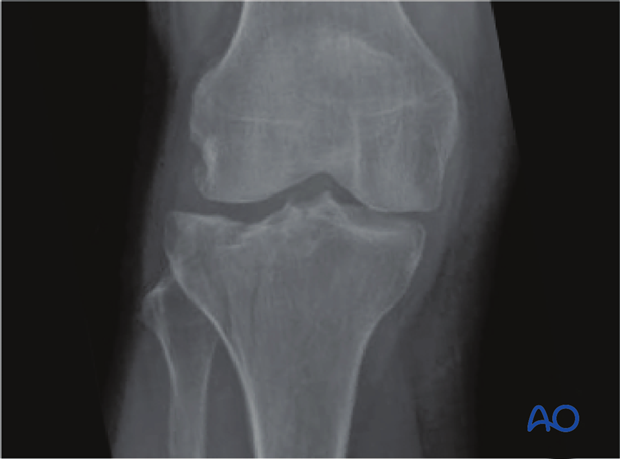
This case represents a lateral depression fracture with a nondisplaced split in the cortex as seen on the CT transverse cuts.
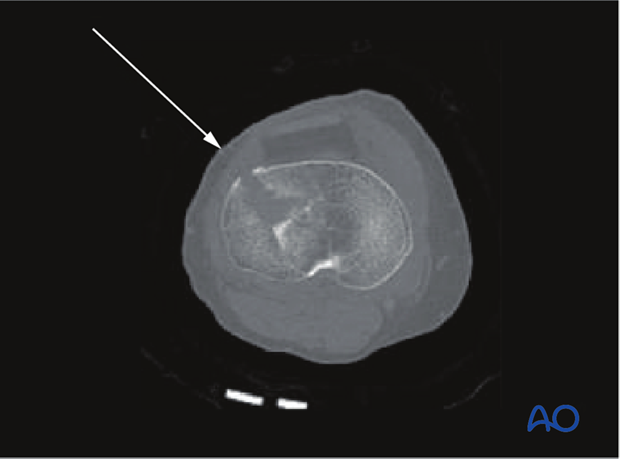
AP X-ray.
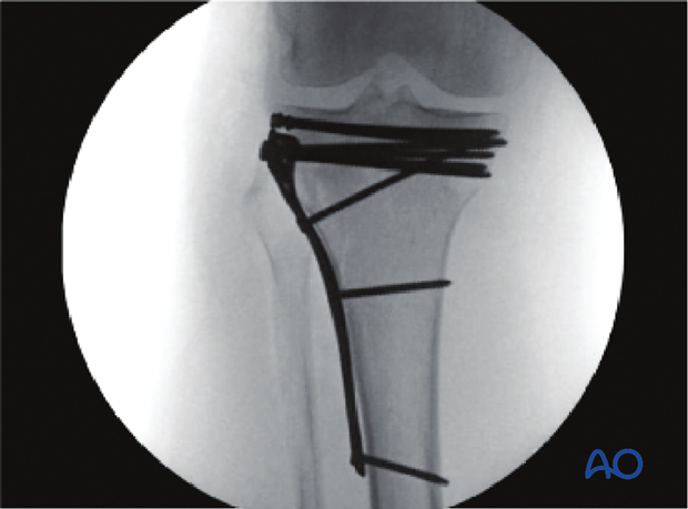
It has been reduced and fixed with both a buttress plate with raft screws and raft function (to protect the cortical window) and subchondral raft screws immediately below the joint to supplement the fixation as well. This construct of hardware is thought to be the best to maintain reduction in these pure impaction injuries.
6. Aftercare
Compartment syndrome and nerve injury
Close monitoring of the tibial compartments should be carried out especially during the first 48 hours after surgery to rule out compartment syndrome.
The neurovascular status of the extremity must be carefully monitored. Impaired blood supply or developing neurological loss must be investigated as an emergency and dealt with expediently.
Functional treatment
Unless there are other injuries or complications, mobilization may be performed on post OP day 1. Continuous passive motion (CPM) splints are very helpful in the early phase of rehabilitation. Static quadriceps exercises with passive range of motion of the knee should be encouraged. Afterwards special emphasis should be given to active knee and ankle movement.
Following any injury, and also after surgery, the neurovascular status of the extremity must be carefully monitored. Impaired blood supply or developing neurological loss must be investigated as an emergency and dealt with expediently. The goal of early active and passive range of motion is to achieve as full range of motion as possible within the first 4 - 6 weeks. Optimal stability should be achieved at the time of surgery, in order to allow early range of motion exercises.
Weight bearing
No weight bearing in the treatment of articular fractures for a minimum of 10 – 12 weeks.
Follow up
Wound healing should be assessed on a short term basis within the first two weeks. Subsequently a 6 and 12 week follow-up is usually performed. If a delayed union is recognized, further surgical care will be necessary and should be carried out as soon as possible.
Implant removal
Implant removal is not mandatory and should be discussed with the patient.


