Posterior wall
Definition
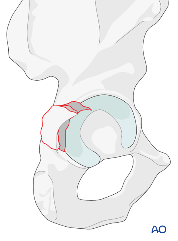
The posterior wall fractures involve the rim of the acetabulum, a portion of the retroacetabular surface, and a variable segment of the articular cartilage. The majority of the posterior column remains intact.
A posterior dislocation is associated in approximately 35% of reported cases.
Posterior wall fractures may occur with femoral head fractures.
Posterior wall fractures commonly involve:
- Multiple fragments
- Marginal impaction
- Incarcerated fragments
- Incomplete/nondisplaced transverse or posterior column fracture lines
Further details on posterior wall fractures are provided in section Characteristics of elemental fracture types.

Radiology
A summary of diagnosing the fracture classification based on x-ray and CT images is presented in section Patient assessment.
The section Radiology of the intact acetabulum provides explanation of the radiologic landmarks.
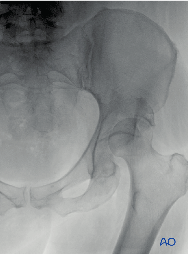
The section Characteristics of elemental fracture types provides further information on the radiology of posterior wall fractures.
X-ray image of posterior wall fracture, courtesy of TA Ferguson


