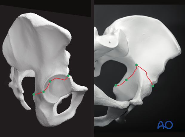
Radiology of the intact acetabulum
1. Standard imaging views
The ultimate goal is for the surgeons to accurately interpret the radiological features of the fractured acetabulum, and to then transform the radiographic data into a 3-dimensional understanding of each specific fracture’s morphology.
For any acetabular fracture, there are three standard imaging views recorded in the following sequence:
- Anteroposterior pelvis
- Obturator oblique view
- Iliac oblique view
Also, most commonly, a CT scan is obtained.
Each radiographic view has specific, reproducible radiological landmarks, and understanding the anatomical origins of the radiographic landmarks is critical in understanding the significance of a disruption of any one of the “lines”.
Letournel demonstrated these lines on radiographs of a dry pelvis specimen with lead wires accentuating the landmarks.
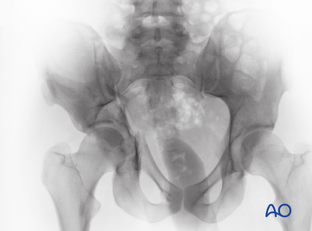
2. Anteroposterior radiograph (AP pelvic radiograph)
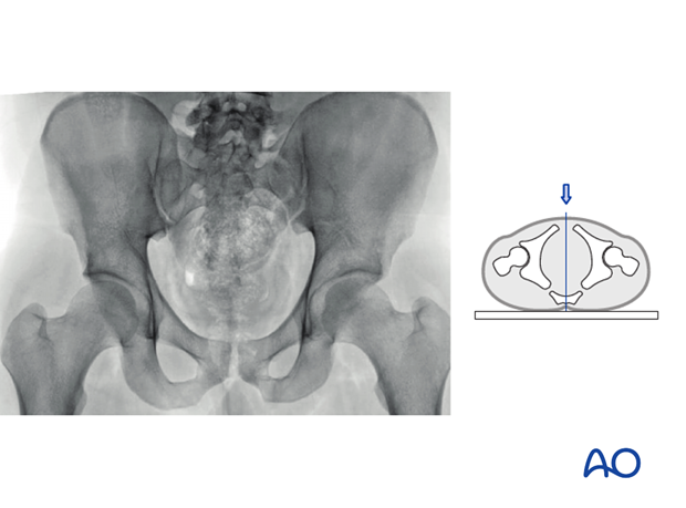
An AP view of the pelvis is required for all patients with a suspected acetabular fracture. The radiograph is obtained with the patient in the supine position and is centered on the symphysis pubis.
The quality of the radiography is assessed based on an evaluation of the rotation of the lumbar spine.
There are six fundamental radiological landmarks that should be identified on the AP pelvic projection. The anatomical correlate to each landmark needs to be understood, so that when a fracture line is identified traversing any one of the landmarks one can know what area of the innominate bone has been violated by the fracture.
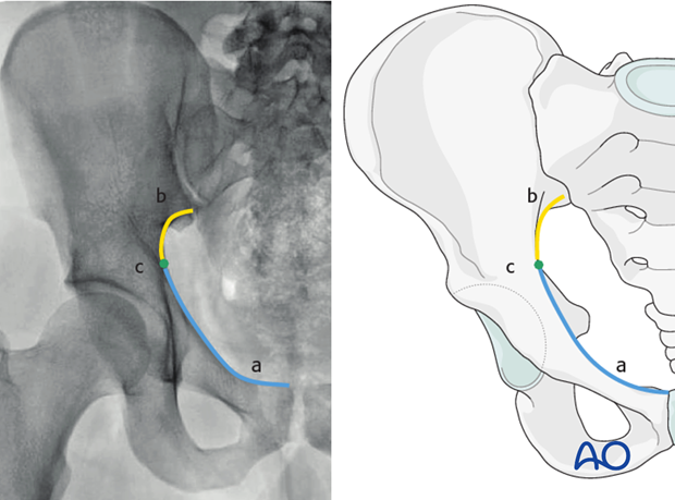
1. Iliopectineal line
The iliopectineal line correlates exactly with the pelvic brim for the anterior three quarters of the line (from the superior border of the symphysis pubis to the region where the beginning of the ilioischial line is identified), as highlighted by blue line segment (a).
The posterior quarter of the iliopectineal line diverges from the pelvic brim and corresponds to the dense bone of the internal surface of the sciatic buttress 1-2 cm below the actual pelvic brim (b, yellow line segment).
The point of divergence where the radiographic iliopectineal line no longer corresponds with the anatomic pelvic brim is indicated by a green dot (c).
The iliopectineal line is representative of the structural integrity of the anterior column of the acetabulum.
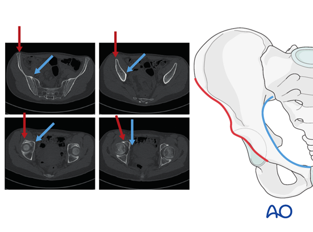
The iliopectineal line (the pelvic brim, blue line/arrow) is found to be the most medial and anterior portion of the innominate bone. This is separate from the anterior border (red line/arrow) of the bone, identified as the most anterior lateral portion of the innominate bone.
Starting cranially, the superior iliac spine can be followed distally to the inferior iliac spine and along the anterior wall of the acetabulum (red line/arrow).
2. Ilioischial line
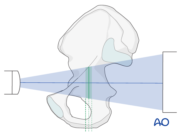
The ilioischial line is the result of the X-ray beam occurring in tangent across a segment of the surface of the quadrilateral plate.
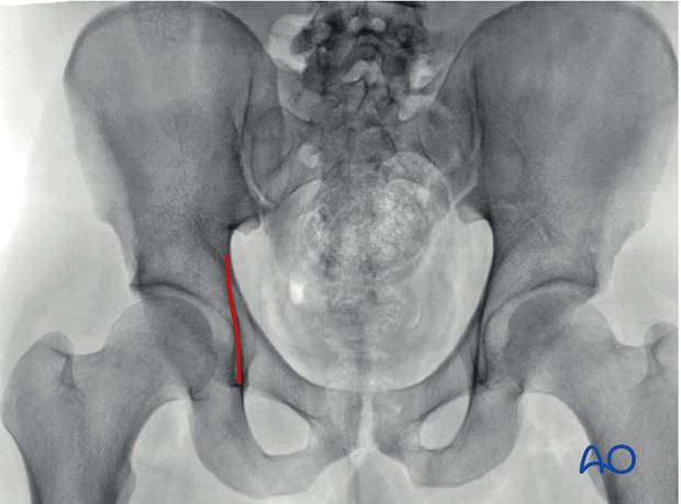
This occurs because the X-ray beam is parallel with the quadrilateral surface when the patient is in the supine position.
The anterior limit coincides with the anterior border of the obturator foramen approximately 1 cm in front of the tip of the ischial spine. It extends superiorly to approximately 1 cm below the apex of the greater sciatic notch.
The ilioischial line represents the structural posterior column of the acetabulum.
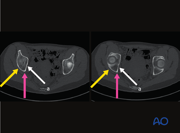
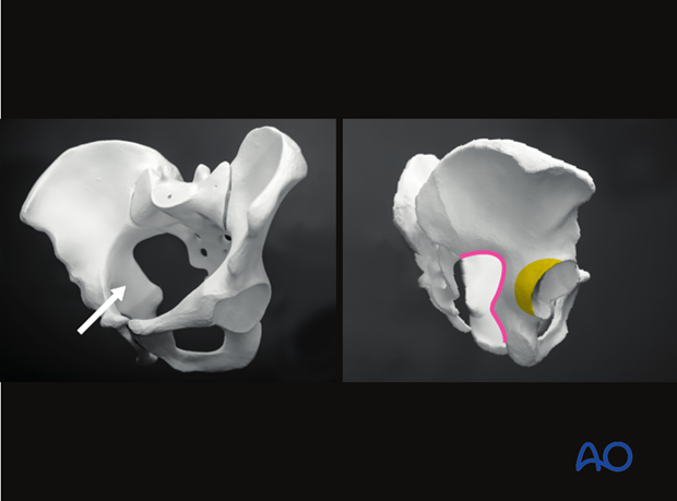
On the CT scan, the area of bone creating the ilioischial line is the portion of the quadrilateral plate indicated by the white arrow.
Notably, it is distinct from the posterior border of the bone (pink arrow). It is also distinct from the retro-acetabular surface (yellow arrow).
Here the same anatomical structures are highlighted on a bone model:
- White arrow - area of bone creating the ilioischial line
- Pink line - posterior border
- Yellow area - retro-acetabular surface
3. Radiological roof
The radiological roof, or sourcil, is a line produced by the X-ray beam tangential to the most cranial portion of the acetabulum.
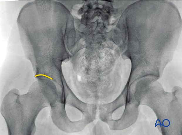
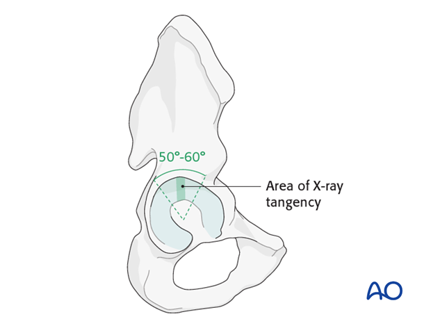
The outline of the roof represents only a small 2-3 mm region of the acetabulum region.
The radiological roof is simply a radiological line. It does not represent the anatomical roof or the portion of the acetabulum responsible for the weight bearing responsibilities of the acetabulum. The anatomical roof is provided by a 50-60° arch of the articular surface of the acetabulum as demonstrated in the illustration.
4. Anterior rim
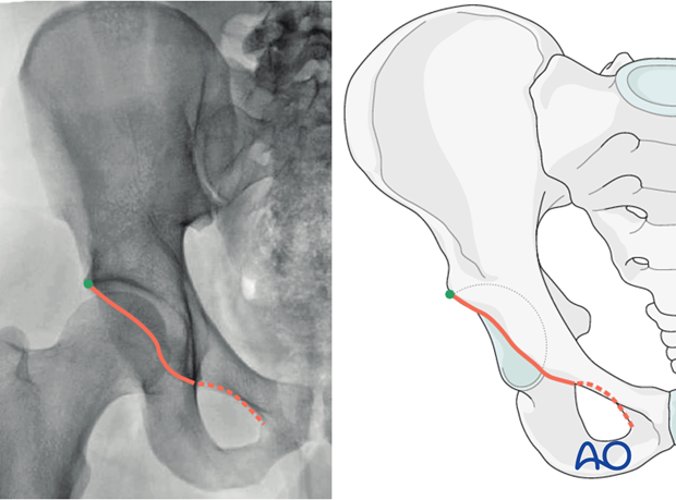
The anterior rim, or border, (orange line) of the acetabulum begins at the superior lateral aspect of the radiological roof (green dot). It descends caudally and medially to meet the superior border of the obturator foramen (dashed line).
Due to the anteversion of the acetabulum, the profile of the anterior rim is significantly more medial than its posterior counterpart. Thus, on the AP projection, it is superimposed on the shadow of the posterior wall, and difficult to see.
If one follows the superior border to the obturator foramen from medial to lateral (orange), then looks to the lateral point of the roof (dot), one will nearly always be able to identify the anterior rim as it connects these two points (orange). Judet and Letournel refer to this whole line as the acetabulo-obturator line.
The radiographic line of the anterior rim corresponds to the lateral border of the anterior wall.
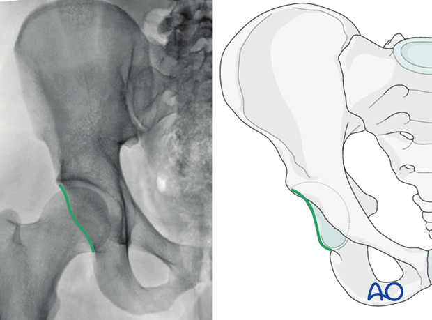
5. Posterior rim
The outline of the posterior rim is visualized starting caudally at the lateral most aspect of the radiological roof and proceeds caudally to the subcotyloid groove above the ischial tuberosity.
The radiographic line of the posterior rim represents the lateral aspect of the posterior wall.
6. Teardrop
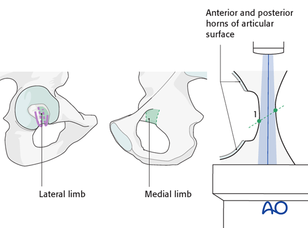
The radiological teardrop is created by the superimposition of the distal anterior and posterior horns of the articular surface as well as the tangent of the inferior surface of the cotyloid fossa.
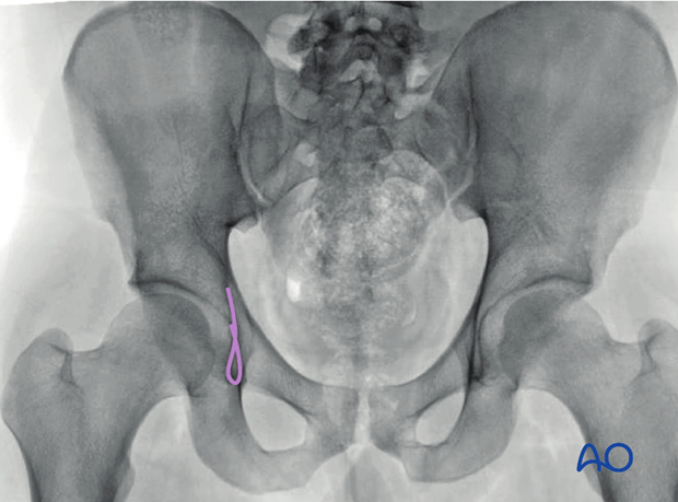
The radiological teardrop is produced by a U-shaped continuous surface of bone.
The lateral limb is created by an X-ray tangent of the inferior cotyloid fossa (1).
The medial limb is formed by the obturator canal as it merges with the quadrilateral surface.
Case 1
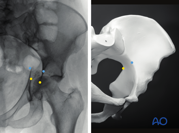
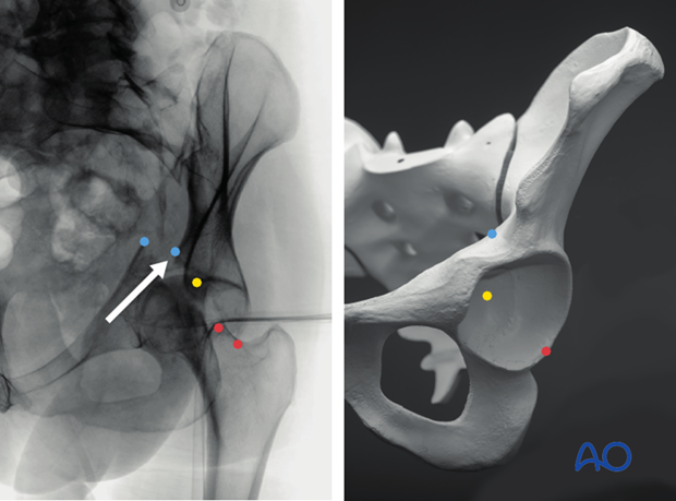
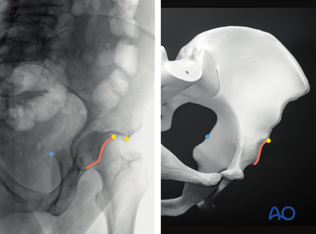
Once the landmarks are understood, one can evaluate an AP pelvis of an injury and translate the observed disruptions of the landmark lines onto a 3-dimensional pelvic model as shown.
The blue dots in the X-ray demonstrate the disruption of the iliopectineal line, which represents a fracture through the pelvic brim at the point demonstrated by a blue dot on the model.
The yellow dots in the X-ray demonstrates the disruption of the ilioischial line at its mid-portion, which can now be drawn as demonstrated to represent a fracture midway through the region of the quadrilateral surface which creates the ilioischial line (yellow dot on model).
This exercise should be performed for the six standard landmarks before moving on to the oblique views to augment and validate the interpreted injury based on the information provided by the AP pelvis.
3. Obturator oblique radiograph
The obturator-oblique radiograph is obtained at 45° to the coronal plane, with the injured hip raised and with the center of the beam on the center of the femoral head.
In the perfect view, the anterior superior iliac spine and the posterior superior iliac spine is superimposed, such that the iliac cross section is a small as possible and thus the obturator foramen as large as possible. If the iliac cross section appears wide, it is due to insufficient rotation of the patient.
The coccyx should be at the center of the femoral head.
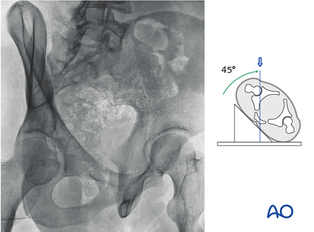
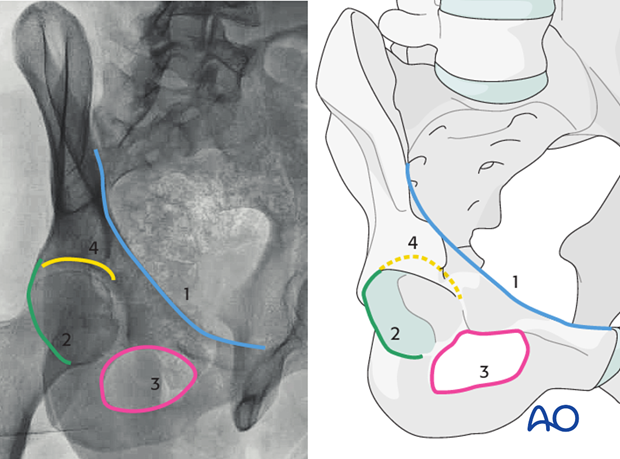
There are four consistent landmarks that should be inspected on the obturator oblique in each case:
- The pelvic brim and the represented periarticular anterior column
- The posterior border of the acetabulum
- The obturator foramen
- The acetabular roof
Additionally, the iliac wing can be visualized in cross section and most of the landmarks associated with the internal and external surface identified.
Blue: The pelvic brim is demonstrated in blue. The periarticular region of the anterior column is seen clearly. Additionally, time should be spent identifying the anterior superior iliac spine, the anterior inferior spine, the psoas gutter, and the pubic spine.
Green: The posterior border of the acetabulum is easily visible due to the rotation of the anterior rim away and thus out of line for super-imposition. The posterior border can be followed cranial to identify the supra-acetabular surface, the gluteus medius pilar and the landmarks associated with the external surface of the innominate bone.
Pink: The outline of the obturator foramen is completely visualized.
Yellow: The acetabular roof can be easily visualized.
The obturator oblique shows clearly this large posterior wall fracture involving a substantial region of the roof and the supra-acetabular surface. The anterior column is seen to be intact.
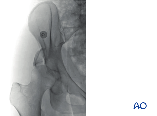
The T-shaped fracture is easily classified as the vertical limb can be seen traversing the obturator foramen, and the fracture is seen to involve the roof indicating a transtectal transverse component.
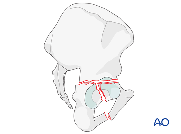
The spur sign is seen on the obturator oblique and defines this fracture as an associated both column fracture.
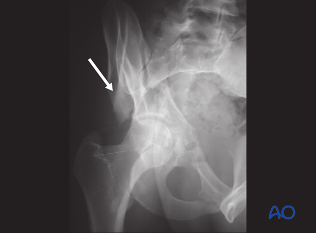
Case 1
The obturator oblique of the same case as shown above in AP section:
- Confirms the region of the pelvic brim involvement and the anterior column (blue dots).
- Indicates that the transverse fracture violates a small area of the roof at its medial most region. It appears as though a very small area of the roof remains on the displaced fragment. We will use the CT scan to verify the fracture’s position with respect to the roof to determine if this injury is a trans- or juxtatectal fracture (yellow dot).
- Demonstrates the displacement through the posterior rim but lack of a posterior wall fragment (red dots).
- Demonstrates the integrity of the obturator foramen, excluding the diagnosis of a T-shaped fracture. (This will also be verified using a CT scan).
- Demonstrates a large segment of roof remaining on the intact region of the acetabulum excluding the associated both column fracture from the differential diagnosis.
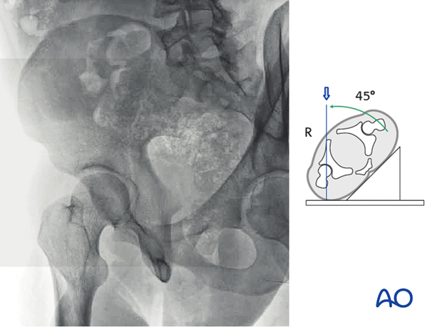
4. Iliac oblique radiograph
The Iliac oblique radiograph is obtained at 45° to the coronal plane, with the contralateral hip raised and with the center of the beam on the center of the femoral head. This view perfectly exposes the entire surface of the iliac wing and the obturator foramen is as thin as possible in section.
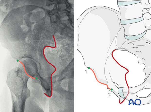
The three fundamental landmarks of the iliac oblique radiograph include:
- The posterior border of the iliac bone and thus the posterior column
- The anterior border of the acetabular rim
- The iliac wing in profile
The posterior border of the entire innominate bone can be identified, as well as the posterior spines, greater sciatic notch, ischial spine and tuberosity (as illustrated in red). Any fractures violating the posterior column will be identified here.
The anterior rim can be challenging to identify.
A line can be identified between the superior anterior articular margin of the acetabulum (green dot 1) and caudally the root of the superior ramus (green dot 2). This line is typically seen to have an inflection point, or a point of concavity midway as illustrated by the green line.
The remaining iliac wing can be identified from the posterior superior spine to the anterior superior iliac spine, to the anterior inferior spine, and is thus an excellent view for characterizing fractures in the iliac fossa.
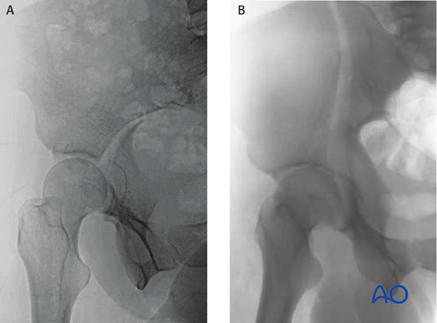
The iliac oblique is useful in identifying the level of involvement of the posterior column at the apex of the sciatic notch in this transverse fracture (A).
The iliac oblique image is also useful in ruling out fractures crossing the posterior of the bone, as in this example (B). The intact posterior column in a case such as this eliminates the associated both column and the anterior posterior hemi-transverse fractures from the differential diagnosis. The cranial extent of the anterior column fracture can also be characterized, making this a high elementary anterior column fracture.
Case 1
The iliac oblique view of the same case as shown above in AP and obturator oblique section.
Evaluating the iliac oblique of our current example, we can see the level at which the fracture disrupts the posterior border (blue) of the bone is at the ischial spine. Also, if we trace the anterior border of the acetabulum (orange) we see that there is a wide displacement very cranially, almost at the level of the roof (yellow).
5. CT Scan
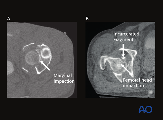
The CT scan is an important part of the radiographic protocol and can identify many aspects commonly missed on plain radiography. Examples include
- Marginal impaction as demonstrated in picture A
- Femoral head impaction as demonstrated in picture B
- Retained/incarcerated intraarticular fragments as demonstrated in B
- Non-displaced fractures (particularly transverse or posterior hemi-transverse fractures)
The CT scan can miss fractures lines, particularly those in the plane of the CT data acquisition. While the topic is heavily debated, the authors continue to obtain conventional orthogonal radiographs in conjunction with the CT scan to best characterize fractures of the acetabulum.
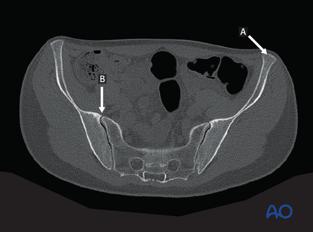
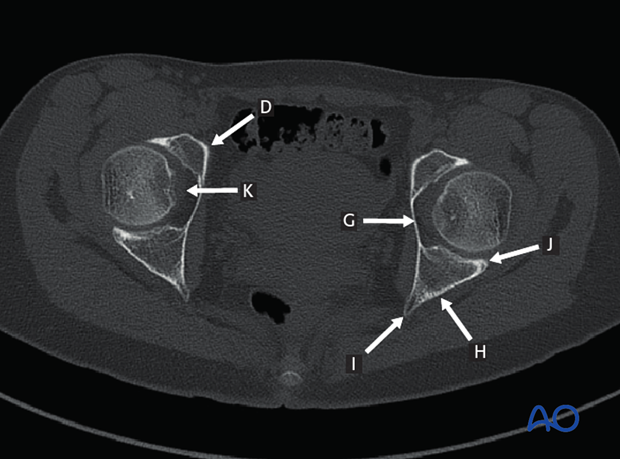
The CT scan of the intact acetabulum and pelvis should be understood completely.
A: Anterior superior iliac spine
B: SI joint
C: Sciatic notch
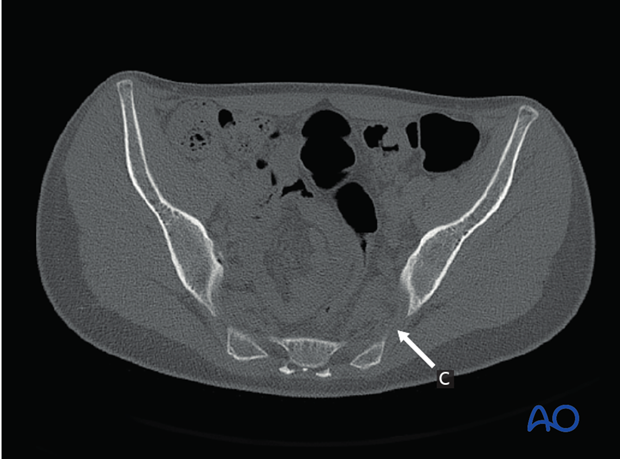
C: Sciatic notch.
D: Pelvic brim (source of iliopectineal line)
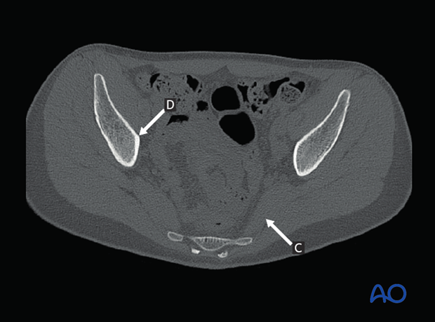
D: Pelvic brim (source of iliopectineal line)
E: Subchondral bone of acetabular roof
F: Anterior inferior iliac spine
G: Quadrilateral surface
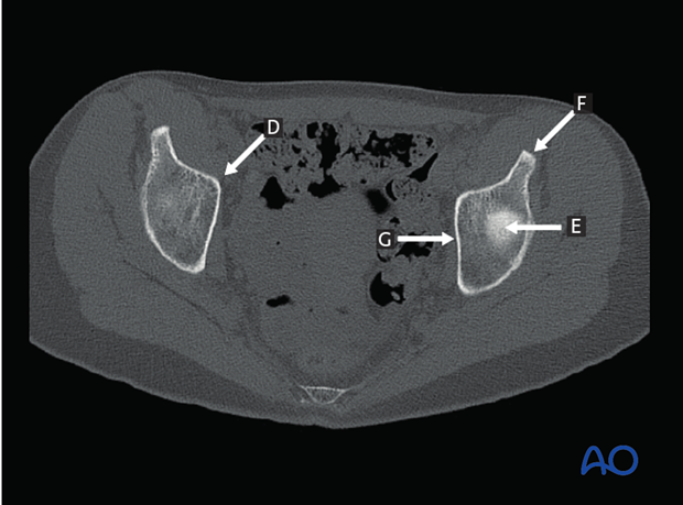
D: Pelvic brim (source of iliopectineal line)
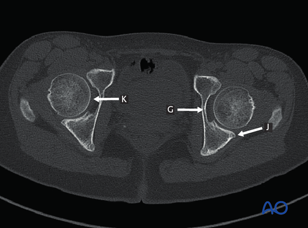
G: Quadrilateral surface
H: Retroacetabular surface
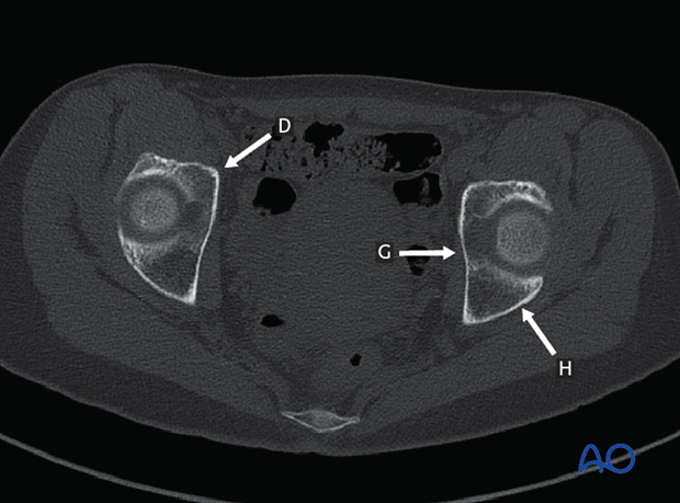
D: Pelvic brim (source of iliopectineal line)
G: Quadrilateral surface
H: Retroacetabular surface
I: Ischial spine
J: Posterior rim
K: Cotyloid fossa
G: Quadrilateral surface
J: Posterior rim
K: Cotyloid fossa
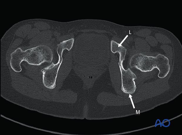
L: Pubic ramus
M: Ischial tuberosity
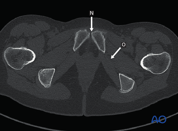
N: Pubic symphysis
O: Obturator foramen
Example 1
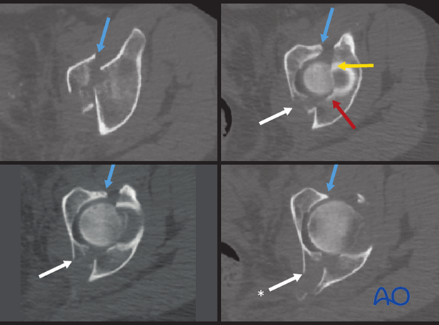
The CT scan confirms the assessment of the location and obliquity of this fracture. It is a single fracture in a single (sagittal) plane. The fracture orientation begins cranially anteriorly (blue) and descends the quadrilateral plate (white), then traverses the posterior border of bone through the ischial spine low (*). Thus, the obliquity of the fracture is as expected based on the oblique images.
The CT confirms violation of the acetabular roof (yellow). Also, an area of marginal impaction is identified (red). The CT scan confirms:
- Absence of an independent posterior wall fragment
- Integrity of the iliac segment
- Displacement of the distal (ischiopubic) segment, and
- Lack of a vertical stem or violation of the obturator foramen
Notes
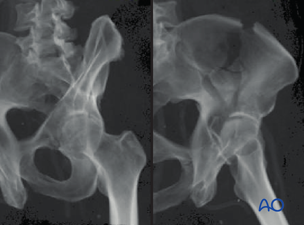
Volume rendering
Volume rendering is a set of techniques used to display 2D images from the 3D acquisition CT. In acetabular fracture surgery, many surgeons have explored the use of volume rendered images that mimic the plain images.
Some surgeons and hospitals prefer to use these images in place of the traditional oblique views.
The image quality is heavily dependent on the algorithm for the rendering, the software version, and the technique of the image generation. Poor quality volume rendered images should not act as a substitute for the conventional AP Pelvis and Judet views as important fracture morphological features can be missed.
3D surface rendering
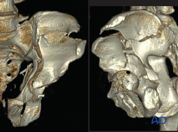
3D surface rendering images can augment the surgeon’s three dimensional understanding of the fracture.
Again, these images are dependent on the quality and quantity of the data acquisition of the CT scan, on the rendering algorithm and on the technique. The rendering protocols are improving worldwide and accessibility is becoming less of an issue.
Nevertheless, the authors continue to use these modalities in concert with the conventional orthogonal Judet views.
Example 2
The entire radiological inventory (AP pelvis, Obturator oblique, Iliac oblique, CT) confirm a transtectal transverse fracture, with an obliquity that is cranial anteriorly and very distal posteriorly.
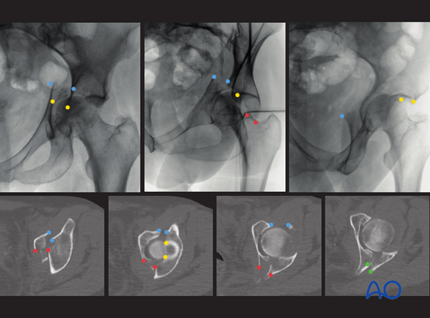
This fracture’s characteristics will be considered in detail in the section dedicated to elementary transverse fractures
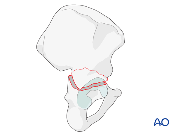
Practicing the exercise of interpreting the images from the conventional radiographs at axial CT scan and translating this information to a 3D model is invaluable in learning to understand the intricate morphological characteristics of each specific fracture.
