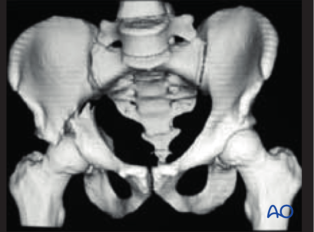
Characteristics of elemental fracture types
1. Posterior wall
Introduction
Elementary posterior wall fractures are the most common acetabular fractures and account for approximately 24% of acetabular fractures.
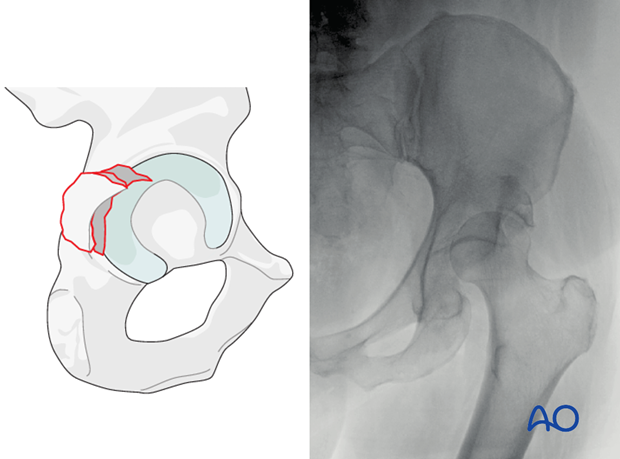
The posterior wall fractures involve the rim of the acetabulum, a portion of the retroacetabular surface, and a variable segment of the articular cartilage. The majority of the posterior column remains intact.
A posterior dislocation is associated in approximately 35% of reported cases.
Posterior wall fractures may occur with femoral head fractures.
Epidemiology
High-energy trauma is the primary cause in younger individuals and association with other fractures and pelvic ring disruptions are common. Fractures secondary to moderate or minimal trauma are increasingly of concern in those over 60 years because of osteoporotic changes.
Mechanism of injury
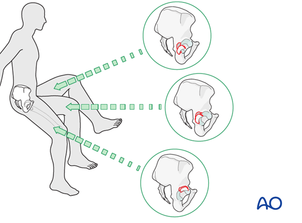
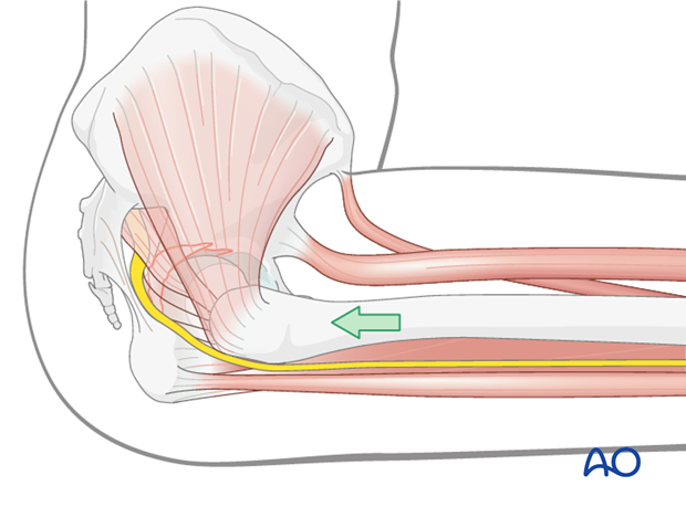
The posterior wall becomes most vulnerable when the hip is flexed and a force is directed along the axis of the femur. Thus, this injury is very commonly associated with a motor vehicle accident. The seated occupant’s flexed knee strikes the dashboard, for example, and the force is transmitted through the femoral head directly into the posterior wall, the exact location of which depends upon the position of the hip at the time of impact (as illustrated).
- If the hip is also in abduction, a posterior column posterior wall associated fracture may occur due to the more medially directed force.
- If the hip is also in adduction, a hip dislocation may occur.
- If the hip is flexed less than 90°, the superior region (roof) may become involved.
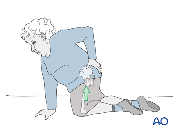
The worldwide increased incidence of fragility fractures of the acetabulum has resulted in a substantial number of posterior wall fractures and fracture dislocations secondary to minor trauma. Fragility fractures that result in a posterior wall fracture involve a fall onto the knee with the hip in flexion (in contrast to the more common geriatric fractures, the anterior column family, which occur secondary to a fall onto the greater trochanter).
Hip dislocation
When a posterior dislocation is associated with a posterior wall fracture, open reduction and internal fixation is virtually always indicated because the posterior wall defect renders the hip unstable.
Posterior dislocations also increase the risks of sciatic nerve injury and of avascular necrosis of the femoral head.
The more time that passes before the hip is relocated, the higher the risk of avascular necrosis.
Greater displacement of the dislocated hip also adversely affects the outcome.
Several characteristics render a posterior wall fracture dislocation irreducible:
- Incarcerated fragment
- Involvement of a substantial area of the roof, such that there is not a stable area to maintain the reduced femoral head beneath
- Soft tissue incarceration or “button-hole” of the femoral head through the capsular labral complex
In these cases, the surgeon risks femoral neck fracture with repeated, forceful attempts at reduction.
Comminution
If the posterior wall fragment is comminuted, reduction and fixation will be more difficult, and the prognosis is poorer.
Intraarticular fracture fragments
Incarcerated fragments produce incongruity and instability. If interposed between the joint surfaces, they cause arthritis. The presence of incarcerated fragments is thus an indication for acetabular surgery.
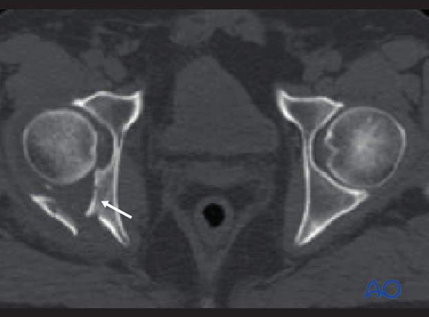
Marginal impaction
Crushing of the posterior acetabular surface (see arrow) creates a condition of incongruity and instability. Reduction requires elevation of the impacted articular surface and bone grafting of the resulting defect.
The reduced femoral head is a helpful template.
If a satisfactory and stable reduction can be achieved, the final outcome may be better. Otherwise, prognosis is poor. Unreduced impaction, which not only leaves an incongruent area of joint surface but also prevents proper reduction of adjacent displaced wall fragments, adversely affects prognosis.
Femoral head contusion and abrasion
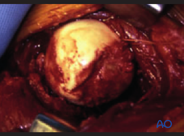
Femoral head contusions and abrasions are both extremely difficult to repair and increase the risk of posttraumatic arthritis.
Radiology
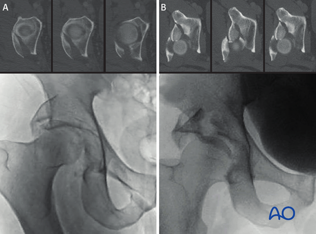
Fractures of the posterior wall involve a separation of a segment of the posterior articular surface from the remaining acetabulum. The fracture leaves the majority of the posterior column structure undisturbed. In the most typical form (60-70%) the fractures of the posterior wall are below the level of the roof (A); 25-30% of posterior wall fractures are considered posterosuperior fractures in which a portion of the roof is separated with the posterior wall fragments (B). Less commonly the fracture may be extended or transitional (as described below).
Regardless of form, the posterior wall fractures commonly involve and should be evaluated for:
- Multiple fragments
- Marginal impaction
- Incarcerated fragments
- Incomplete/nondisplaced transverse or posterior column fracture lines
- Quality of reduction (in cases of dislocation)
- Soft tissue injury (particularly the sciatic nerve)
X-ray imaging
The elementary posterior wall fracture involves only the articular surface. Therefore, the only radiological landmark on the AP pelvis disrupted is the posterior rim of the acetabulum. The other five landmarks (iliopectineal and ilioischial lines, radiological roof, anterior rim and tear drop) are not violated in the typical posterior wall fractures. In this example, the posterior wall fracture involves a posterior dislocation. The posterior rim is seen to be disrupted through the vacant acetabulum (as illustrated with the green dots).
In this example, the femoral head is seen to remain in a congruent relationship with the displaced portion of the posterior wall.
Note
Superior posterior wall variants may involve a portion of the radiological roof as shown in B above.
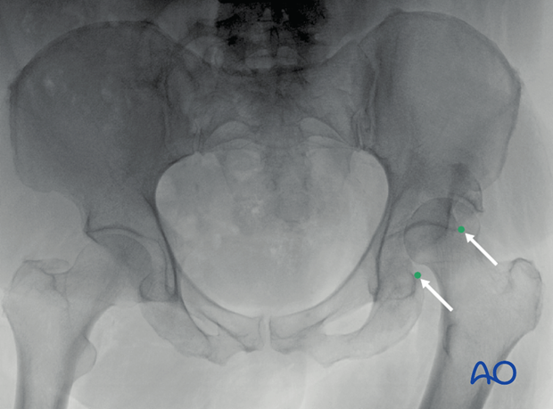
Reduction of hip makes the evaluation of the disruption of the posterior rim more difficult; however, a trained eye will identify the disruption of this primary radiological landmark.
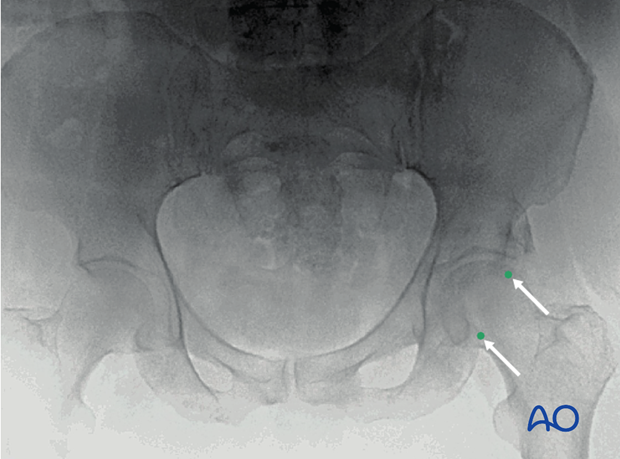
The obturator oblique radiograph is the best source of information of the size, character, and displacement of the posterior wall fragment because the femoral head and ilium are rotated to allow exposure of the retro- and supraacetabular surfaces. The integrity of the anterior column and the obturator foramen is evaluated on this view. If the hip has been reduced, the congruence of the reduction should be inspected on this view, and the involvement (or lack thereof) of the acetabular roof should be considered.
- In this example, a large piece involving a small portion of the radiological roof is seen. It extends cranially above the acetabulum. It appears as a large fragment; however, there may be a perforation of the cortex based on this image. The anterior column is intact, as is the obturator foramen. The femoral head is reduced and comparison to the other side would demonstrate near congruence of the reduction. Certainly, the CT scan will be inspected for incarcerated fragments but the reduction is appropriate at this stage.
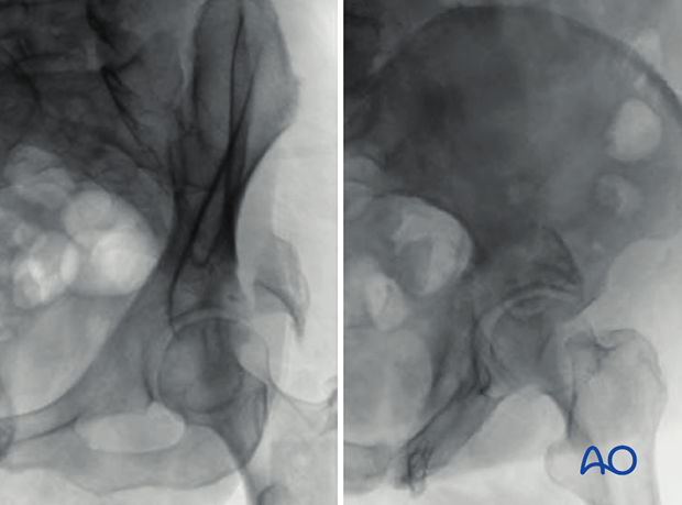
The iliac oblique radiograph demonstrates the integrity of the posterior column, the iliac wing, and the anterior border of the bone. The displaced posterior wall fragment(s) is superimposed on the ilium and thus difficult to see.
- In this example, the posterior column is seen to be intact as is the anterior rim.
CT imaging
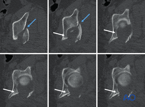
With posterior wall fractures, one should always evaluate the CT scan for:
- The size of the fragment and the region of the posterior wall involved. In this example, the fracture begins well cephalad to the acetabular roof and extends along the supraacetabular surface approximately half way to the anterior inferior iliac spine (blue arrow). Thus, this is a posterosuperior wall variant.
- The number of fragments associated with the fracture: In this example, the CT scan demonstrates that the posterior wall fragment indeed involves at least two cortical fragments.
- Marginal impaction: In this example, we see severe impaction along the entire posterior acetabular surface (white arrow).
- Incarcerated fragments: There are no incarcerated fragments in this example.
- The quality of the hip reduction: This requires comparison to the other side but appears congruent in this example.
- Incomplete or nondisplaced transverse fracture line (not present in this example).
- Incomplete or nondisplaced posterior column fracture line (not present in this example).
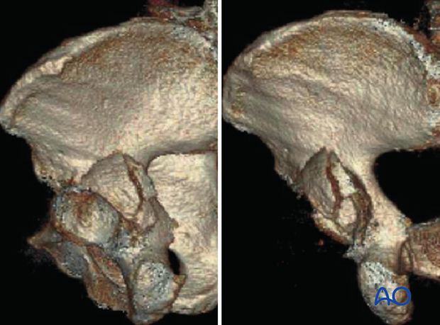
A 3D rendered CT scan can be obtained to confirm the assessment made with the plain radiographs and the axial CT scans.
2. Posterior column
Fracture characteristics
Isolated posterior column fractures are rare (2.4-3.2% of acetabular fractures [Matta, Letournel]) and are usually associated with a posterior dislocation of the hip. Even associated with a posterior wall fragment, the prevalence of this lesion is not so high (3.4% [Matta, Letournel]).
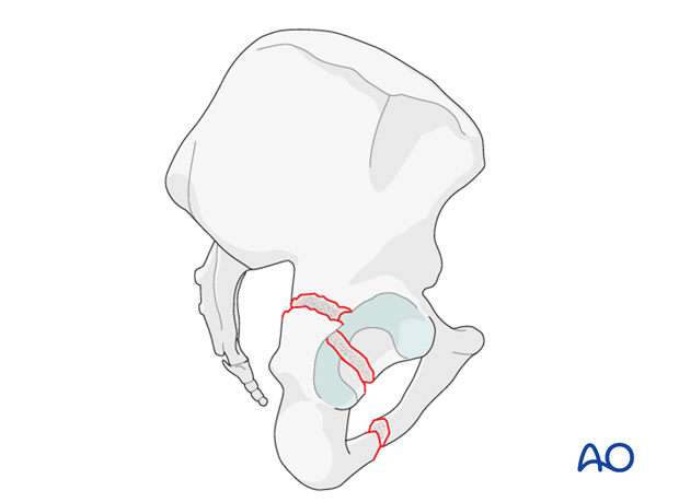
Posterior column fractures originate at the greater sciatic notch, pass through the roof or weight bearing dome and exit through the obturator ring. The result is a complete detachment of the posterior column.
The fracture is usually displaced posteriorly, medially, and in internal rotation, as the posterior column rotates about the ischial tuberosity.
As the femoral head is driven through the posterior column and fractures it, it tends to open up the posterior column like a swinging door, moving posteriorly into the pelvis.
The superior gluteal vessels and nerves can be at risk from displaced fragments.
Radiology
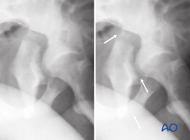
In x-ray images one will see the disruption in the ilioischial line and a break in the obturator ring.
On the axial CT the posterior column fracture line is from medial to lateral through the posterior column.
3. Anterior wall
Introduction
The anterior wall fracture is a rare fracture pattern. It is common in elderly patients and usually a result of low-energy injury. The bone involved is commonly osteoporotic.
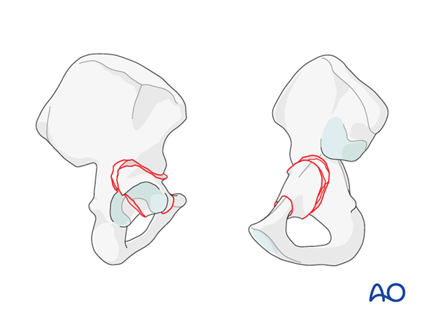
Fractures of the anterior wall are segmental fractures of the mid anterior column, are trapezoidal in shape and typically involve the anterior segment of the acetabulum.
Cranially, the fracture begins at the anterior border of the acetabulum, just below the anterior-inferior iliac spine. It crosses the articular surface, detaching the anterior articular facet and a variable portion of the acetabular fossa. Distally, the fracture traverses the superior margin of the obturator foramen transecting the pubic ramus.
On the internal surface, the fracture crosses the iliopectineal line 3-4 cm anterior to the SI joint. It descends along the quadrilateral surface below the pelvic brim, to the upper border of the obturator foramen and transecting the pubic ramus.
Dislocation
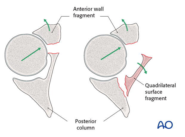
The femoral head is displaced anteriorly and medially, pushing the anterior wall fragment(s) in the same direction. The anterior wall fragment is typically rotated externally around its long axis.
In the vast majority of cases, a fragment of the quadrilateral surface fractures as the femoral head displaced medially. This plate of bone usually maintains a posterior hinge, but it may also be a free fragment. This fragment hinges around its posterior margin separating from the anterior wall fragment to allow subluxation of the femoral head.
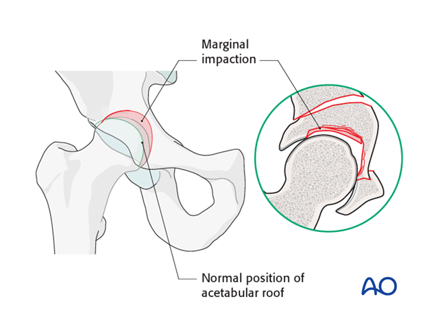
Marginal impaction
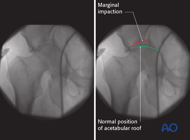
Marginal impaction of the acetabular roof articular cartilage is commonly associated with fractures of the anterior wall, particularly in elderly patients.
X-ray image of marginal impaction
Radiology
For an anterior wall fracture the iliac oblique x-ray image best demonstrates the separation of the anterior wall line.
On the axial CT the anterior wall fracture line runs obliquely at the level of the joint.
4. Anterior column
Definition
Anterior column fractures separate a segment of anterior acetabulum from the rest of the innominate bone. The fracture starts from the middle of the ischiopubic ramus below, and then passes through the anterior acetabulum. The proximal extension of this fracture passes variably through the innominate bone, at different levels above the acetabulum, as far upwards as the middle third of the iliac crest.
Anterior column fractures are described by the level of their proximal extent. They may also be segmental.
These fractures are commonly associated with a fracture along the pelvic brim and medialization of the quadrilateral surface.
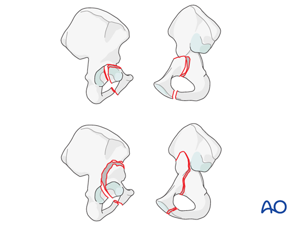
Displacement of the femoral head
Displacement of the anterior column fragment always is the result of anterior femoral head displacement. The head of the femur pushes the fragments anteriorly and medially, and remains displaced, impacted into the articular surface.
The head is easily visible in the fracture gap, particularly if seen from the modified Stoppa approach. The pelvic brim fragment is rotated externally around its long axis. The quadrilateral plate is rotated internally.
Femoral head damage
The femoral head may show areas of contusion, abrasion, or indentation. These conditions cannot be reversed and worsen the prognosis.

Intraarticular fracture fragments and marginal impaction
Part of the internal portion of the roof may appear isolated or be impacted into the underlying cancellous bone. Intraarticular fracture fragments and marginal impaction create a condition of incongruity and instability.

X-ray imaging
On this plain film one can see the femoral head remains congruent with the displaced anterior column (red). There is an area of marginal impaction (green) which has been impacted into the intact posterior column. The intact portion of the acetabulum remains in its native location laterally (white).
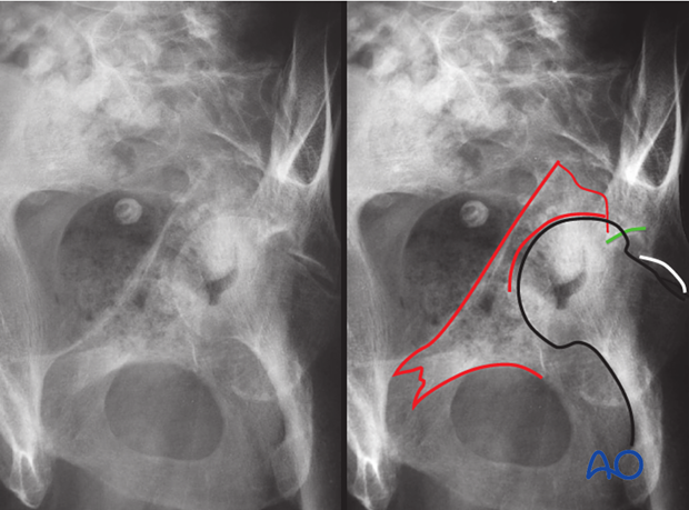
CT imaging
The CT scan demonstrates that the femoral head remains congruent with the anterior wall fragment. The quadrilateral surface has hinged around its posterior margin. The area of marginal impaction is seen to be impacted into the posterior column. Importantly, the intact portion of the acetabular roof is seen to cause an impaction injury to the femoral head.
The lack of a fracture line disrupting the retroacetabular surface qualifies this fracture as an elementary anterior column fracture. If there was violation of the retroacetabular surface, the fracture would be classified as an anterior column and posterior hemitransverse fracture.
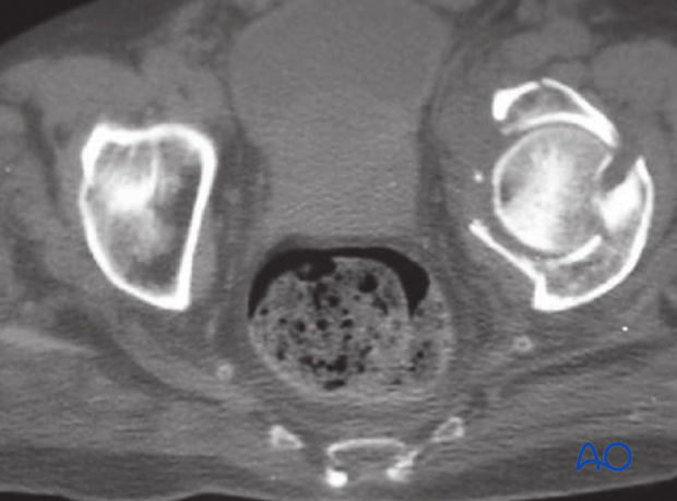
5. Transverse
Definition
Transverse acetabular fractures involve a single fracture line which crosses the acetabulum through both posterior and anterior columns. Such fractures divide the acetabulum into an upper portion (ilium with the roof), and a lower portion (ischium and pubis).
The transverse fracture can cross the columns at different levels, and is variable in its inclination in sagittal and coronal planes. Occasionally, comminution is present.
The lower portion of a transverse fracture and femoral head are typically displaced medially, with more or less rotational malalignment. It is usually more displaced posteriorly, but occasionally the greater displacement is anterior.
If the fracture line involves the weight bearing superior dome, a perfect reduction is required for a good result. However, transverse fractures may be difficult to reduce. Through the Kocher-Langenbeck approach, the surgeon has direct access only to the posterior end of the fracture.
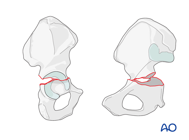
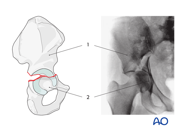
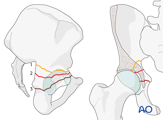
Such fractures divide the acetabulum into an upper segment (1, ilium with the roof), and a lower segment (2, ischiopubic).
It is the lower, ischiopubic segment that displaces, while the iliac segment remains in situ with a normal relationship at the SI-joint. The typical ischiopubic displacement involves an inward rotation (medialization) around the vertical axis, hinging at the symphysis.
Transverse fractures may run at different levels through the acetabulum.
Transverse fractures are classified according to their relationship to the roof of the acetabulum:
- Transtectal - dividing the roof of the acetabulum (1, yellow fracture line)
- Juxtatectal - dividing the fossa acetabuli and the roof (2, red fracture line)
- Infratectal - cutting the fossa acetabuli in the middle (3, brown fracture line)
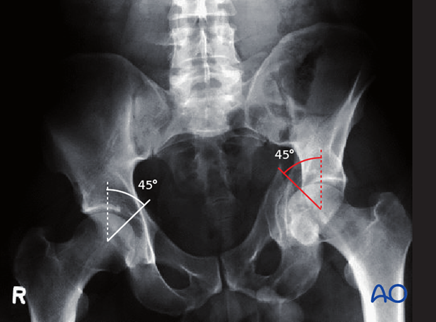
The roof arc angle can be used to further describe the location of the fracture line to guide the likely requirement for surgery.
Note
The transtectal transverse fracture has the worst prognosis because it involves the most critical part of the weight bearing dome.
Roof arc angle
The roof arc angle is determined in the following way:
- Three radiographic views are needed (AP view, iliac and obturator oblique views).
- Begin by drawing a vertical line through the center of the acetabulum (or the center of the reduced femoral head) on each radiographic view.
- Now add a second line, intersecting the first at a 45° angle at the level of the femoral head’s center.
If the second line is outside of the fracture zone in each of the three radiographic views, the fracture is considered stable.
Our example shows a fractured weight bearing dome of the left acetabulum, with the second line well within the fracture zone, indicating instability.
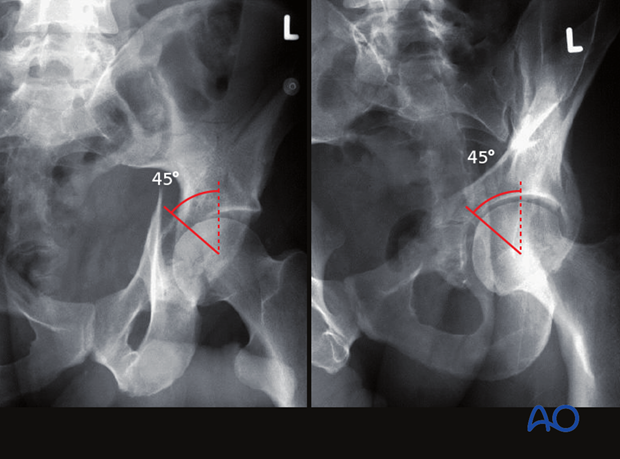
These images show corresponding measurement of the roof arc on iliac oblique (left) and obturator oblique (right) views.
X-ray imaging
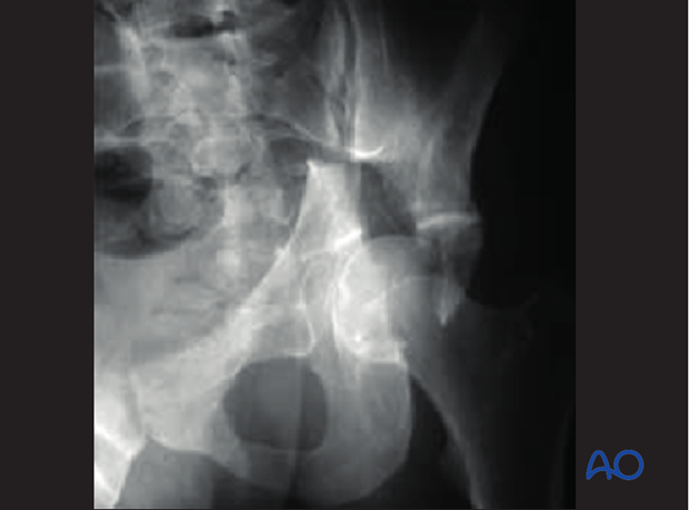
On the AP view, all the vertical lines of the acetabulum are disrupted by the fracture: the anterior and posterior borders, and the iliopectineal and the ilioischial lines, as well as the roof.
With transverse fractures involving the medial roof and juxtatectal area, the femoral head may be dislocated into the pelvis, or medially displaced with articular incongruity.
The inferior acetabular segment is displaced medially and rotated internally around an anteroposterior axis, hinging on the pubic symphysis.
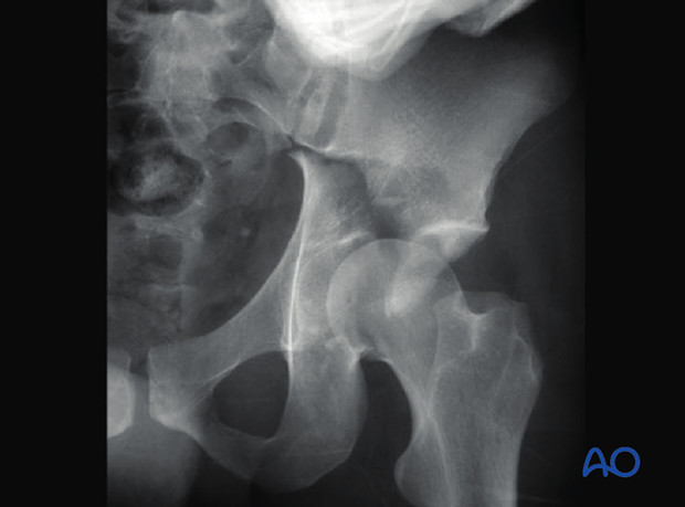
This image demonstrates the transtectal fracture on the iliac oblique view.
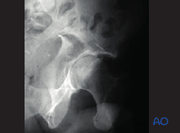
The obturator oblique view demonstrates the location of the fracture relative to the acetabular roof and confirms the integrity of the obturator foramen, and thus excludes the possibility of a T-type fracture.
Notice the medial dislocation of the femoral head along with the lower portion of the acetabulum or ischiopubic segment.
Also, note that the obturator oblique view demonstrates the posterior acetabular wall. It is intact on this image. However, posterior wall fractures may be associated with transverse fractures.
CT imaging
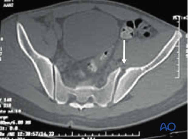
In transverse fractures, the ilium can be slightly externally rotated, with medial dislocation of the femoral head. In this example (CT image on the level of the lower SI joint), anterior SI ligament tearing is present, with SI joint widening (arrow).
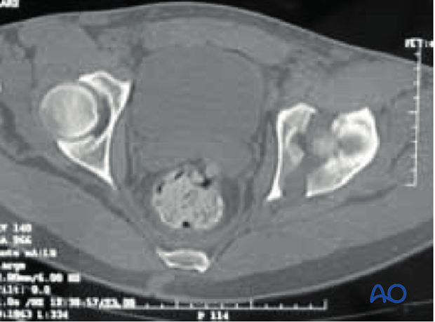
On the level of the acetabular roof, the transverse fracture can be seen crossing anterior and posterior columns, dividing the acetabulum into lateral-superior and medial-inferior portions.
The anteroposterior orientation on CT scan is a diagnostic feature of transverse fractures.
This 3D CT reconstruction image demonstrates medial displacement of femoral head and medial displacement with internal rotation of the distal acetabular segment, as well as slight opening of the ipsilateral SI joint.
