ORIF through extended iliofemoral approach
1. General considerations
Sequence of the treatment
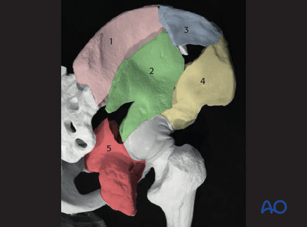
The sequence of reduction of the typical associated both column fracture reduced via the extended iliofemoral approach begins with the reconstruction of the ilium.
- Reduction of the SI joint dislocation component when applicable (1-2)
- Iliac crest (3) to reconstructed intact ilium (1-2)
- Anterior column (4) to reconstructed ilium (1-2-3)
- Posterior column (5) to retroacetabular surface (2)
The reduction sequence will vary depending on the fracture morphology to some extent. For example, if there is a posterior wall component, it may require reduction prior to the reduction of the posterior column (5).
Additionally, there may be cases in which the posterior column reconstruction to the intact ilium precedes the anterior column reduction.
The associated both column fractures operated by the extended iliofemoral approach are the most complex and difficult fracture patterns. Though the sequence may vary from case to case, it is critical to have a well thought out surgical strategy prior to surgery.
Planning/templating
Preoperative templating is essential for understanding the complexity of an acetabular fracture.
When using implants on the innominate bone, it is important to know the best starting points for obtaining optimal screw anchorage (see General stabilization principles and screw directions).
Patient positioning
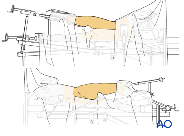
The patient is positioned lateral on a flat top radiolucent table or a fracture table. Typically, a distal femoral traction pin is applied to allow the application of traction and also to stabilize the extremity. The knee is flexed 45°.
Capsulotomy
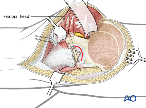
The extended iliofemoral approach allows the hip joint to be opened with a capsulotomy, just distal to the labrum. Lateral and/or distal retraction of the femoral head makes it easier to see into the joint.
It also allows the fracture to be seen well on the external surfaces of both the anterior and the posterior columns.
The internal surface of the pelvis can be palpated with a finger in the greater sciatic notch, for further assessment of the reduction.
Indirect visualization
Unusually for a significant joint, articular reduction of acetabular fractures is indirect. The articular surface of the hip joint is not seen directly. Reduction must be assessed by the appearance of the extraarticular fracture lines and intraoperative fluoroscopic assessment. Some fracture lines are palpated manually but not seen directly such as transverse fracture lines on the quadrilateral plate.
Marginal impaction in the posterior wall fracture can be seen directly prior to final closure of the cortical fragment.
Quality of reduction
Posttraumatic arthrosis is directly related to the quality of reduction - the better the reduction, the greater the chance of a good or excellent result.
Teaching video
AO teaching video: Both column fracture through the extended iliofemoral approach
2. Joint distraction
Femoral head subluxation
If the femoral head has been reduced in a closed fashion, subluxation of the joint to clear bony fragments would be required.
If the head is still dislocated then the joint can be cleaned before reduction.
Application of traction
Lateral traction is applied to extract the displaced femoral head from the pelvis. This also allows some indirect reduction of the medially displaced fracture fragments by ligamentotaxis. Lateral traction is typically achieved with a Schanz screw applied through the lateral greater trochanter region of the femur. Because the patient is in the lateral position, this typically requires a manual force.
Longitudinal traction is also required to correct proximal displacement. Longitudinal traction can be obtained with a fracture table applied to a distal femoral traction pin (A), a femoral distractor (B), or with manual traction on the affected extremity.
In some surgeon’s experience, the use of a traction table post or other traction frame is helpful during this operation.
Note
Excessive longitudinal or lateral traction may result in the locking of the fracture, inhibiting further reduction maneuvers. Furthermore, the position of the femur may affect reduction and visualization. Commonly, one must reposition the involved extremity and change the vector of traction several times during the course of the operation. Thus, it must be easy to adjust the traction and the limb position intraoperatively.

Teaching video
AO teaching video: Use of the distractor on the pelvis
3. Case example
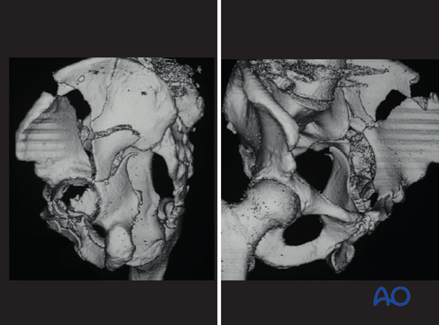
We will take, as an example, this case of an associated both column fracture, with a fracture dislocation of the SI joint complicating the posterior column moiety. This fragment (2) is not accessible for manipulation, reduction, or fixation through the ilioinguinal exposure. This is a common fracture variant requiring the extended iliofemoral approach.
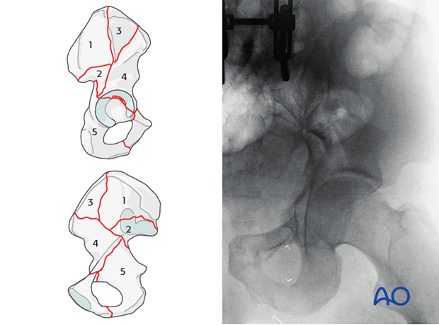
Evaluation of the Judet obliques and the CT scans confirms the various fragments of this complicated fracture requiring attention. While the iliac crest and anterior column reconstruction can be accomplished via the ilioinguinal exposure, the reduction of the posterior column and the intercalary fragment of the SI joint are not accessible from the internal pelvis.
4. SI joint fracture dislocation: reduction and fixation
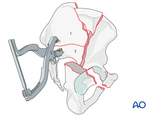
After the exposure has been completed, the first step in the reconstruction involves the reduction and fixation of the SI joint fracture dislocation.
The intact segment of the ilium (1 - that which has maintained its normal relationship with the sacrum) acts as the cornerstone for the reduction. The intercalary fragment of the SI joint/sciatic buttress (2) is reduced to the intact iliac segment. This is commonly accomplished with a two-screw technique utilizing a Farabeuf or Jungbluth clamp. The read of the fracture reduction on the outer ilium guides the reduction. Reaching though the sciatic notch, one can palpate the internal reduction of the SI joint, which will occur as this fragment is reduced.
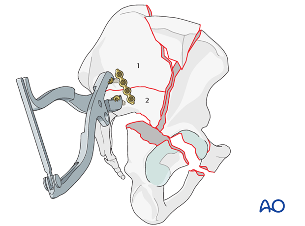
Once the reduction has been obtained, a reconstruction plate is applied to maintain the reduction. Care must be taken to avoid placing these screws into the SI joint. At times, based on the patient’s bone quality, one may have to place these screws into the SI body.
Additional screws can be placed between the internal and external tables to augment this fixation. This should be done only after careful planning as space in the iliac corridor must be left for the iliac screws required for the anterior column stabilization.
5. Iliac crest: reduction and fixation
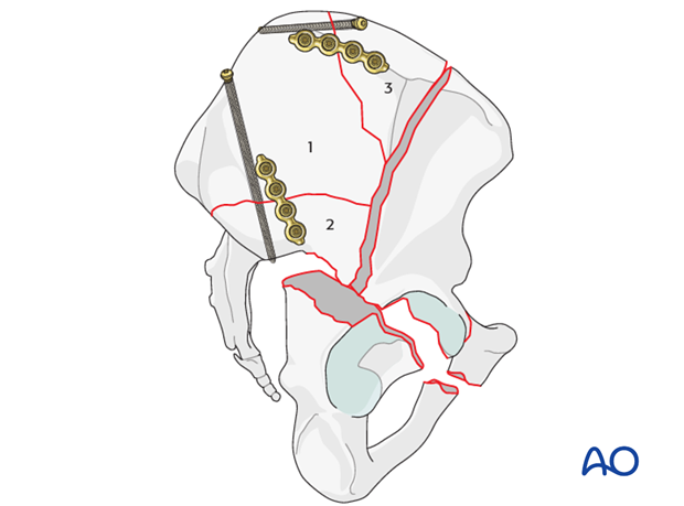
Very commonly the iliac crest involvement is complex and requires reduction and fixation of accessory fragments (3).
Apply a pointed reduction forceps onto the iliac crest as a reduction aid and for temporarily fixation.
Alternatively, apply a Farabeuf or Jungbluth clamp over screws placed into the crest on each side of the fracture.
Care must be taken to avoid wedge opening of the fracture line on the internal aspect of the crest.
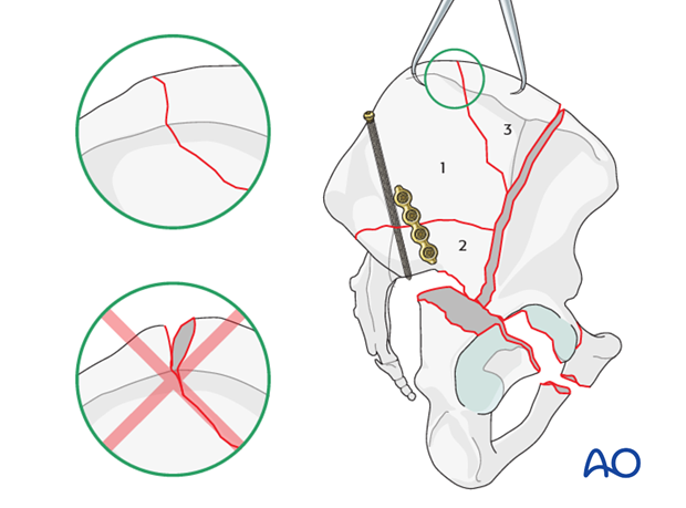
Fixation of the intercalary iliac crest fragments is accomplished with interfragmentary 3.5 mm lag screws, 3.5 mm low profile pelvic reconstruction plates, or with a combination of these implants.
Note
The area on the pelvis for fixation of the anterior column needs to be considered. Room for implants securing the anterior column to the newly reconstructed ilium needs to be prioritized. When choosing fixation for the intercalary fragments of the iliac crest, be cautious not to use all available corridor space.
Occasionally, short reconstruction plates or screws may require removal or replacement with longer implants to accommodate the anterior column fixation.
6. Anterior column: reduction and fixation
Derotation, alignment and reduction of the anterior column
The reduction of the anterior column (4) takes multiple adjuncts. Hip flexion can relax the pull of the rectus and greatly assist at this stage in the surgery.
The anterior column should be derotated with help of a Farabeuf clamp and/or a Schanz screw applied to the anterior innominate bone.
The iliac crest portion of the fracture can be manipulated and the reduction maintained with the help of pointed reduction forceps.
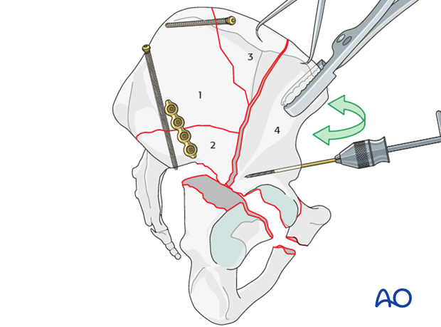
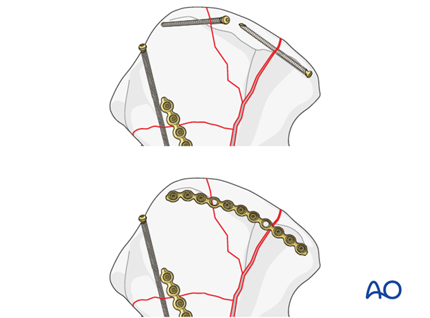
Once the cranial extend of the anterior column reduction has been achieved, the iliac wing can be fixed with intramedullary screw fixation or pelvic reconstruction plates or a combination of both.
If, however, the reduction remains imperfect, adjustment of the deeper portions of the anterior column is required before definitive fixation.
After provisional or even definitive fixation of the cranial anterior column, it is possible to adjust the distal anterior column reduction (green arrows). Thus, final alignment and periarticular fixation are the essential final steps of anterior column reduction and fixation.
Multiple clamps can aid in this final reduction. A point to point Weber clamp applied to the anterior column at the interspinous notch to the newly reconstructed ilium can achieve excellent compression and maintain provisional reduction of the anterior column. Commonly, one must augment this reduction maneuver with a Schanz screw or additional adjuncts.
Alternatively, a two-screw technique utilizing a Farabeuf or Jungbluth clamp can effectively derotate the anterior column while compressing its fracture line to the reconstructed ilium.
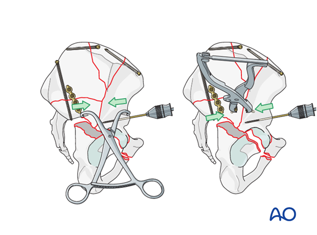
Depending on the fracture obliquity, an asymmetric clamp may be utilized, with the internal point applied within the pelvis and the external point applied on the outer surface of the anterior column. This clamp greatly facilitates the derotation of the anterior column when there is sufficient obliquity to the fracture line.
The internal point is applied through the interspinous notch with a small additional exposure of the internal surface of the ilium.
An additional clamp (Jungbluth, pointed reduction forceps) or Schanz screw in the anterior column may be required to complete this reduction.
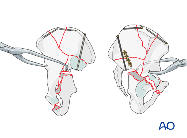
Fixation of the anterior column
Multiple points of fixation would be necessary to sufficiently stabilize the reduction of the anterior column. Starting peripherally, augmentation of the iliac crest reconstruction may require a long reconstruction plate. While intramedullary screws are preferred, complexities of the iliac crest fracture may threaten the quality of this fixation. The iliac crest plate has a disadvantage of being prominent on the outer ilium and dependent on very short screws. However, it may be necessary.
Fixation of the deep anterior column relies on both intramedullary screw fixation and reconstruction plate application. One or two screws applied along the iliac corridor are the mainstays of this fixation. Screws placed from the AIIS to the posterior ilium are required. These screws typically are greater than 150 mm in length and commonly can be 4.5 mm screws. They can be placed from anteriorly to posteriorly or vice versa.
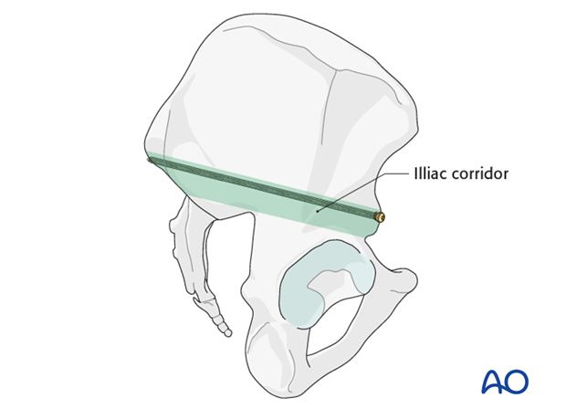
Anterior column screw
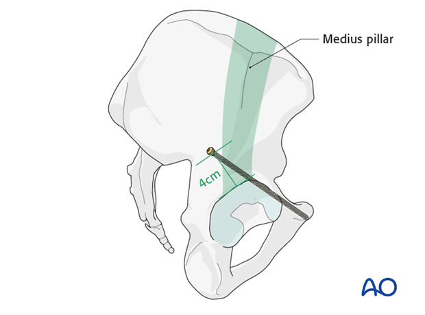
Further fixation of the anterior column to the distal part of the pubis can also be achieved through lag screws that run from the medius gluteal pillar to the pubis (depending on fracture configuration and necessity).
One or two 3.5 mm lag screws are used to stabilize the inferior part of the anterior column fracture.
The starting point for the anterior column screw applied through the extended iliofemoral exposure is different than that applied through the Kocher-Langenbeck. The starting point begins 3-4 cm above the lateral rim of the acetabulum, just posterior to the gluteus medius pillar.
The trajectory follows the axis of the anterior column which is easily seen along the pectineal eminence through this exposure. For the correct direction, use the index finger along the pubic ramus and try to target it with the drill bit.
The correct inclination and direction of the screw must be confirmed with fluoroscopic imaging (modified Judet views).
Take great care not to penetrate the hip joint.
Use an oscillating drill and progress slowly. Remember that the external iliac vessels lie immediately adjacent to the superior pubic ramus.
Reconstruction plate stabilization of the anterior column
It may be necessary to supplement the fixation with the application of a reconstruction plate, undercontoured to support the rotational reduction of the anterior column. However, it is commonly difficult to apply this definitive plate prior to the reduction of the posterior column fracture. Furthermore, if there is a posterior wall component, it will need to be reduced and stabilized with the same plate. Therefore, plate fixation of the anterior column commonly occurs after reduction and provisional fixation of the posterior fragments.
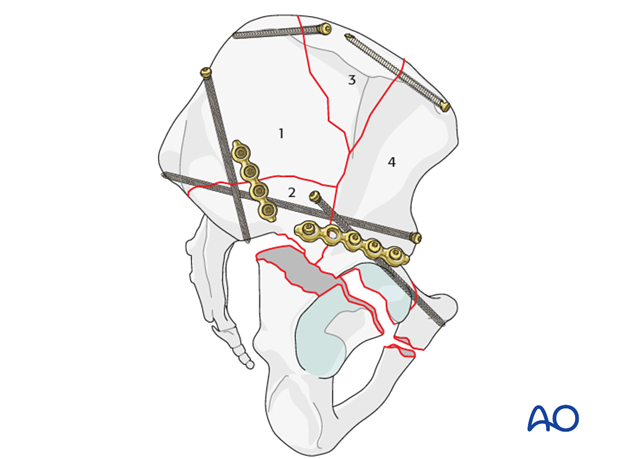
7. Posterior column: reduction
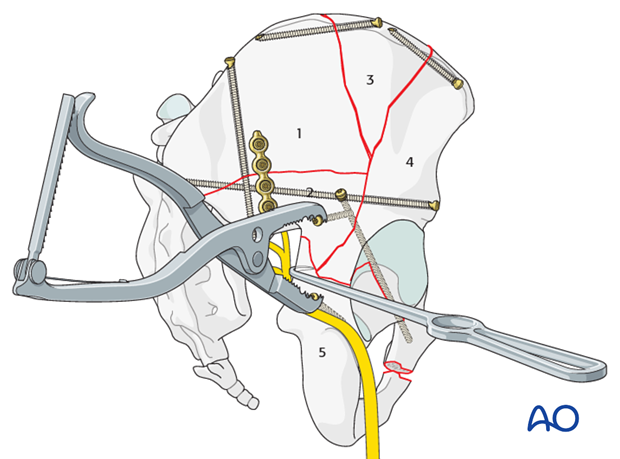
Multiple reduction adjuncts will be required to successfully reduce the posterior column (5). Furthermore, lateral traction is critical at this stage to allow manipulating of the posterior column around the femoral head.
In this case, a two-screw reduction technique manipulating the posterior column (5) to the reconstructed ilium and anterior column (1-4) is accomplished using a Farabeuf clamp. Another possibility is to use a pointed reduction forceps across the fracture, or a Jungbluth clamp.
A bone hook has been applied through the notch to adjust the rotational reduction. While the hook applies the force from medial to lateral, the clamp will close the fracture line and derotate the posterior column fragment. Take great care not to damage the sciatic nerve. A Schanz screw in the ischial tuberosity is also useful as a joystick.
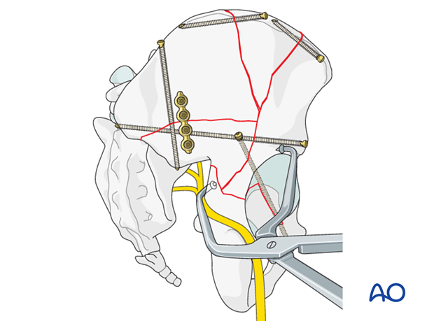
An asymmetrical angled clamp can also be used to translate and derotate the posterior column. The internal point is placed carefully through the sciatic notch and applied to the quadrilateral surface of the displaced posterior column. The external point is applied to the retroacetabular surface of the stable fragment of the anterior column.
8. Posterior column: fixation
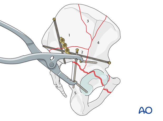
Lag screw fixation
Fix the reduced posterior column with one or more interfragmentary screws.
The screw orientation will vary depending on the obliquity of the fracture line.
The starting point for the insertion of this screw is located in the reconstructed ilium (1-2).
This screw should not interfere with the desired position of a posterior column plate if this is planned for the final construct.
Alternatively, a retrograde lag screw, also called "butt screw", may be used from the ischial tuberosity.
Application of a neutralization plate
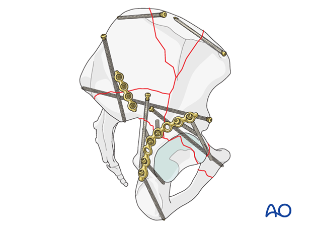
In this case, a posterior column plate has been contoured to reinforce the rotational reduction accomplished. The most distal hole of the plate should rest on the ischium. Aim the first screw through this hole, so that it travels through the ischium for a length of 40-60 mm.
Proximally, this plate utilizes the sciatic buttress and excellent purchase can be expected.
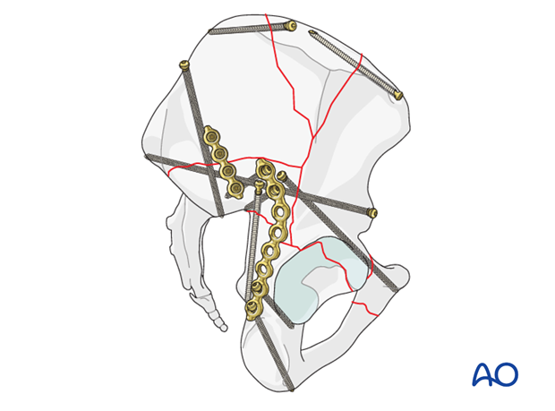
Another option, especially when a posterior wall fragment is present, is to apply a plate that starts from the ischial tuberosity and ends with at least two holes in the anterior column.
Note
When the plate is positioned close to the acetabular rim, the risk of joint penetration is increased.
9. Posterior wall: reduction and fixation
Reduction
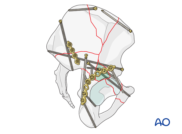
Commonly, the associated both column fracture requiring reduction via the extended iliofemoral approach has an associated posterior wall fracture. The morphology and location of the posterior wall components are highly variable as shown in these three examples. Very commonly, this fragment is located in between the fracture lines of the anterior and posterior column fragments.
It is important that the fragment remains anatomically positioned between posterior and anterior columns, without gaps or step offs of the joint surface.
The sequence of reduction of the posterior wall fragments will vary based on its morphology and spacial relationship to the columns. In cases of very cranial wall fragments (A), the posterior wall may be reduced to the anterior column prior to reduction and fixation of the posterior column. In other cases, if the wall fragment is distal for example (B), the posterior wall may be reduced after the posterior and anterior columns have been reconstructed.

Fixation
Regardless of location and morphology, the posterior wall fragment will be stabilized with independent lag screws and buttressed with plate fixation.
Most commonly, the plate will span the anterior and posterior columns as well. However, commonly, more than one plate may be necessary.
10. Final radiographic assessment
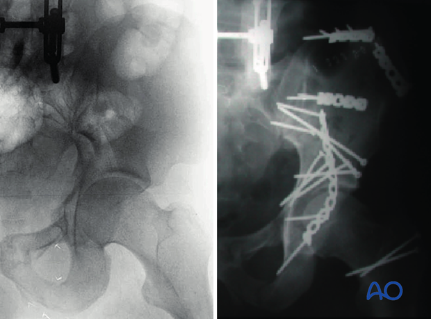
Careful evaluation of the postoperative radiograph demonstrates:
- Reduction and fixation of the SI joint fracture dislocation. The intercalary fragment of the sciatic buttress is reduced and fixed to reconstruct the intact ilium.
- The iliac crest has been reduced and fixed to the intact ilium by plate osteosynthesis.
- The anterior column fragment has been fixed to the reconstructed intact ilium with two 160 mm anterior to posterior screws in the iliac corridor.
- The posterior column has been reduced to the anterior column with anatomic restoration of the radiological roof. A lag screw has been placed across this fracture line from anterior to posterior.
- The posterior column reduction has been stabilized with independent posterior column interfragmentary screws.
- The posterior column plate has been applied. Note the position of the distal screw down the ischium.
- This surgeon utilized a trochanteric osteotomy option to perform the extended iliofemoral exposure. Two screws stabilize the trochanteric fragment.
- All screws are extraarticular (though this evaluation requires the obturator and the iliac oblique views).
11. Postoperative care
During the first 24-48 hours, antibiotics are administered intravenously, according to hospital prophylaxis protocol. In order to avoid heterotopic ossification in high-risk patients, the use of indomethacin or single low dose radiation should be considered. Every patient needs DVT treatment. There is no universal protocol, but 6 weeks of anticoagulation is a common strategy.
Wound drains are rarely used. Local protocols should be followed if used, aiming to remove the drain as soon as possible and balancing output with infection risk.
Specialized therapy input is essential.
Following the extended iliofemoral approach, the patient’s leg may be positioned in abduction in order to reduce tension on the muscles of the reconstructed area.
Follow up
X-rays are taken for immediate postoperative control, and at 8 weeks prior to full weight bearing.
Postoperative CT scans are used routinely in some units, and only obtained if there are concerns regarding the quality of reduction or intraarticular hardware in others.
With satisfactory healing, sutures are removed around 10-14 days after surgery.
Mobilization
Early mobilization should be stressed and patients encouraged to sit up within the first 24-48 hours following surgery.
Mobilization touch weight bearing for 8 weeks is advised.
Weight bearing
The patient should remain on crutches touch weight bearing (up to 20 kg) for 8 weeks. This is preferable to complete non-weight bearing because forces across the hip joint are higher when the leg is held off the floor. Weight bearing can be progressively increased to full weight after 8 weeks.
With osteoporotic bone or comminuted fractures, delay until 12 weeks may be considered.
Implant removal
Generally, implants are left in situ indefinitely. For acute infections with stable fixation, implants should usually be retained until the fracture is healed. Typically, by then a treated acute infection has become quiescent. Should it recur, hardware removal may help prevent further recurrences. Remember that a recurrent infection may involve the hip joint, which must be assessed in such patients with arthrocentesis. For patients with a history of wound infection who become candidates for total hip replacement, a two-stage reconstruction may be appropriate.
Sciatic nerve palsy
Posterior hip dislocation associated with posterior wall, posterior column, transverse, and T-shaped fractures can be associated with sciatic nerve palsy. At the time of surgical exploration, it is very rare to find a completely disrupted nerve and there are no treatment options beyond fracture reduction, hip stabilization and hemostasis. Neurologic recovery may take up to 2 years. Peroneal division involvement is more common than tibial. Sensory recovery precedes motor recovery and it is not unusual to see clinical improvement in the setting of grossly abnormal electrodiagnostic findings.


