Anterior column and posterior hemitransverse
Definition
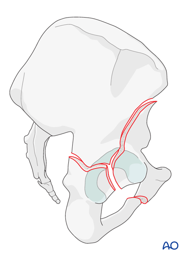
In this group of acetabular fractures, an anterior column or wall fracture is associated with a posterior hemitransverse fracture.
In addition, the femoral head is usually medially subluxed. Variations in this fracture pattern are based on whether the anterior column fracture exits at the iliac crest or inferior to the anterior inferior iliac spine.
The posterior column component varies on the basis of level of involvement in the greater sciatic notch.
The posterior column fracture morphology usually follows an oblique sagittal orientation unlike the posterior column involvement in a T-type fracture.
Further details on anterior column and posterior hemitransverse fractures are provided in section Characteristics of associated fracture types.

Classification of anterior column and posterior hemitransverse fractures
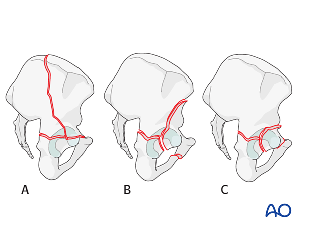
The anterior column and posterior hemitransverse classification is based on the cranial exit of the fracture in the ilium, with high fractures (A) exiting above the iliac crest, intermediate fractures crossing between the spines (B), and low or very low fractures exit below the anterior inferior iliac spine. Also, the anterior portion may be an anterior wall fragment (C).

Radiology
A summary of diagnosing the fracture classification based on x-ray and CT images is presented in section Patient assessment.
The section Radiology of the intact acetabulum provides explanation of the radiologic landmarks.
The section Characteristics of associated fracture types provides further information on the radiology of anterior column and posterior hemitransverse fractures.
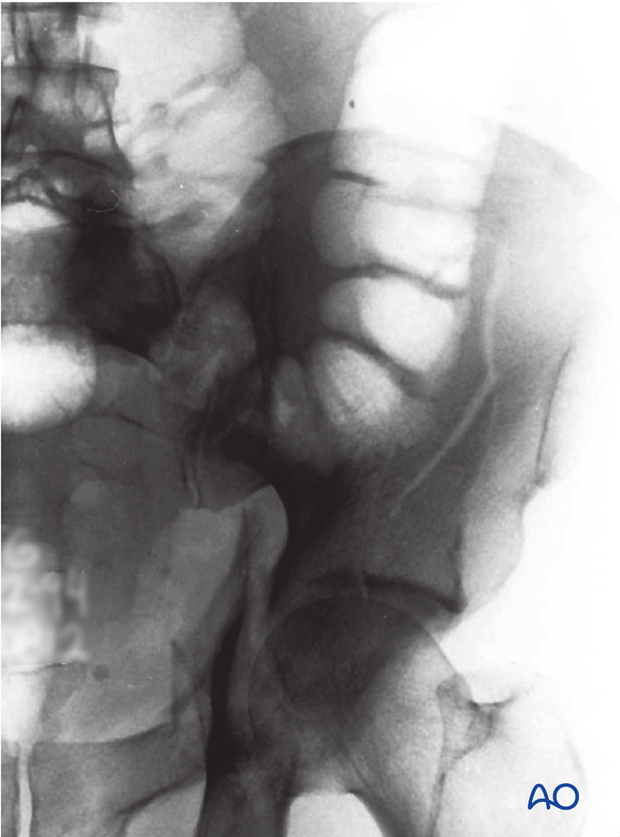
X-ray image of anterior column and posterior hemitransverse fracture, courtesy of TA Ferguson


