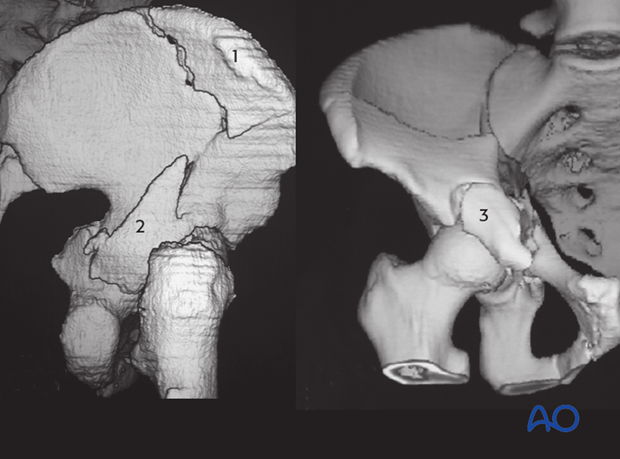
Characteristics of associated fracture types
1. Posterior column and wall
Definition
These fractures are a combination of the two elemental fracture patterns: posterior column and posterior wall. The posterior column component is often nondisplaced, and the wall fracture is the more obvious component. The prevalence of this fracture is low (3.4% [Matta, Letournel]).
The posterior wall fractures involve the rim of the acetabulum, a portion of the retroacetabular surface, and a variable segment of the articular cartilage.
Posterior column fractures originate at the greater sciatic notch, pass through the roof or weight bearing dome and exit through the obturator ring. The result is a complete detachment of the posterior column, in this case associated with fractures of the posterior wall. The fracture is usually displaced posteriorly, medially, and in internal rotation, as the posterior column rotates about the ischial tuberosity. As the femoral head is driven through the posterior column and fractures it, it tends to open up the posterior column like a swinging door, moving posteriorly into the pelvis.
Posterior column and wall fractures may be associated with femoral head fractures.
The superior gluteal vessels and nerves can be at risk from displaced fragments.
Epidemiology
High-energy trauma is the primary cause in younger individuals and association with other fractures and pelvic ring disruptions are common. Fractures secondary to moderate or minimal trauma are increasingly of concern in those over 60 years because of osteoporotic changes.
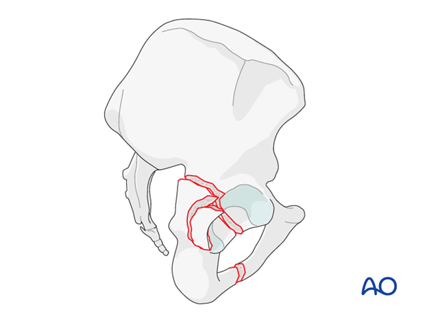
Posterior dislocation
When a posterior dislocation of the femoral head is associated with a posterior column and wall fracture, open reduction and internal fixation is virtually always indicated because the posterior wall defect renders the hip unstable.
Posterior dislocations also increase the risks of sciatic nerve injury and of avascular necrosis of the femoral head.
Any dislocation should have been reduced as soon as possible after the patient’s arrival in hospital. This reduction should be maintained, with traction if necessary, until open reduction internal fixation.
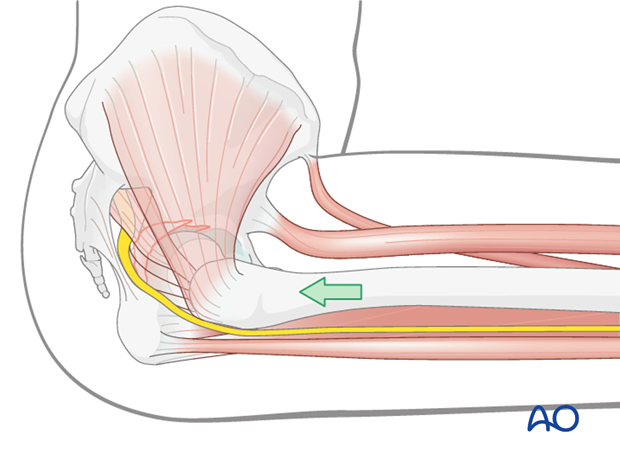
Mechanism of injury
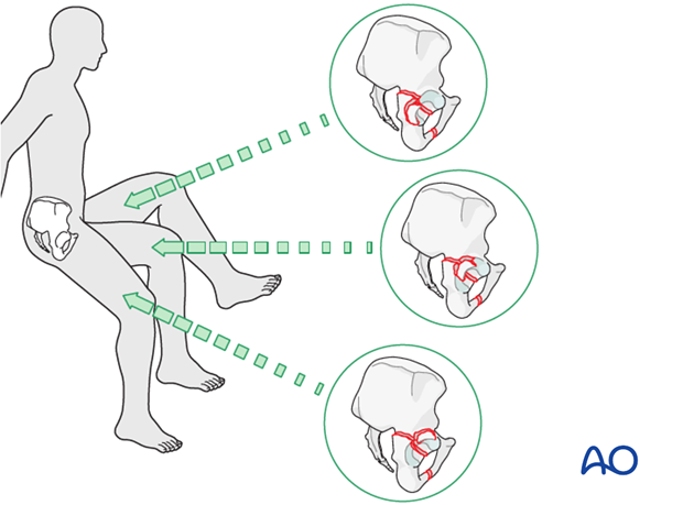
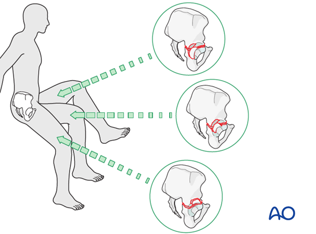
All posterior wall fracture types, elemental and associated, are commonly caused by a blow to the flexed knee on the dashboard in a car accident. The illustration shows a changing posterior wall injury that is dependent upon position of the knee.
Note
The knee is also at risk of injury, such as patellar fracture or posterior cruciate ligament tear.
Radiology
For posterior column and wall fractures the x-ray images show the disruption in the ilioischial line and a break in the obturator ring as well as the wall fragment on the obturator oblique view.
On the axial CT the posterior column fracture line is from medial to lateral through the posterior column. The posterior wall fracture line at the level of the joint is seen obliquely and the fragment size as well as any marginal impaction can be seen.
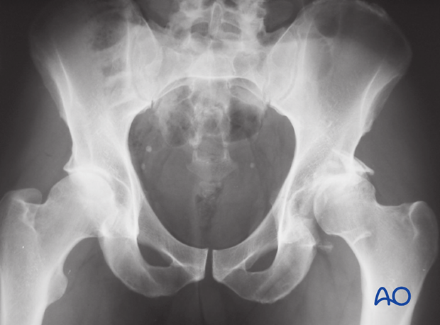
Case example 1: complex wall with undisplaced column
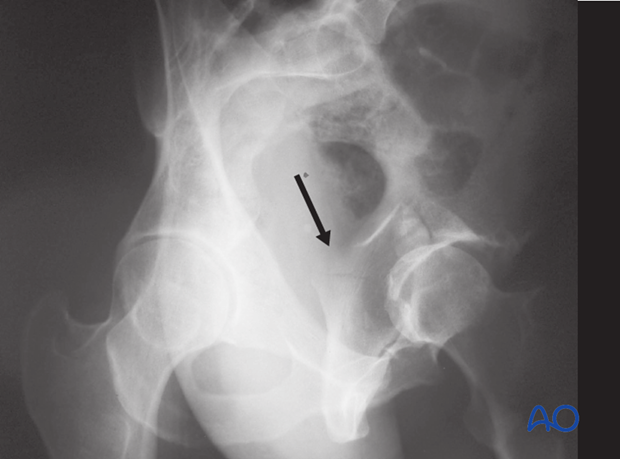
Complex fracture of the posterior wall and dislocation of the femoral head associated with nondisplaced fracture of the posterior column.
In the iliac oblique view, the disruption of the posterior column is shown by the arrow.
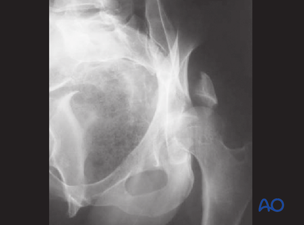
Case example 2: Displacement of both components
Associated posterior column and wall fracture pattern with displacement of both components.
Note that the posterior column is comminuted and the femoral head is dislocated.
2. Transverse and posterior wall
Definition
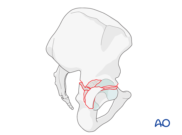
The association of the transverse plus posterior wall fracture combines a normal transverse configuration with one or more separate posterior wall fragments.
The transverse fracture could be transtectal, juxtatectal or infratectal.
Associated transverse plus posterior wall form the second largest group of associated fractures. There are two different patterns of injury.
In the first, the posterior wall is much more displaced than the transverse component, with likely posterior dislocation.
In the second, the transverse component is much more displaced than the posterior wall, with typically associated “central” dislocation.
Mechanism of the injury
Transverse and posterior wall fractures are commonly caused by a blow to the flexed knee on the dashboard in a car accident. The illustration shows a changing posterior wall injury that is dependent upon position of the knee.
Associated knee injuries should be excluded.
X-ray imaging
For transverse and posterior wall fractures one will see the disruption of both the ilioischial (red) and the iliopectineal lines (blue) but no break in the obturator ring. The wall fragment will be seen on the obturator oblique view.
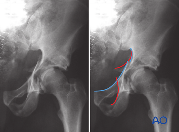
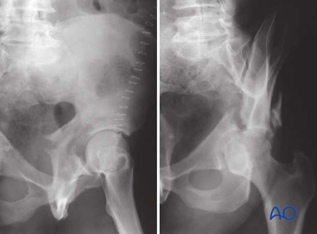
Iliac oblique and obturator oblique views
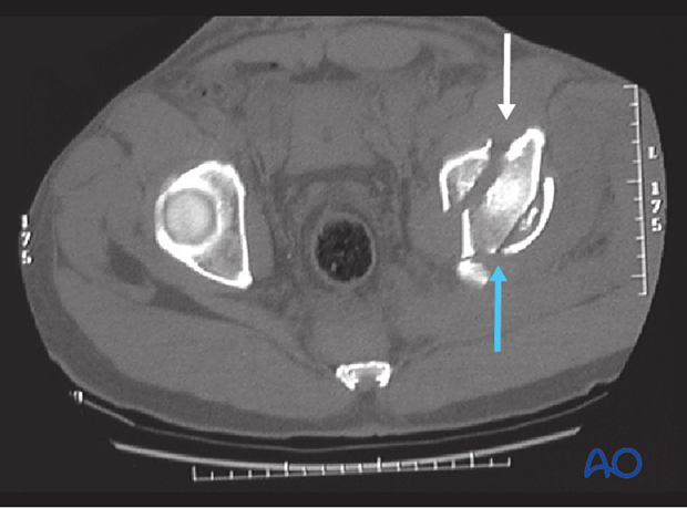
CT imaging
On the axial CT the transverse fracture line is from anterior to posterior (white arrow). The posterior wall fracture line (blue arrow) at the level of the joint is seen obliquely and the fragment size as well as any marginal impaction can be seen.
3. T-type
Definition
T-type fractures, though relatively uncommon, present unique surgical challenges.
This pattern combines a transverse component with a stem which exits either through the obturator ring or - in unusual cases - at various levels through the ischium.
Fractures in this group may also include a posterior wall component.
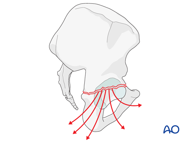
In contrast to the pure transverse fracture, the posterior and anterior column fractures frequently do not share a common orientation.
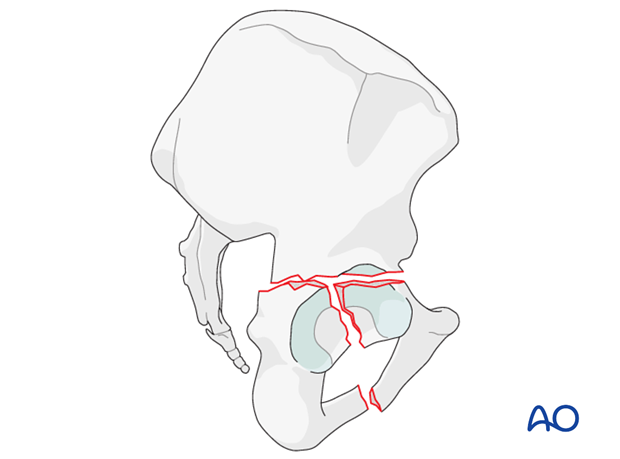
The downward limb of the T can be highly variable in its orientation.
X-ray imaging
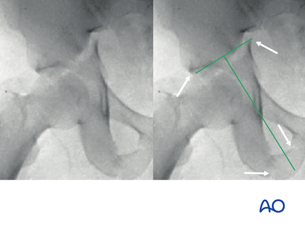
For T-type fractures, the standard x-ray images indicate disruption of both the ilioischial and the ilioinguinal lines as well as a break in the obturator ring.
CT imaging
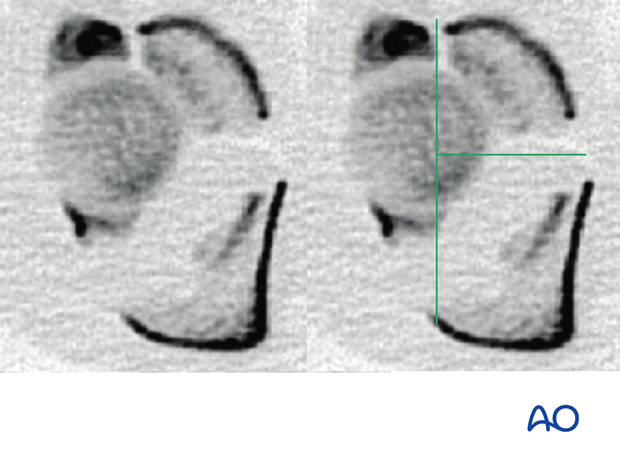
On the axial CT there is a transverse fracture line from anterior to posterior as well as separation of the two columns extending in the obturator ring.
4. Anterior column and posterior hemitransverse
Definition
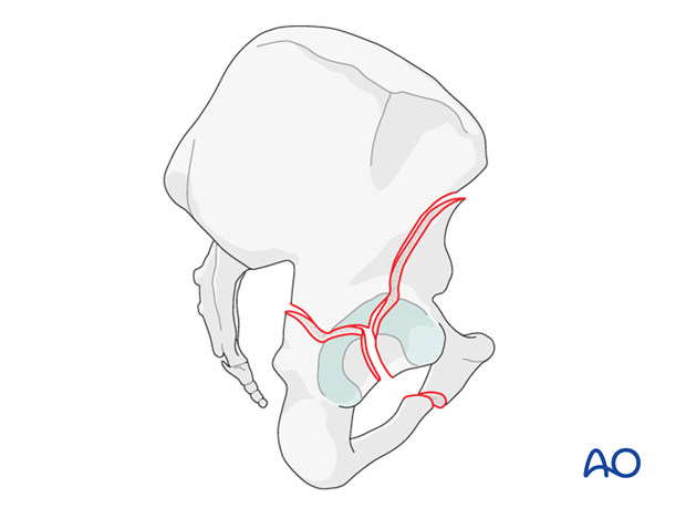
In this group of acetabular fractures, an anterior column or wall fracture is associated with a posterior hemitransverse fracture.
In addition, the femoral head is usually medially subluxed. Variations in this fracture pattern are based on whether the anterior column fracture exits at the iliac crest or inferior to the anterior inferior iliac spine.
The posterior column component varies on the basis of level of involvement in the greater sciatic notch.
The posterior column fracture morphology usually follows an oblique sagittal orientation unlike the posterior column involvement in a T-type fracture.
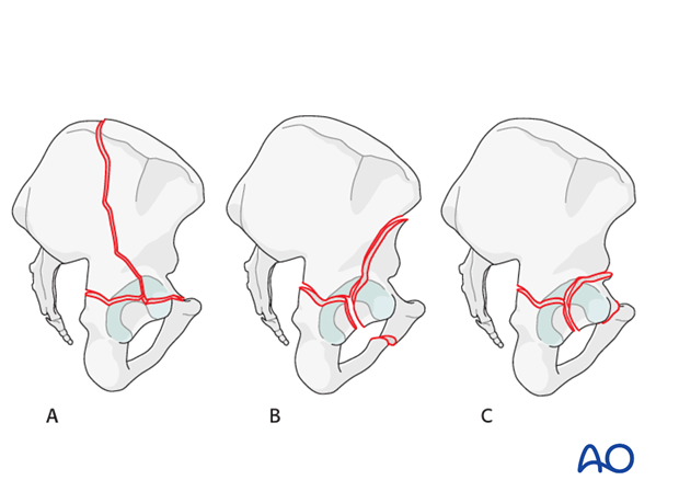
Fracture classification
The anterior column and posterior hemitransverse classification is based on the cranial exit of the fracture in the ilium, with high fractures (A) exiting above the iliac crest, intermediate fractures crossing between the spines (B), and low or very low fractures exit below the anterior inferior iliac spine. Also, the anterior portion may be an anterior wall fragment (C).
The posterior hemitransverse fracture may cross the greater sciatic notch at a variable level.
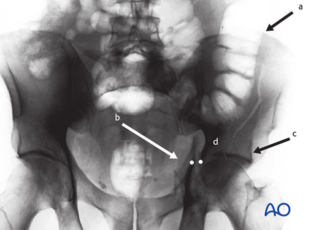
X-ray imaging
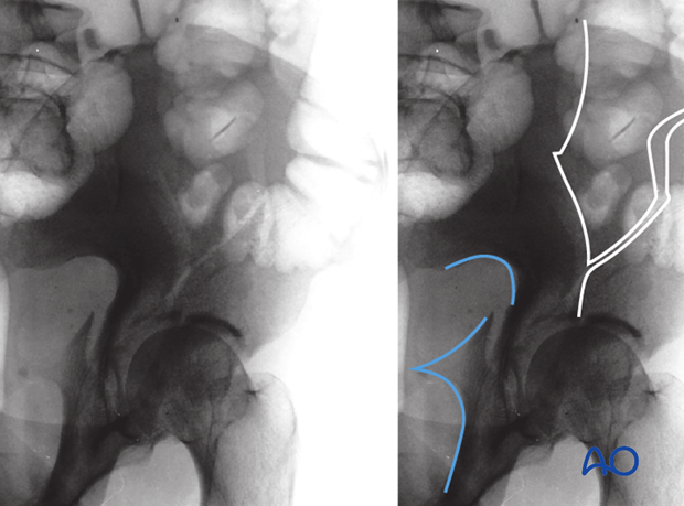
The AP radiograph demonstrates a high anterior column and posterior hemitransverse fracture. There is a disruption of the iliopectineal and the ilioischial lines at their confluence (dots). The cranial extent of the fracture exits the iliac crest (a). One can see significant displacement of the posterior column (b). A significant portion of the radiological roof is associated with the displaced anterior column (c). However, there is a portion of the radiological roof which remains associated with the intact iliac segment (d), distinguishing this fracture from an associated both column facture.
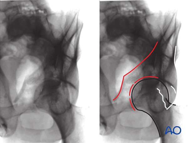
This can be seen very clearly on the obturator oblique. The femoral head remains congruent with the displaced anterior column (red). The intact portion of the acetabulum remains in its native location laterally (white).
The iliac oblique demonstrates a disruption between the posterior border of the bone and displacement of the posterior column. It also shows the entire profile of the fracture through the iliac wing and the portion of the articular surface remaining with the intact ilium.
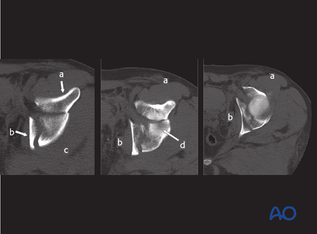
CT imaging
This CT scan demonstrates the anterior column fracture which is similar in character to that elementary anterior column fracture (fragment a). The posterior hemitransverse fracture follows an oblique sagittal orientation and there is a displacement of the posterior column fragment (b). This fracture line causes disruption of the retroacetabular surface distinguishing it from an elementary anterior column or wall fracture (fragment c). There is a segment of the radiological roof remaining intact with the uninjured innominate bone (d).
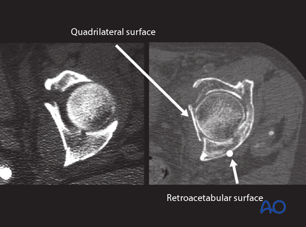
This family of fractures is commonly encountered in the geriatric patient population. In osteoporotic bone, the anterior column and posterior hemitransverse fracture is commonly associated with an individual fragment of the quadrilateral surface, and impaction injury involving the acetabular roof. Distinct to anterior column and posterior hemitransverse the fracture through posterior column violates the retroacetabular surface.
5. Both column
Definition
Although five of the ten fracture patterns involve both anterior and posterior columns, there is only one associated both column fracture.
The defining characteristic is that no part of the joint surface remains attached to the stable proximal part of the iliac wing and axial skeleton, such that it represents a complete articular fracture, in contrast to the other nine categories which are all partial articular.
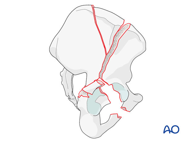
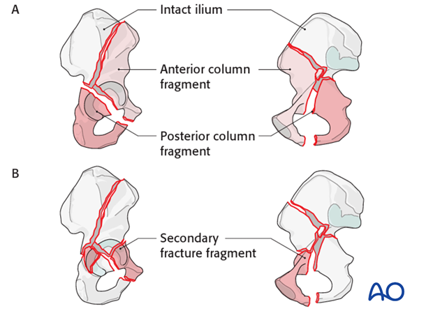
Associated both column fractures are formed by the association of two elementary fractures, the posterior column and the anterior column fractures (A).
In the majority of cases, the fractures become much more complex because the principle fragments are split by secondary fractures (B).
Some both column fractures may behave and be treated like more simple patterns such as an anterior column fracture due to the lack of displacement of the posterior elements.
The anterior column component
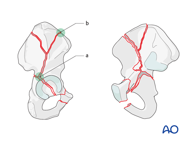
The anterior column is separated by a fracture, which begins at a variable point on the retroacetabular surface (a).
The fracture line then travels across the ilium. Like the elementary anterior column fractures, this fracture may exit the ilium at any point and defines the anterior fragment (high, medium, low, very low). The anterior column fractures exit cranial to the anterior superior iliac spine (b) in this example.
In type I both column fractures, per Letournel, the anterior column fracture line crosses the iliac crest.
There may be a separate fragment of the iliac crest (Y-shaped fracture).
On the internal surface, the fracture descends towards the pelvic brim. Very commonly, there is a separate fragment just above the pelvic brim where the anterior column fracture line crosses.
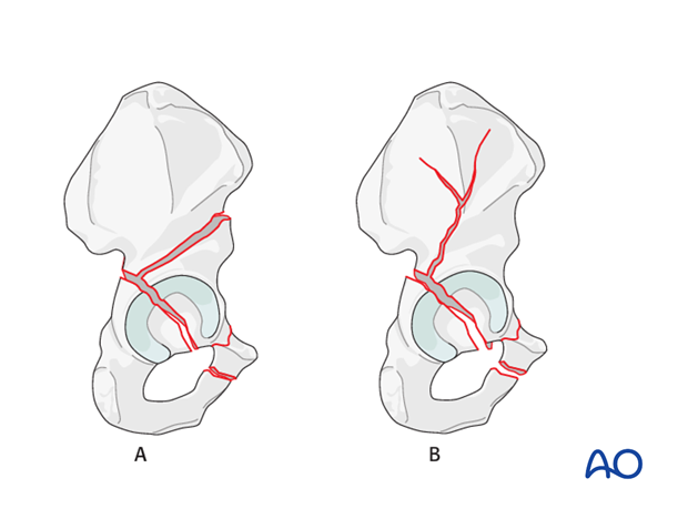
In type II both column fractures, the anterior column fracture line exits through the anterior portion of the iliac wing, at variable levels (A).
As in type I fractures, the posterior column fracture may be comminuted and/or at different levels.
The anterior column fracture may be incomplete and not exit the anterior ilium (B). This is particularly common in the elderly patient.
There may be an associated fragment of the quadrilateral surface, also common in the elderly patient.
Most of the acetabular roof stays with the anterior column fragments. As with posterior column and anterior wall fractures, marginal impaction may occur.
The posterior column component
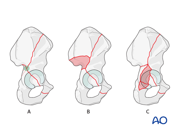
The posterior column is detached cranially by a fracture that begins at a variable point along the posterior border of the bone (A). Most commonly, the fracture line begins about the apex of the greater sciatic notch.
Complexities in the posterior column fragment include extension into the SI joint (B), segmental involvement of the posterior column, and associated fractures of the posterior wall (C).
These complexities of the posterior column fragment and additional involvement of the retroacetabular surface may render the fracture irreducible through the ilioinguinal approach, which does not give extensive access to the posterior column and external surface of the bone.
Based on the complexity of the posterior column fragment, one may consider using the extended iliofemoral approach for reduction.
X-ray imaging - AP view
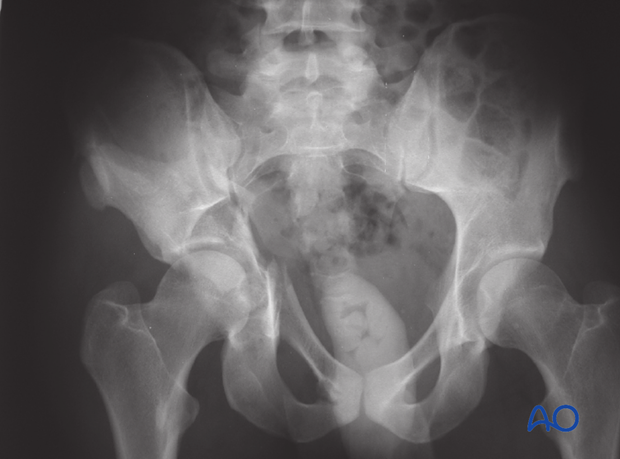
The AP view shows fracture lines crossing the iliopectineal line and anterior wall, iliac crest, and posterior wall and posterior column. There is medialization of the whole joint, with tilting of the free roof segment. There is also a fracture line through the obturator foramen.
Legend
- blue - iliopectineal line
- green - ilioischial line
- black - teardrop
- red - roof of acetabulum
- yellow - anterior border of acetabulum
- orange - posterior border of acetabulum
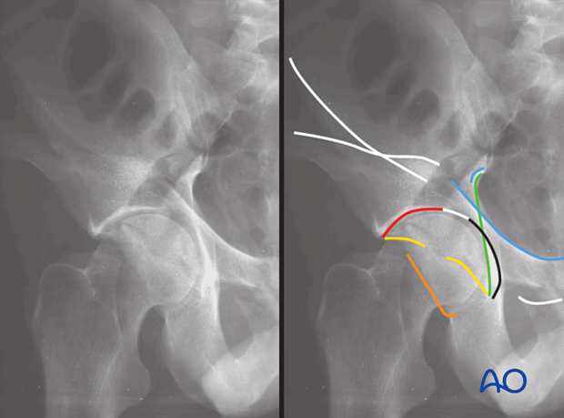
AP view of type I - variation: The iliopectineal line can be broken as well as the ilioischial line in several places. This indicates comminution with additional fractures along the anterior column or posterior column respectively.
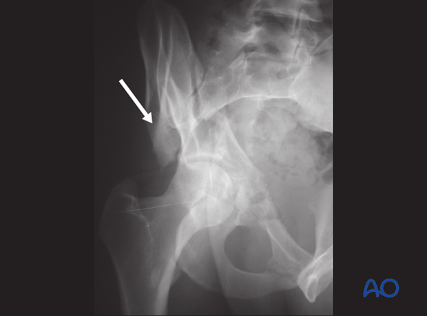
X-ray imaging - Obturator oblique view
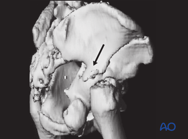
The obturator oblique view typically reveals the spur sign ( arrow).
Typical both column fractures displace the hip joint medially. The spur sign is the radiographic projection of the most distal (stable) part of the iliac wing, which remains attached to the axial skeleton through the SI joint.
The obturator oblique view clearly shows anterior column disruption along the iliopectineal line.
Fractures of the posterior wall, producing free fragments of the wall can usually be visualized best in the obturator oblique radiograph.
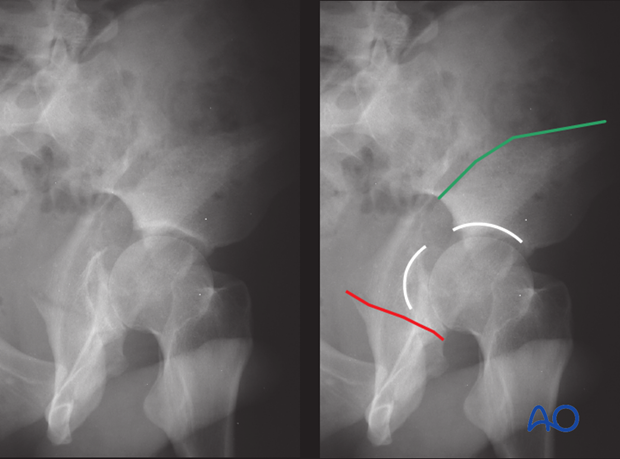
X-ray imaging - Iliac oblique view
The iliac oblique view shows where the anterior column fracture ends along the iliac crest or anterior border (green).
It demonstrates the location and orientation of fracture lines (red) within the posterior column.
Disruption of the acetabular roof (white) can be seen with a widely displaced fracture line.
CT imaging
On CT the full extent of the fracture lines can be seen. Some of these may be undisplaced and not require fixation such that although classified as a both column fracture the fixation may resemble as a less severe category.
CT imaging with multiplanar views or 3D reconstructions also aid evaluation of the spur sign ( arrow).
Variations of both column fracture patterns
The most common variants are as follows:
- Additional V-shaped fragment (1) proximally in the iliac wing
- Posterior wall fragment or additional fractures along the posterior column (2)
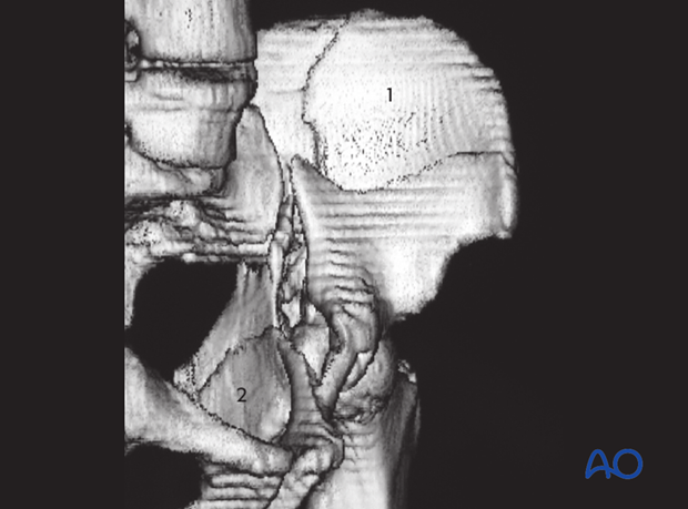
- Fracture line through the anterior wall that can result in a separated anterior wall fragment (3)
